JAMA Oncology ( IF 22.5 ) Pub Date : 2017-11-01 , DOI: 10.1001/jamaoncol.2017.1256 Nariya Cho 1 , Wonshik Han 2 , Boo-Kyung Han 3 , Min Sun Bae 1 , Eun Sook Ko 3 , Seok Jin Nam 4 , Eun Young Chae 5 , Jong Won Lee 6 , Sung Hun Kim 7 , Bong Joo Kang 7 , Byung Joo Song 8 , Eun-Kyung Kim 9 , Hee Jung Moon 9 , Seung Il Kim 10 , Sun Mi Kim 11 , Eunyoung Kang 12 , Yunhee Choi 13 , Hak Hee Kim 5 , Woo Kyung Moon 1
|
Importance Younger women (aged ≤50 years) who underwent breast conservation therapy may benefit from breast magnetic resonance imaging (MRI) screening as an adjunct to mammography.
Objective To prospectively determine the cancer yield and tumor characteristics of combined mammography with MRI or ultrasonography screening in women who underwent breast conservation therapy for breast cancers and who were 50 years or younger at initial diagnosis.
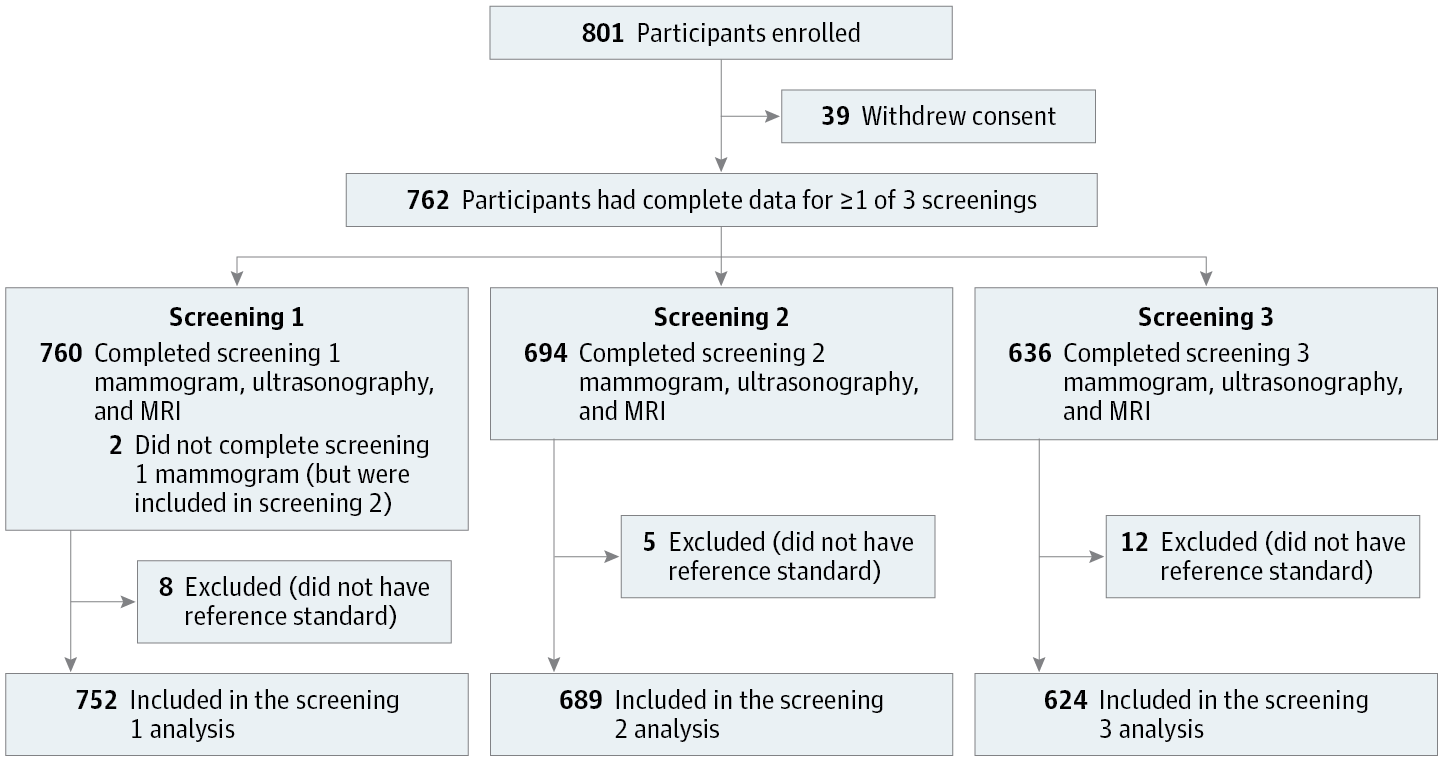
Design, Setting, and Participants This multicenter, prospective, nonrandomized study was conducted from December 1, 2010, to January 31, 2016, at 6 academic institutions. Seven hundred fifty-four women who were 50 years or younger at initial diagnosis and who had undergone breast conservation therapy for breast cancer were recruited to participate in the study. Reference standard was defined as a combination of pathology and 12-month follow-up.
Interventions Participants underwent 3 annual MRI screenings of the conserved and contralateral breasts in addition to mammography and ultrasonography, with independent readings.
Main Outcomes and Measures Cancer detection rate, sensitivity, specificity, interval cancer rate, and characteristics of detected cancers.
Results A total of 754 women underwent 2065 mammograms, ultrasonography, and MRI screenings. Seventeen cancers were diagnosed, and most of the detected cancers (13 of 17 [76%]) were stage 0 or stage 1. Overall cancer detection rate (8.2 vs 4.4 per 1000; P = .003) or sensitivity (100% vs 53%; P = .01) of mammography with MRI was higher than that of mammography alone. After the addition of ultrasonography, the cancer detection rate was higher than that by mammography alone (6.8 vs 4.4 per 1000; P = .03). The specificity of mammography with MRI or ultrasonography was lower than that by mammography alone (87% or 88% vs 96%; P < .001). No interval cancer was found.
Conclusions and Relevance After breast conservation therapy in women 50 years or younger, the addition of MRI to annual mammography screening improves detection of early-stage but biologically aggressive breast cancers at acceptable specificity. Results from this study can inform patient decision making on screening methods after breast conservation therapy.
中文翻译:
在诊断为 50 岁或以下并接受保乳治疗的女性中,使用乳房 X 线摄影加超声或磁共振成像进行乳腺癌筛查
重要性 接受保乳治疗的年轻女性(年龄≤50 岁)可能会受益于乳腺磁共振成像 (MRI) 筛查作为乳房 X 线摄影的辅助手段。
目的 前瞻性确定接受乳房保留治疗乳腺癌且初诊时年龄在 50 岁或以下的女性,联合乳房 X 线摄影与 MRI 或超声筛查的癌症检出率和肿瘤特征。
设计、地点和参与者 这项多中心、前瞻性、非随机研究于 2010 年 12 月 1 日至 2016 年 1 月 31 日在 6 个学术机构进行。招募了 754 名初始诊断为 50 岁或以下且因乳腺癌接受保乳治疗的女性参与研究。参考标准被定义为病理学和 12 个月随访的组合。
干预 除了乳房 X 光检查和超声检查外,参与者还接受了 3 次年度 MRI 筛查,包括乳房 X 光检查和超声检查,并进行独立读数。
主要成果和措施 癌症检出率、敏感性、特异性、间期癌症发生率和检出癌症的特征。
结果 共有 754 名女性接受了 2065 次乳房 X 线检查、超声检查和 MRI 筛查。诊断出 17 种癌症,大多数检测到的癌症(17 种中的 13 种 [76%])为 0 期或 1 期。总体癌症检出率(8.2 对 4.4/1000;P = .003)或敏感性(100% 对 53 %;P = .01) 与 MRI 的乳房 X 线摄影比单独的乳房 X 线摄影更高。加入超声检查后,癌症检出率高于单独乳房 X 线检查(6.8 vs 4.4 per 1000;P = .03)。用 MRI 或超声进行乳房 X 线摄影的特异性低于单独使用乳房 X 线摄影的特异性(87% 或 88% 对 96%;P < .001)。未发现间期癌。
结论和相关性 对 50 岁或以下女性进行保乳治疗后,在每年一次的乳房 X 线摄影筛查中增加 MRI 可以提高对早期但具有生物学侵袭性的乳腺癌的检出率,并且具有可接受的特异性。这项研究的结果可以为患者在保乳治疗后做出筛查方法的决策提供信息。


 京公网安备 11010802027423号