当前位置:
X-MOL 学术
›
J. Am. Coll. Cardiol.
›
论文详情
Our official English website, www.x-mol.net, welcomes your
feedback! (Note: you will need to create a separate account there.)
Cardiac MR With Late Gadolinium Enhancement in Acute Myocarditis With Preserved Systolic Function
Journal of the American College of Cardiology ( IF 21.7 ) Pub Date : 2017-10-01 , DOI: 10.1016/j.jacc.2017.08.044 Giovanni Donato Aquaro , Matteo Perfetti , Giovanni Camastra , Lorenzo Monti , Santo Dellegrottaglie , Claudio Moro , Alessia Pepe , Giancarlo Todiere , Chiara Lanzillo , Alessandra Scatteia , Mauro Di Roma , Gianluca Pontone , Martina Perazzolo Marra , Andrea Barison , Gianluca Di Bella
Journal of the American College of Cardiology ( IF 21.7 ) Pub Date : 2017-10-01 , DOI: 10.1016/j.jacc.2017.08.044 Giovanni Donato Aquaro , Matteo Perfetti , Giovanni Camastra , Lorenzo Monti , Santo Dellegrottaglie , Claudio Moro , Alessia Pepe , Giancarlo Todiere , Chiara Lanzillo , Alessandra Scatteia , Mauro Di Roma , Gianluca Pontone , Martina Perazzolo Marra , Andrea Barison , Gianluca Di Bella
|
BACKGROUND
The prognostic role of cardiac magnetic resonance (CMR) and late gadolinium enhancement (LGE) has not been clarified in acute myocarditis (AM) with preserved left ventricular (LV) ejection fraction (EF). OBJECTIVES
This study sought to evaluate the role of CMR and LGE in the prognosis of AM with preserved LVEF. METHODS
This study analyzed data from ITAMY (ITalian multicenter study on Acute MYocarditis) and evaluated CMR results from 386 patients (299 male; mean age 35 ± 15 years) with AM and preserved LVEF. Clinical follow-up was performed for a median of 1,572 days. A clinical combined endpoint of cardiac death, appropriate implantable cardioverter-defibrillator firing, resuscitated cardiac arrest, and hospitalization for heart failure was used. RESULTS
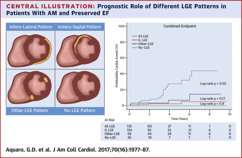
Among the 374 patients with suitable images, LGE involved the subepicardial layer inferior and lateral wall in 154 patients (41%; IL group), the midwall layer of the anteroseptal wall in 135 patients (36%; AS [anteroseptal] group), and other segments in 59 patients (16%; other-LGE group), and it was absent in 26 patients (no-LGE group). The AS group had a greater extent of LGE and a higher LV end-diastolic volume index than other groups, but levels of inflammatory markers were lower than in the other groups. Kaplan-Meier curve analysis indicated that the AS group had a worse prognosis than the other groups (p < 0.0001). Finally, in multivariable analysis, AS LGE was the best independent CMR predictor of the combined endpoint (odds ratio: 2.73; 95% confidence interval: 1.2 to 5.9; p = 0.01). CONCLUSIONS
In patients with AM and preserved LVEF, LGE in the midwall layer of the AS myocardial segment is associated with a worse prognosis than other patterns of presentation.
中文翻译:

保留收缩功能的急性心肌炎晚期钆增强的心脏 MR
背景 心脏磁共振 (CMR) 和晚期钆增强 (LGE) 在左心室 (LV) 射血分数 (EF) 保留的急性心肌炎 (AM) 中的预后作用尚未阐明。目的 本研究旨在评估 CMR 和 LGE 在保留 LVEF 的 AM 预后中的作用。方法 本研究分析了来自 ITAMY(意大利急性心肌炎多中心研究)的数据,并评估了 386 名患有 AM 和保留 LVEF 的患者(299 名男性;平均年龄 35 ± 15 岁)的 CMR 结果。临床随访的中位数为 1,572 天。使用了心源性死亡、适当的植入式心律转复除颤器触发、心脏骤停复苏和心力衰竭住院的临床综合终点。结果 在 374 例图像合适的患者中,LGE 累及心外膜下壁和侧壁 154 例(41%;IL 组),135 例患者(36%;AS [前间隔] 组)前间隔壁中壁层,59 例(16%)累及其他部分;其他-LGE组),26例患者(无LGE组)不存在。与其他组相比,AS 组具有更大程度的 LGE 和更高的 LV 舒张末期容积指数,但炎症标志物的水平低于其他组。Kaplan-Meier 曲线分析表明,AS 组的预后比其他组差(p < 0.0001)。最后,在多变量分析中,AS LGE 是联合终点的最佳独立 CMR 预测因子(优势比:2.73;95% 置信区间:1.2 至 5.9;p = 0.01)。结论 在 AM 和 LVEF 保留的患者中,
更新日期:2017-10-01
中文翻译:
保留收缩功能的急性心肌炎晚期钆增强的心脏 MR
背景 心脏磁共振 (CMR) 和晚期钆增强 (LGE) 在左心室 (LV) 射血分数 (EF) 保留的急性心肌炎 (AM) 中的预后作用尚未阐明。目的 本研究旨在评估 CMR 和 LGE 在保留 LVEF 的 AM 预后中的作用。方法 本研究分析了来自 ITAMY(意大利急性心肌炎多中心研究)的数据,并评估了 386 名患有 AM 和保留 LVEF 的患者(299 名男性;平均年龄 35 ± 15 岁)的 CMR 结果。临床随访的中位数为 1,572 天。使用了心源性死亡、适当的植入式心律转复除颤器触发、心脏骤停复苏和心力衰竭住院的临床综合终点。结果 在 374 例图像合适的患者中,LGE 累及心外膜下壁和侧壁 154 例(41%;IL 组),135 例患者(36%;AS [前间隔] 组)前间隔壁中壁层,59 例(16%)累及其他部分;其他-LGE组),26例患者(无LGE组)不存在。与其他组相比,AS 组具有更大程度的 LGE 和更高的 LV 舒张末期容积指数,但炎症标志物的水平低于其他组。Kaplan-Meier 曲线分析表明,AS 组的预后比其他组差(p < 0.0001)。最后,在多变量分析中,AS LGE 是联合终点的最佳独立 CMR 预测因子(优势比:2.73;95% 置信区间:1.2 至 5.9;p = 0.01)。结论 在 AM 和 LVEF 保留的患者中,


 京公网安备 11010802027423号