Our official English website, www.x-mol.net, welcomes your
feedback! (Note: you will need to create a separate account there.)
Three-dimensional microtissues as an in vitro model for personalized radiation therapy
Analyst ( IF 3.6 ) Pub Date : 2017-07-26 00:00:00 , DOI: 10.1039/c7an00794a Yuting Qiu 1, 2, 3, 4 , Dandan Ning 5, 6, 7, 8, 9 , Peipei Zhang 5, 6, 7, 8, 9 , Stephanie Curly 4, 10, 11, 12 , Yong Qiao 4, 10, 11, 12 , Liyuan Ma 1, 2, 3, 4, 5 , Ming Su 1, 2, 3, 4, 5
Analyst ( IF 3.6 ) Pub Date : 2017-07-26 00:00:00 , DOI: 10.1039/c7an00794a Yuting Qiu 1, 2, 3, 4 , Dandan Ning 5, 6, 7, 8, 9 , Peipei Zhang 5, 6, 7, 8, 9 , Stephanie Curly 4, 10, 11, 12 , Yong Qiao 4, 10, 11, 12 , Liyuan Ma 1, 2, 3, 4, 5 , Ming Su 1, 2, 3, 4, 5
Affiliation
|
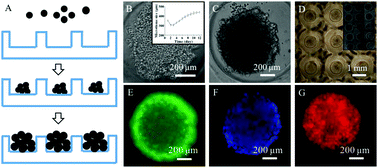
This paper describes the use of 3D microtissues as an intermediate model between the 2D cell culture and the animal model to assess radiation-induced cellular and DNA damage in the context of personalized radiation therapy. An agarose microwell array was used to generate 3D microtissues with controlled size and shape. The microtissues were exposed to X-ray radiation of various doses, and the radiation damage to cells was examined using a variety of techniques with different end points. Damage to cell membranes and reduction in metabolic activity were examined with the MTT assay and dye inclusion assay. DNA damage was tested with the micronucleus assay, γ-H2AX immunostaining, and HaloChip assay. 3D microtissues exposed to X-rays are smaller compared to unexposed ones in extended cultures, indicating that X-ray radiation can retard the growth of cells in 3D microtissues, where cells at the outer shells of microtissues can prevent further damage to those inside.
中文翻译:

三维微组织作为个性化放射治疗的体外模型
本文介绍了使用3D微组织作为2D细胞培养与动物模型之间的中间模型,以在个性化放射治疗的背景下评估辐射诱导的细胞和DNA损伤的方法。琼脂糖微孔阵列用于产生大小和形状可控的3D微组织。将微组织暴露于各种剂量的X射线辐射,并使用具有不同终点的多种技术检查对细胞的辐射损伤。用MTT测定法和染料包合测定法检查对细胞膜的损伤和代谢活性的降低。用微核试验,γ-H2AX免疫染色和HaloChip试验检测DNA损伤。与扩展培养中未暴露的3D微组织相比,暴露于X射线的3D微组织更小,
更新日期:2017-09-25
中文翻译:
三维微组织作为个性化放射治疗的体外模型
本文介绍了使用3D微组织作为2D细胞培养与动物模型之间的中间模型,以在个性化放射治疗的背景下评估辐射诱导的细胞和DNA损伤的方法。琼脂糖微孔阵列用于产生大小和形状可控的3D微组织。将微组织暴露于各种剂量的X射线辐射,并使用具有不同终点的多种技术检查对细胞的辐射损伤。用MTT测定法和染料包合测定法检查对细胞膜的损伤和代谢活性的降低。用微核试验,γ-H2AX免疫染色和HaloChip试验检测DNA损伤。与扩展培养中未暴露的3D微组织相比,暴露于X射线的3D微组织更小,


 京公网安备 11010802027423号