Our official English website, www.x-mol.net, welcomes your
feedback! (Note: you will need to create a separate account there.)
Tailored release of triiodothyronine and retinoic acid from a spatio-temporally fabricated nanofiber composite instigating neuronal differentiation
Nanoscale ( IF 5.8 ) Pub Date : 2017-08-28 00:00:00 , DOI: 10.1039/c7nr05918c Aishwarya Satish 1, 2, 3, 4 , Purna Sai Korrapati 1, 2, 3, 4
Nanoscale ( IF 5.8 ) Pub Date : 2017-08-28 00:00:00 , DOI: 10.1039/c7nr05918c Aishwarya Satish 1, 2, 3, 4 , Purna Sai Korrapati 1, 2, 3, 4
Affiliation

|
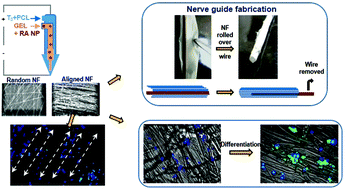
Regeneration of the central and peripheral nervous system is challenging since the functional restoration of injured nerves is an incredible task. The fabrication of an ideal nerve guide that fulfills the requirement to regenerate nerve tissue is a herculean challenge requiring a combination of both biochemical and topographical cues. The present study explores the combinatorial effect of aligned nanofibers and the regulated delivery of triiodothyronine and retinoic acid on nerve regeneration. A sequential release mechanism is adopted in fabricating the nanofiber scaffold, with triiodothyronine incorporated into the nanofiber shell ensuring its prior release, followed by retinoic acid (entrapped within zein nanoparticles) from the core. The composite nanofibers thus fabricated possess excellent mechanical, physical and thermal properties and good topographical morphology and were highly biocompatible. The nanofibers were scrutinized for their efficacy in stimulating differentiation to a neuronal phenotype. The elongation factor (E-factor) of the neural cells had doubled in the bioactive incorporated composite compared to other scaffolds, as observed on phalloidin staining of their cytoskeleton, which endorsed enhanced neural differentiation on the fabricated nanofiber scaffold. There was a significant increase in the expression of neural-lineage specific markers on investigation of mRNA by real time PCR, showing a 10 fold increase in the gene expression of β-III-tubulin, a 5.5 fold increase for microtubule associated protein 2 gene and 3.5 fold for neurofilament M gene in the cells cultured over bioactive incorporated aligned nanofiber composites. Similarly protein expression was analyzed by immunofluorescence and flow cytometry studies, which showed an increase in the expression of β-III-tubulin in the composite nanofiber. This corroborates that neuronal differentiation is enhanced by the aligned nanotopography and spatio-temporal delivery of triiodothyronine and retinoic acid, opening avenues for nerve regenerative graft fabrication.
中文翻译:

从时空制造的纳米纤维复合物中量身定制的三碘甲状腺氨酸和视黄酸释放,促进神经元分化
中枢和周围神经系统的再生具有挑战性,因为受损神经的功能恢复是一项不可思议的任务。满足再生神经组织要求的理想神经导管的制造是一项艰巨的挑战,需要结合生化和地形学提示。本研究探讨了排列的纳米纤维和三碘甲状腺素和视黄酸对神经再生的调节作用的组合作用。在制造纳米纤维支架中采用了顺序释放机制,将三碘甲腺氨酸掺入纳米纤维壳中以确保其事先释放,然后从核心中吸收视黄酸(被包裹在玉米醇溶蛋白纳米颗粒中)。这样制成的复合纳米纤维具有优异的机械性能,物理和热学性质以及良好的地形形态,并且具有高度的生物相容性。仔细检查纳米纤维在刺激分化为神经元表型方面的功效。伸长率(E与其他支架相比,生物活性掺入的复合物中神经细胞的因子增加了一倍,这是通过对它们的细胞骨架的鬼笔环肽染色观察到的,这认可了在制造的纳米纤维支架上神经分化的增强。实时PCR检测mRNA时,神经谱系特异性标志物的表达显着增加,表明β-III-微管蛋白的基因表达增加了10倍,微管相关蛋白2基因的表达增加了5.5倍,在通过生物活性并入排列的纳米纤维复合材料上培养的细胞中,神经丝M基因的3.5倍。类似地,通过免疫荧光和流式细胞术研究分析了蛋白质表达,这表明复合纳米纤维中β-III-微管蛋白的表达增加。
更新日期:2017-09-21
中文翻译:

从时空制造的纳米纤维复合物中量身定制的三碘甲状腺氨酸和视黄酸释放,促进神经元分化
中枢和周围神经系统的再生具有挑战性,因为受损神经的功能恢复是一项不可思议的任务。满足再生神经组织要求的理想神经导管的制造是一项艰巨的挑战,需要结合生化和地形学提示。本研究探讨了排列的纳米纤维和三碘甲状腺素和视黄酸对神经再生的调节作用的组合作用。在制造纳米纤维支架中采用了顺序释放机制,将三碘甲腺氨酸掺入纳米纤维壳中以确保其事先释放,然后从核心中吸收视黄酸(被包裹在玉米醇溶蛋白纳米颗粒中)。这样制成的复合纳米纤维具有优异的机械性能,物理和热学性质以及良好的地形形态,并且具有高度的生物相容性。仔细检查纳米纤维在刺激分化为神经元表型方面的功效。伸长率(E与其他支架相比,生物活性掺入的复合物中神经细胞的因子增加了一倍,这是通过对它们的细胞骨架的鬼笔环肽染色观察到的,这认可了在制造的纳米纤维支架上神经分化的增强。实时PCR检测mRNA时,神经谱系特异性标志物的表达显着增加,表明β-III-微管蛋白的基因表达增加了10倍,微管相关蛋白2基因的表达增加了5.5倍,在通过生物活性并入排列的纳米纤维复合材料上培养的细胞中,神经丝M基因的3.5倍。类似地,通过免疫荧光和流式细胞术研究分析了蛋白质表达,这表明复合纳米纤维中β-III-微管蛋白的表达增加。


 京公网安备 11010802027423号