当前位置:
X-MOL 学术
›
J. Mater. Chem. B
›
论文详情
Our official English website, www.x-mol.net, welcomes your
feedback! (Note: you will need to create a separate account there.)
Fluorescent microspheres for one-photon and two-photon imaging of mesenchymal stem cells
Journal of Materials Chemistry B ( IF 6.1 ) Pub Date : 2017-09-12 00:00:00 , DOI: 10.1039/c7tb01942d Qi Zhang 1, 2, 3, 4, 5 , Jihua Nie 1, 2, 3, 4, 5 , Hong Xu 5, 6, 7, 8, 9 , Yuyou Qiu 5, 6, 7, 8, 9 , Xiaoran Li 4, 10, 11, 12, 13 , Wei Gu 1, 2, 3, 4, 5 , Guangyu Tang 5, 6, 7, 8, 9 , Judong Luo 5, 14, 15, 16
Journal of Materials Chemistry B ( IF 6.1 ) Pub Date : 2017-09-12 00:00:00 , DOI: 10.1039/c7tb01942d Qi Zhang 1, 2, 3, 4, 5 , Jihua Nie 1, 2, 3, 4, 5 , Hong Xu 5, 6, 7, 8, 9 , Yuyou Qiu 5, 6, 7, 8, 9 , Xiaoran Li 4, 10, 11, 12, 13 , Wei Gu 1, 2, 3, 4, 5 , Guangyu Tang 5, 6, 7, 8, 9 , Judong Luo 5, 14, 15, 16
Affiliation
|
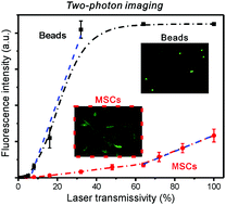
Stem cell-mediated therapy has emerged as a novel regenerative treatment for tissue repair in the last decade. However, noninvasive monitoring of stem cells remains a grand challenge for stem cell-based regenerative medicine for the complete understanding of the function and the behaviors of cells. Herein, acridine orange, a fluorescent dye was encapsulated into polymer nanospheres based on double emulsions. The fluorescent nanospheres with a diameter of around 200 nm were incubated with human mesenchymal stem cells (hMSCs) to produce nanosphere-endocytosed hMSCs. The fluorescent imaging of hMSCs can be directly and clearly captured using confocal microscopy in the one-photon and two-photon modes. The results indicated that the hMSCs adhered and spread well on the surface of the scaffolds with a high population and good distribution. Meanwhile, polystyrene microspheres were stained with dye complexes by using a gradual solvent evaporation method. The as-prepared fluorescent beads exhibited bright and multiple colors and a broad emission spectrum ranging from 500 to 800 nm, which was further used to quantitatively evaluate the one-photon and two-photon fluorescent signals of the hMSCs. Our study offers the possibility of direct monitoring of stem cells with high resolution, and encourages future quantitative clinical assessment in imaging-guided cell therapies.
中文翻译:

荧光微球用于间充质干细胞的单光子和双光子成像
在过去的十年中,干细胞介导的治疗已成为一种新的组织修复再生疗法。然而,对干细胞的无创性监测对于完全了解细胞的功能和行为仍然是基于干细胞的再生医学的巨大挑战。在此,基于双重乳液将荧光染料a啶橙封装到聚合物纳米球中。将直径约200 nm的荧光纳米球与人间充质干细胞(hMSC)孵育,以产生纳米球内吞的hMSC。hMSC的荧光成像可以使用共聚焦显微镜在单光子和双光子模式下直接清晰地捕获。结果表明,hMSCs在支架表面上粘附并良好地分布,具有高种群和良好分布。同时,聚苯乙烯微球通过使用逐步溶剂蒸发法用染料配合物染色。所制备的荧光珠表现出明亮和多种颜色以及在500至800 nm范围内的宽发射光谱,该光谱进一步用于定量评估hMSC的单光子和双光子荧光信号。我们的研究提供了以高分辨率直接监测干细胞的可能性,并鼓励将来在影像引导的细胞疗法中进行定量的临床评估。进一步用于定量评估hMSC的单光子和双光子荧光信号。我们的研究提供了以高分辨率直接监测干细胞的可能性,并鼓励将来在影像引导的细胞疗法中进行定量的临床评估。进一步用于定量评估hMSC的单光子和双光子荧光信号。我们的研究提供了以高分辨率直接监测干细胞的可能性,并鼓励将来在影像引导的细胞疗法中进行定量的临床评估。
更新日期:2017-09-20
中文翻译:
荧光微球用于间充质干细胞的单光子和双光子成像
在过去的十年中,干细胞介导的治疗已成为一种新的组织修复再生疗法。然而,对干细胞的无创性监测对于完全了解细胞的功能和行为仍然是基于干细胞的再生医学的巨大挑战。在此,基于双重乳液将荧光染料a啶橙封装到聚合物纳米球中。将直径约200 nm的荧光纳米球与人间充质干细胞(hMSC)孵育,以产生纳米球内吞的hMSC。hMSC的荧光成像可以使用共聚焦显微镜在单光子和双光子模式下直接清晰地捕获。结果表明,hMSCs在支架表面上粘附并良好地分布,具有高种群和良好分布。同时,聚苯乙烯微球通过使用逐步溶剂蒸发法用染料配合物染色。所制备的荧光珠表现出明亮和多种颜色以及在500至800 nm范围内的宽发射光谱,该光谱进一步用于定量评估hMSC的单光子和双光子荧光信号。我们的研究提供了以高分辨率直接监测干细胞的可能性,并鼓励将来在影像引导的细胞疗法中进行定量的临床评估。进一步用于定量评估hMSC的单光子和双光子荧光信号。我们的研究提供了以高分辨率直接监测干细胞的可能性,并鼓励将来在影像引导的细胞疗法中进行定量的临床评估。进一步用于定量评估hMSC的单光子和双光子荧光信号。我们的研究提供了以高分辨率直接监测干细胞的可能性,并鼓励将来在影像引导的细胞疗法中进行定量的临床评估。


 京公网安备 11010802027423号