当前位置:
X-MOL 学术
›
J. Mater. Chem. B
›
论文详情
Our official English website, www.x-mol.net, welcomes your
feedback! (Note: you will need to create a separate account there.)
Heterogeneity of mesenchymal and pluripotent stem cell populations grown on nanogrooves and nanopillars
Journal of Materials Chemistry B ( IF 6.1 ) Pub Date : 2017-08-23 00:00:00 , DOI: 10.1039/c7tb01878a Peng-Yuan Wang 1, 2, 3, 4, 5 , Sheryl Ding 1, 2, 3, 4, 5 , Huseyin Sumer 1, 2, 3, 4, 5 , Raymond Ching-Bong Wong 6, 7, 8, 9 , Peter Kingshott 1, 2, 3, 4, 5
Journal of Materials Chemistry B ( IF 6.1 ) Pub Date : 2017-08-23 00:00:00 , DOI: 10.1039/c7tb01878a Peng-Yuan Wang 1, 2, 3, 4, 5 , Sheryl Ding 1, 2, 3, 4, 5 , Huseyin Sumer 1, 2, 3, 4, 5 , Raymond Ching-Bong Wong 6, 7, 8, 9 , Peter Kingshott 1, 2, 3, 4, 5
Affiliation
|
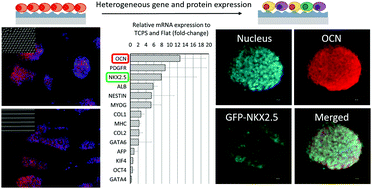
Surface nanotopographies are an important way of mimicking the stem cell niche on biomaterial surfaces. Previous studies have focused on the differentiation of stem cells into a defined lineage using nanotopographies, but they have rarely considered the homogeneity of cell populations produced. We examined the impact of two types of substrates (i.e. nanogrooves and nanopillars made by soft lithography) on the surface-induced differentiation of human amniotic membrane-derived mesenchymal stem cells (hAM-MSCs) and mouse embryonic stem cells (mESCs) without the use of additional chemical induction medium components. Cell morphology and proliferation were analysed at day 1 and day 3. Gene expression was analysed at day 14 for hAM-MSCs and at day 7 for mESC-derived embryoid bodies (mEBs) using quantitative real-time polymerase chain reaction (qPCR). The substrates with nanogrooves had a noticeable effect on cell alignment in a depth dependent manner with both cell types showing strong alignment along the deep grooves. On the other hand, the nanopillar substrates showed inhibition of cell spreading for both cell types. The nanogrooves showed inhibition of hAM-MSC growth but enhanced mEB proliferation, especially on the deeper grooves. The nanopillars did not significantly affect hAM-MSC growth, but can modulate mEB growth depending on the pillar density, indicating that mEBs are more sensitive to nanotopographies in terms of proliferation, while hAM-MSCs are only sensitive to specific structures and sizes. Genes associated with bone, cartilage, and fat were investigated for hAM-MSCs, whereas genes of the endoderm, mesoderm, ectoderm, and pluripotency were investigated for mEBs. In general, gene expression for hAM-MSCs was not enhanced significantly by the nanotopographies compared to the flat control. On the other hand, genes of bone, cartilage, skeletal muscle, heart, and liver were up-regulated on both nanopillars and nanogrooves, especially OP65 (ordered pillars with 65% density) and SG40 (shallow grooves with 40 nm depth) in a feature size dependent manner. We found that a small portion of mEBs was composed of cardiac-like beating cells (i.e. GFP-NKX2.5 positive) and a bone cell marker (i.e. OCN) indicating a heterogeneous cell population being generated on those types of surfaces. This work highlights the importance of nanotopographies in stem cell differentiation and how studying multiple properties of the substrate and cells is needed as we strive to generate homogeneous and mature cell populations using biomaterials.
中文翻译:

在纳米槽和纳米柱上生长的间充质和多能干细胞群体的异质性
表面纳米形貌是模仿生物材料表面上干细胞生态位的重要方法。先前的研究集中于使用纳米形貌学将干细胞分化为确定的谱系,但他们很少考虑所产生细胞群体的均质性。我们研究了两种基材的影响(即纳米凹槽和通过软光刻法制成的纳米柱)在不使用其他化学诱导培养基成分的情况下,对人羊膜来源的间充质干细胞(hAM-MSC)和小鼠胚胎干细胞(mESCs)的表面诱导分化产生了影响。在第1天和第3天分析细胞的形态和增殖。使用定量实时聚合酶链反应(qPCR),在第14天分析hAM-MSC的基因表达,在第7天分析mESC来源的胚状体(mEBs)的基因表达。具有纳米凹槽的基底以深度依赖的方式对细胞排列具有显着影响,两种细胞类型均显示出沿深槽的强烈排列。另一方面,对于两种细胞类型,纳米柱基质均显示出对细胞扩散的抑制作用。纳米槽显示出抑制hAM-MSC的生长,但增强了mEB的增殖,特别是在深沟处。纳米柱不会显着影响hAM-MSC的生长,但可以根据柱密度来调节mEB的生长,表明mEB在增殖方面对纳米形貌更敏感,而hAM-MSC仅对特定的结构和大小敏感。对于hAM-MSC,研究了与骨骼,软骨和脂肪相关的基因,而对于mEB,研究了内胚层,中胚层,外胚层和多能性的基因。通常,与平面对照相比,纳米形貌并未显着增强hAM-MSC的基因表达。另一方面,纳米柱和纳米槽上的骨骼,软骨,骨骼肌,心脏和肝脏的基因均上调,尤其是OP65(密度为65%的有序柱子)和SG40(深度为40 nm的浅沟槽)的特征尺寸依赖方式。我们发现一小部分mEB由心脏样搏动细胞组成(例如GFP-NKX2.5阳性)和骨细胞标记(即OCN),表明在这些类型的表面上生成了异种细胞群。这项工作强调了纳米形貌在干细胞分化中的重要性,以及在我们努力使用生物材料生成均质且成熟的细胞群体时,如何研究底物和细胞的多种特性的重要性。
更新日期:2017-09-20
中文翻译:
在纳米槽和纳米柱上生长的间充质和多能干细胞群体的异质性
表面纳米形貌是模仿生物材料表面上干细胞生态位的重要方法。先前的研究集中于使用纳米形貌学将干细胞分化为确定的谱系,但他们很少考虑所产生细胞群体的均质性。我们研究了两种基材的影响(即纳米凹槽和通过软光刻法制成的纳米柱)在不使用其他化学诱导培养基成分的情况下,对人羊膜来源的间充质干细胞(hAM-MSC)和小鼠胚胎干细胞(mESCs)的表面诱导分化产生了影响。在第1天和第3天分析细胞的形态和增殖。使用定量实时聚合酶链反应(qPCR),在第14天分析hAM-MSC的基因表达,在第7天分析mESC来源的胚状体(mEBs)的基因表达。具有纳米凹槽的基底以深度依赖的方式对细胞排列具有显着影响,两种细胞类型均显示出沿深槽的强烈排列。另一方面,对于两种细胞类型,纳米柱基质均显示出对细胞扩散的抑制作用。纳米槽显示出抑制hAM-MSC的生长,但增强了mEB的增殖,特别是在深沟处。纳米柱不会显着影响hAM-MSC的生长,但可以根据柱密度来调节mEB的生长,表明mEB在增殖方面对纳米形貌更敏感,而hAM-MSC仅对特定的结构和大小敏感。对于hAM-MSC,研究了与骨骼,软骨和脂肪相关的基因,而对于mEB,研究了内胚层,中胚层,外胚层和多能性的基因。通常,与平面对照相比,纳米形貌并未显着增强hAM-MSC的基因表达。另一方面,纳米柱和纳米槽上的骨骼,软骨,骨骼肌,心脏和肝脏的基因均上调,尤其是OP65(密度为65%的有序柱子)和SG40(深度为40 nm的浅沟槽)的特征尺寸依赖方式。我们发现一小部分mEB由心脏样搏动细胞组成(例如GFP-NKX2.5阳性)和骨细胞标记(即OCN),表明在这些类型的表面上生成了异种细胞群。这项工作强调了纳米形貌在干细胞分化中的重要性,以及在我们努力使用生物材料生成均质且成熟的细胞群体时,如何研究底物和细胞的多种特性的重要性。


 京公网安备 11010802027423号