PLoS Pathogens ( IF 5.5 ) Pub Date : 2017-09-19 , DOI: 10.1371/journal.ppat.1006609 Linya Wang , Ja Yeon Kim , Helene Minyi Liu , Michael M. C. Lai , Jing-hsiung James Ou
|
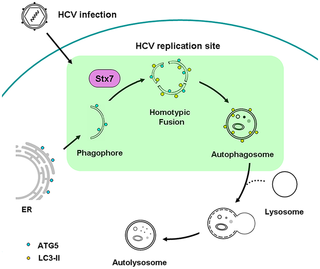
Hepatitis C virus (HCV) induces autophagy to promote its replication, including its RNA replication, which can take place on double-membrane vesicles known as autophagosomes. However, how HCV induces the biogenesis of autophagosomes and how HCV RNA replication complex may be assembled on autophagosomes were largely unknown. During autophagy, crescent membrane structures known as phagophores first appear in the cytoplasm, which then progress to become autophagosomes. By conducting electron microscopy and in vitro membrane fusion assay, we found that phagophores induced by HCV underwent homotypic fusion to generate autophagosomes in a process dependent on the SNARE protein syntaxin 7 (STX7). Further analyses by live-cell imaging and fluorescence microscopy indicated that HCV-induced phagophores originated from the endoplasmic reticulum (ER). Interestingly, comparing with autophagy induced by nutrient starvation, the progression of phagophores to autophagosomes induced by HCV took significantly longer time, indicating fundamental differences in the biogenesis of autophagosomes induced by these two different stimuli. As the knockdown of STX7 to inhibit the formation of autophagosomes did not affect HCV RNA replication, and purified phagophores could mediate HCV RNA replication, the assembly of the HCV RNA replication complex on autophagosomes apparently took place during the formative stage of phagophores. These findings provided important information for understanding how HCV controlled and modified this important cellular pathway for its own replication.
中文翻译:
HCV诱导的自噬体是通过介导HCV RNA复制的吞噬细胞的同型融合产生的
丙型肝炎病毒(HCV)诱导自噬以促进其复制,包括其RNA复制,这种复制可以发生在称为自噬体的双膜囊泡上。但是,HCV如何诱导自噬体的生物发生以及HCV RNA复制复合物如何在自噬体上组装尚不清楚。在自噬过程中,被称为吞噬细胞的新月形膜结构首先出现在细胞质中,然后发展成为自噬体。通过电子显微镜和体外膜融合测定法中,我们发现HCV诱导的吞噬细胞在依赖SNARE蛋白语法7(STX7)的过程中经历了同型融合产生自噬体。通过活细胞成像和荧光显微镜进行的进一步分析表明,HCV诱导的吞噬细胞起源于内质网(ER)。有趣的是,与营养饥饿引起的自噬相比,HCV诱导的噬菌体向自噬体的发展花费了更长的时间,这表明这两种不同刺激所诱导的自噬体在生物发生中的根本差异。由于抑制抑制自噬小体形成的STX7不会影响HCV RNA复制,纯化的噬菌体可以介导HCV RNA复制,HCV RNA复制复合物在自噬体上的组装显然发生在吞噬细胞的形成阶段。这些发现为理解HCV如何控制和修饰这一重要的细胞途径以使其自身复制提供了重要信息。


 京公网安备 11010802027423号