当前位置:
X-MOL 学术
›
ACS Biomater. Sci. Eng.
›
论文详情
Our official English website, www.x-mol.net, welcomes your
feedback! (Note: you will need to create a separate account there.)
A Bioinformatics 3D Cellular Morphotyping Strategy for Assessing Biomaterial Scaffold Niches
ACS Biomaterials Science & Engineering ( IF 5.4 ) Pub Date : 2017-09-19 00:00:00 , DOI: 10.1021/acsbiomaterials.7b00473 Stephen J. Florczyk , Mylene Simon , Derek Juba , P. Scott Pine , Sumona Sarkar , Desu Chen 1 , Paula J. Baker , Subhadip Bodhak , Antonio Cardone , Mary C. Brady , Peter Bajcsy , Carl G. Simon
ACS Biomaterials Science & Engineering ( IF 5.4 ) Pub Date : 2017-09-19 00:00:00 , DOI: 10.1021/acsbiomaterials.7b00473 Stephen J. Florczyk , Mylene Simon , Derek Juba , P. Scott Pine , Sumona Sarkar , Desu Chen 1 , Paula J. Baker , Subhadip Bodhak , Antonio Cardone , Mary C. Brady , Peter Bajcsy , Carl G. Simon
Affiliation
|
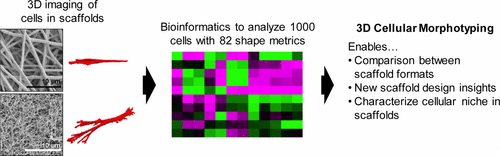
Many biomaterial scaffolds have been advanced to provide synthetic cell niches for tissue engineering and drug screening applications; however, current methods for comparing scaffold niches focus on cell functional outcomes or attempt to normalize materials properties between different scaffold formats. We demonstrate a three-dimensional (3D) cellular morphotyping strategy for comparing biomaterial scaffold cell niches between different biomaterial scaffold formats. Primary human bone marrow stromal cells (hBMSCs) were cultured on 8 different biomaterial scaffolds, including fibrous scaffolds, hydrogels, and porous sponges, in 10 treatment groups to compare a variety of biomaterial scaffolds and cell morphologies. A bioinformatics approach was used to determine the 3D cellular morphotype for each treatment group by using 82 shape metrics to analyze approximately 1000 cells. We found that hBMSCs cultured on planar substrates yielded planar cell morphotypes, while those cultured in 3D scaffolds had elongated or equiaxial cellular morphotypes with greater height. Multivariate analysis was effective at distinguishing mean shapes of cells in flat substrates from cells in scaffolds, as was the metric L1-depth (the cell height along its shortest axis after aligning cells with a characteristic ellipsoid). The 3D cellular morphotyping technique enables direct comparison of cellular microenvironments between widely different types of scaffolds and design of scaffolds based on cell structure–function relationships.
中文翻译:

用于评估生物材料支架壁ches的生物信息学3D细胞形态学策略
已经开发了许多生物材料支架,以提供用于组织工程和药物筛选应用的合成细胞壁ni。然而,目前用于比较支架壁ches的方法集中于细胞功能结果或试图标准化不同支架形式之间的材料性质。我们展示了一种三维(3D)细胞形态分型策略,用于比较不同生物材料支架形式之间的生物材料支架细胞壁ches。将人类原代骨髓基质细胞(hBMSC)培养在8个不同的生物材料支架上,包括纤维支架,水凝胶和多孔海绵,在10个治疗组中进行比较,以比较各种生物材料支架和细胞形态。通过使用82种形状指标来分析大约1000个细胞,使用了生物信息学方法来确定每个治疗组的3D细胞形态。我们发现,在平面基底上培养的hBMSC产生平面细胞形态,而在3D支架中培养的hBMSC具有细长或等轴的细胞形态,且高度更高。多变量分析可有效区分平坦基质中细胞的平均形状和支架中细胞的平均形状,如度量L1深度(将单元格与特征椭球对齐后沿其最短轴的单元格高度)。3D细胞形态分型技术可直接比较广泛不同类型的支架之间的细胞微环境,并基于细胞结构与功能的关系设计支架。
更新日期:2017-09-19
中文翻译:
用于评估生物材料支架壁ches的生物信息学3D细胞形态学策略
已经开发了许多生物材料支架,以提供用于组织工程和药物筛选应用的合成细胞壁ni。然而,目前用于比较支架壁ches的方法集中于细胞功能结果或试图标准化不同支架形式之间的材料性质。我们展示了一种三维(3D)细胞形态分型策略,用于比较不同生物材料支架形式之间的生物材料支架细胞壁ches。将人类原代骨髓基质细胞(hBMSC)培养在8个不同的生物材料支架上,包括纤维支架,水凝胶和多孔海绵,在10个治疗组中进行比较,以比较各种生物材料支架和细胞形态。通过使用82种形状指标来分析大约1000个细胞,使用了生物信息学方法来确定每个治疗组的3D细胞形态。我们发现,在平面基底上培养的hBMSC产生平面细胞形态,而在3D支架中培养的hBMSC具有细长或等轴的细胞形态,且高度更高。多变量分析可有效区分平坦基质中细胞的平均形状和支架中细胞的平均形状,如度量L1深度(将单元格与特征椭球对齐后沿其最短轴的单元格高度)。3D细胞形态分型技术可直接比较广泛不同类型的支架之间的细胞微环境,并基于细胞结构与功能的关系设计支架。


 京公网安备 11010802027423号