Our official English website, www.x-mol.net, welcomes your
feedback! (Note: you will need to create a separate account there.)
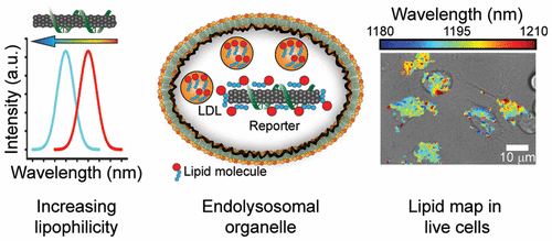
A Carbon Nanotube Optical Reporter Maps Endolysosomal Lipid Flux
ACS Nano ( IF 15.8 ) Pub Date : 2017-09-12 00:00:00 , DOI: 10.1021/acsnano.7b04743 Prakrit V Jena 1 , Daniel Roxbury 2 , Thomas V Galassi 1, 3 , Leila Akkari 1, 4 , Christopher P Horoszko 1, 3 , David B Iaea 3 , Januka Budhathoki-Uprety 1 , Nina Pipalia 3 , Abigail S Haka 3 , Jackson D Harvey 1, 3 , Jeetain Mittal 5 , Frederick R Maxfield 3 , Johanna A Joyce 1, 3, 6 , Daniel A Heller 1, 3
ACS Nano ( IF 15.8 ) Pub Date : 2017-09-12 00:00:00 , DOI: 10.1021/acsnano.7b04743 Prakrit V Jena 1 , Daniel Roxbury 2 , Thomas V Galassi 1, 3 , Leila Akkari 1, 4 , Christopher P Horoszko 1, 3 , David B Iaea 3 , Januka Budhathoki-Uprety 1 , Nina Pipalia 3 , Abigail S Haka 3 , Jackson D Harvey 1, 3 , Jeetain Mittal 5 , Frederick R Maxfield 3 , Johanna A Joyce 1, 3, 6 , Daniel A Heller 1, 3
Affiliation
|
Lipid accumulation within the lumen of endolysosomal vesicles is observed in various pathologies including atherosclerosis, liver disease, neurological disorders, lysosomal storage disorders, and cancer. Current methods cannot measure lipid flux specifically within the lysosomal lumen of live cells. We developed an optical reporter, composed of a photoluminescent carbon nanotube of a single chirality, that responds to lipid accumulation via modulation of the nanotube’s optical band gap. The engineered nanomaterial, composed of short, single-stranded DNA and a single nanotube chirality, localizes exclusively to the lumen of endolysosomal organelles without adversely affecting cell viability or proliferation or organelle morphology, integrity, or function. The emission wavelength of the reporter can be spatially resolved from within the endolysosomal lumen to generate quantitative maps of lipid content in live cells. Endolysosomal lipid accumulation in cell lines, an example of drug-induced phospholipidosis, was observed for multiple drugs in macrophages, and measurements of patient-derived Niemann–Pick type C fibroblasts identified lipid accumulation and phenotypic reversal of this lysosomal storage disease. Single-cell measurements using the reporter discerned subcellular differences in equilibrium lipid content, illuminating significant intracellular heterogeneity among endolysosomal organelles of differentiating bone-marrow-derived monocytes. Single-cell kinetics of lipoprotein-derived cholesterol accumulation within macrophages revealed rates that differed among cells by an order of magnitude. This carbon nanotube optical reporter of endolysosomal lipid content in live cells confers additional capabilities for drug development processes and the investigation of lipid-linked diseases.
中文翻译:

碳纳米管光学报告基因绘制内溶酶体脂质通量图
内溶酶体囊泡腔内的脂质积累在多种病理学中观察到,包括动脉粥样硬化、肝脏疾病、神经系统疾病、溶酶体贮积症和癌症。目前的方法无法测量活细胞溶酶体腔内的脂质通量。我们开发了一种光学报告器,由具有单一手性的光致发光碳纳米管组成,通过调节纳米管的光学带隙来响应脂质积累。这种工程纳米材料由短单链 DNA 和单纳米管手性组成,仅定位于溶酶体细胞器的内腔,不会对细胞活力或增殖或细胞器形态、完整性或功能产生不利影响。报道分子的发射波长可以在内溶酶体腔内进行空间解析,以生成活细胞中脂质含量的定量图。在巨噬细胞中观察到多种药物的细胞系内溶酶体脂质积累(药物诱导的磷脂沉积症的一个例子),并且对患者来源的尼曼-皮克 C 型成纤维细胞的测量鉴定了这种溶酶体贮积病的脂质积累和表型逆转。使用报告器进行的单细胞测量发现了平衡脂质含量的亚细胞差异,阐明了分化骨髓源性单核细胞的内溶酶体细胞器之间显着的细胞内异质性。巨噬细胞内脂蛋白衍生胆固醇积累的单细胞动力学揭示了细胞之间存在一个数量级差异的速率。 这种活细胞内溶酶体脂质含量的碳纳米管光学报告器为药物开发过程和脂质相关疾病的研究提供了额外的功能。
更新日期:2017-09-12
中文翻译:
碳纳米管光学报告基因绘制内溶酶体脂质通量图
内溶酶体囊泡腔内的脂质积累在多种病理学中观察到,包括动脉粥样硬化、肝脏疾病、神经系统疾病、溶酶体贮积症和癌症。目前的方法无法测量活细胞溶酶体腔内的脂质通量。我们开发了一种光学报告器,由具有单一手性的光致发光碳纳米管组成,通过调节纳米管的光学带隙来响应脂质积累。这种工程纳米材料由短单链 DNA 和单纳米管手性组成,仅定位于溶酶体细胞器的内腔,不会对细胞活力或增殖或细胞器形态、完整性或功能产生不利影响。报道分子的发射波长可以在内溶酶体腔内进行空间解析,以生成活细胞中脂质含量的定量图。在巨噬细胞中观察到多种药物的细胞系内溶酶体脂质积累(药物诱导的磷脂沉积症的一个例子),并且对患者来源的尼曼-皮克 C 型成纤维细胞的测量鉴定了这种溶酶体贮积病的脂质积累和表型逆转。使用报告器进行的单细胞测量发现了平衡脂质含量的亚细胞差异,阐明了分化骨髓源性单核细胞的内溶酶体细胞器之间显着的细胞内异质性。巨噬细胞内脂蛋白衍生胆固醇积累的单细胞动力学揭示了细胞之间存在一个数量级差异的速率。 这种活细胞内溶酶体脂质含量的碳纳米管光学报告器为药物开发过程和脂质相关疾病的研究提供了额外的功能。


 京公网安备 11010802027423号