当前位置:
X-MOL 学术
›
Adv. Healthcare Mater.
›
论文详情
Our official English website, www.x-mol.net, welcomes your feedback! (Note: you will need to create a separate account there.)
Fabrication of Trabecular Bone‐Templated Tissue‐Engineered Constructs by 3D Inkjet Printing
Advanced Healthcare Materials ( IF 10.0 ) Pub Date : 2017-09-11 , DOI: 10.1002/adhm.201700369 Joseph P Vanderburgh 1, 2 , Shanik J Fernando 1 , Alyssa R Merkel 2, 3, 4, 5 , Julie A Sterling 2, 3, 4, 5, 6 , Scott A Guelcher 1, 2, 6
Advanced Healthcare Materials ( IF 10.0 ) Pub Date : 2017-09-11 , DOI: 10.1002/adhm.201700369 Joseph P Vanderburgh 1, 2 , Shanik J Fernando 1 , Alyssa R Merkel 2, 3, 4, 5 , Julie A Sterling 2, 3, 4, 5, 6 , Scott A Guelcher 1, 2, 6
Affiliation
|
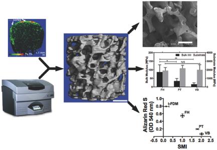
3D printing enables the creation of scaffolds with precisely controlled morphometric properties for multiple tissue types, including musculoskeletal tissues such as cartilage and bone. Computed tomography (CT) imaging has been combined with 3D printing to fabricate anatomically scaled patient‐specific scaffolds for bone regeneration. However, anatomically scaled scaffolds typically lack sufficient resolution to recapitulate the <100 micrometer‐scale trabecular architecture essential for investigating the cellular response to the morphometric properties of bone. In this study, it is hypothesized that the architecture of trabecular bone regulates osteoblast differentiation and mineralization. To test this hypothesis, human bone‐templated 3D constructs are fabricated via a new micro‐CT/3D inkjet printing process. It is shown that this process reproducibly fabricates bone‐templated constructs that recapitulate the anatomic site‐specific morphometric properties of trabecular bone. A significant correlation is observed between the structure model index (a morphometric parameter related to surface curvature) and the degree of mineralization of human mesenchymal stem cells, with more concave surfaces promoting more extensive osteoblast differentiation and mineralization compared to predominately convex surfaces. These findings highlight the significant effects of trabecular architecture on osteoblast function.
中文翻译:

通过 3D 喷墨打印制造小梁骨模板组织工程结构
3D 打印能够为多种组织类型(包括软骨和骨骼等肌肉骨骼组织)创建具有精确控制形态特性的支架。计算机断层扫描 (CT) 成像与 3D 打印相结合,可制造符合解剖学尺寸的患者特异性骨再生支架。然而,解剖学尺度的支架通常缺乏足够的分辨率来概括<100微米尺度的小梁结构,而这对于研究细胞对骨形态测量特性的反应至关重要。在这项研究中,假设骨小梁的结构调节成骨细胞的分化和矿化。为了验证这一假设,通过新的微型 CT/3D 喷墨打印工艺制造了人体骨骼模板 3D 结构。结果表明,该过程可重复地制造骨模板结构,概括骨小梁的解剖部位特定形态测量特性。在结构模型指数(与表面曲率相关的形态参数)和人间充质干细胞的矿化程度之间观察到显着的相关性,与主要为凸面的表面相比,更多凹面促进更广泛的成骨细胞分化和矿化。这些发现强调了小梁结构对成骨细胞功能的显着影响。
更新日期:2017-09-11
中文翻译:
通过 3D 喷墨打印制造小梁骨模板组织工程结构
3D 打印能够为多种组织类型(包括软骨和骨骼等肌肉骨骼组织)创建具有精确控制形态特性的支架。计算机断层扫描 (CT) 成像与 3D 打印相结合,可制造符合解剖学尺寸的患者特异性骨再生支架。然而,解剖学尺度的支架通常缺乏足够的分辨率来概括<100微米尺度的小梁结构,而这对于研究细胞对骨形态测量特性的反应至关重要。在这项研究中,假设骨小梁的结构调节成骨细胞的分化和矿化。为了验证这一假设,通过新的微型 CT/3D 喷墨打印工艺制造了人体骨骼模板 3D 结构。结果表明,该过程可重复地制造骨模板结构,概括骨小梁的解剖部位特定形态测量特性。在结构模型指数(与表面曲率相关的形态参数)和人间充质干细胞的矿化程度之间观察到显着的相关性,与主要为凸面的表面相比,更多凹面促进更广泛的成骨细胞分化和矿化。这些发现强调了小梁结构对成骨细胞功能的显着影响。


 京公网安备 11010802027423号