当前位置:
X-MOL 学术
›
Adv. Funct. Mater.
›
论文详情
Our official English website, www.x-mol.net, welcomes your
feedback! (Note: you will need to create a separate account there.)
Quantum Dots Emitting in the Third Biological Window as Bimodal Contrast Agents for Cardiovascular Imaging
Advanced Functional Materials ( IF 18.5 ) Pub Date : 2017-09-08 , DOI: 10.1002/adfm.201703276 Jie Hu 1 , Dirk H. Ortgies 1, 2 , Rio Aguliar Torres 3 , Nuria Fernández 4 , Lucas Porto 1 , Emma Martín Rodríguez 1, 2 , José García Solé 1 , Daniel Jaque 1, 2 , Fernando Alfonso 3 , Fernando Rivero 3
Advanced Functional Materials ( IF 18.5 ) Pub Date : 2017-09-08 , DOI: 10.1002/adfm.201703276 Jie Hu 1 , Dirk H. Ortgies 1, 2 , Rio Aguliar Torres 3 , Nuria Fernández 4 , Lucas Porto 1 , Emma Martín Rodríguez 1, 2 , José García Solé 1 , Daniel Jaque 1, 2 , Fernando Alfonso 3 , Fernando Rivero 3
Affiliation
|
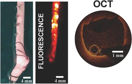
Physicians are demanding innovative technologies for multimodal imaging of the cardiovascular system that would lead to the appearance of advanced diagnosis and therapy procedures. This implies the simultaneous development of new imaging techniques and contrast agents whose synergy would make it possible. Optical coherence tomography (OCT) has recently emerged as a versatile and high‐resolution clinical technique for cardiovascular imaging. Unfortunately, the lack of adequate contrast agents impedes the use of OCT for intracoronary multimodal imaging. In this work, the hitherto unexplored capability of semiconductor quantum dots (IR‐QDs) emitting in the third infrared biological window (1.55–1.87 µm) to act as multimodal agents for intracoronary imaging is demonstrated. Under single line laser excitation at 1.3 µm, IR‐QDs are capable of providing simultaneous backscattering contrast and efficient luminescence at 1.6 µm. In this work, backscattered radiation is successfully employed to construct OCT images in both fluids and tissues whereas the infrared luminescence of the IR‐QDs provides the possibility for simultaneous acquisition of high penetrating fluorescence images. The first multimodal (fluorescence + OCT) imaging of an artery using IR‐QDs as contrast agents is provided herein demonstrating their outstanding potential for future clinical applications.
中文翻译:

在第三个生物窗口中发出的量子点作为心血管成像的双峰造影剂。
医师要求用于心血管系统多峰成像的创新技术,这将导致出现先进的诊断和治疗程序。这意味着同时开发新的成像技术和造影剂,其协同作用将使之成为可能。光学相干断层扫描(OCT)最近已成为一种用于心血管成像的多功能且高分辨率的临床技术。不幸的是,缺乏足够的造影剂阻碍了将OCT用于冠状动脉内多峰成像。在这项工作中,展示了迄今为止在第三红外生物窗口(1.55-1.87 µm)内发射的半导体量子点(IR-QD)充当冠状动脉内成像多峰剂的能力。在1.3 µm的单线激光激发下,IR-QD能够同时提供反向散射对比度和有效的1.6 µm发光。在这项工作中,反向散射辐射被成功地用于在体液和组织中构建OCT图像,而IR-QD的红外发光为同时获取高穿透性荧光图像提供了可能性。本文提供了使用IR-QD作为造影剂对动脉进行的首个多峰(荧光+ OCT)成像,证明了其在未来临床应用中的巨大潜力。反向散射辐射已成功地用于在体液和组织中构建OCT图像,而IR-QD的红外发光为同时获取高穿透性荧光图像提供了可能性。本文提供了使用IR-QD作为造影剂对动脉进行的首个多峰(荧光+ OCT)成像,证明了其在未来临床应用中的巨大潜力。反向散射辐射已成功地用于在体液和组织中构建OCT图像,而IR-QD的红外发光为同时获取高穿透性荧光图像提供了可能性。本文提供了使用IR-QD作为造影剂对动脉进行的首个多峰(荧光+ OCT)成像,证明了其在未来临床应用中的巨大潜力。
更新日期:2017-09-08
中文翻译:
在第三个生物窗口中发出的量子点作为心血管成像的双峰造影剂。
医师要求用于心血管系统多峰成像的创新技术,这将导致出现先进的诊断和治疗程序。这意味着同时开发新的成像技术和造影剂,其协同作用将使之成为可能。光学相干断层扫描(OCT)最近已成为一种用于心血管成像的多功能且高分辨率的临床技术。不幸的是,缺乏足够的造影剂阻碍了将OCT用于冠状动脉内多峰成像。在这项工作中,展示了迄今为止在第三红外生物窗口(1.55-1.87 µm)内发射的半导体量子点(IR-QD)充当冠状动脉内成像多峰剂的能力。在1.3 µm的单线激光激发下,IR-QD能够同时提供反向散射对比度和有效的1.6 µm发光。在这项工作中,反向散射辐射被成功地用于在体液和组织中构建OCT图像,而IR-QD的红外发光为同时获取高穿透性荧光图像提供了可能性。本文提供了使用IR-QD作为造影剂对动脉进行的首个多峰(荧光+ OCT)成像,证明了其在未来临床应用中的巨大潜力。反向散射辐射已成功地用于在体液和组织中构建OCT图像,而IR-QD的红外发光为同时获取高穿透性荧光图像提供了可能性。本文提供了使用IR-QD作为造影剂对动脉进行的首个多峰(荧光+ OCT)成像,证明了其在未来临床应用中的巨大潜力。反向散射辐射已成功地用于在体液和组织中构建OCT图像,而IR-QD的红外发光为同时获取高穿透性荧光图像提供了可能性。本文提供了使用IR-QD作为造影剂对动脉进行的首个多峰(荧光+ OCT)成像,证明了其在未来临床应用中的巨大潜力。


 京公网安备 11010802027423号