当前位置:
X-MOL 学术
›
Chem. Phys.
›
论文详情
Our official English website, www.x-mol.net, welcomes your
feedback! (Note: you will need to create a separate account there.)
Multimodal Hyperspectral Optical Microscopy
Chemical Physics ( IF 2.0 ) Pub Date : 2017-09-02 , DOI: 10.1016/j.chemphys.2017.08.011 Irina V. Novikova , Chuck R. Smallwood , Yu Gong , Dehong Hu , Leif Hendricks , James E. Evans , Ashish Bhattarai , Wayne P. Hess , Patrick Z. El-Khoury
Chemical Physics ( IF 2.0 ) Pub Date : 2017-09-02 , DOI: 10.1016/j.chemphys.2017.08.011 Irina V. Novikova , Chuck R. Smallwood , Yu Gong , Dehong Hu , Leif Hendricks , James E. Evans , Ashish Bhattarai , Wayne P. Hess , Patrick Z. El-Khoury
|
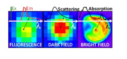
We describe a unique approach to hyperspectral optical microscopy, herein achieved by coupling a hyperspectral imager to various optical microscopes. Hyperspectral fluorescence micrographs of isolated fluorescent beads are first employed to ensure spectral calibration of our detector and to gauge the attainable spatial resolution of our measurements. Different science applications of our instrument are then described. Spatially over-sampled absorption spectroscopy of a single lipid (18:1 Liss Rhod PE) layer reveals that optical densities on the order of 10-3 can be resolved by spatially averaging the recorded optical signatures. This is followed by three applications in the general areas of plasmonics and bioimaging. Notably, we deploy hyperspectral absorption microscopy to identify and image pigments within a simple biological system, namely, a single live Tisochrysis lutea cell. Overall, this work paves the way for multimodal spectral imaging measurements spanning the realms of several scientific disciples.
中文翻译:

多峰高光谱光学显微镜
我们描述了一种高光谱光学显微镜的独特方法,在此通过将高光谱成像仪耦合到各种光学显微镜来实现。首先使用隔离的荧光珠的高光谱荧光显微照片来确保检测器的光谱校准并衡量我们的测量可获得的空间分辨率。然后描述了我们仪器的不同科学应用。单个脂质(18:1 Liss Rhod PE)层的空间过采样吸收光谱显示,可以通过对记录的光学特征进行空间平均来解决10-3数量级的光密度。其次是在等离子和生物成像的一般领域中的三个应用。值得注意的是,我们部署了高光谱吸收显微镜以在简单的生物系统(即,单个活的黄褐线虫黄体细胞。总的来说,这项工作为跨越多个科学门徒领域的多峰光谱成像测量铺平了道路。
更新日期:2017-09-04
中文翻译:
多峰高光谱光学显微镜
我们描述了一种高光谱光学显微镜的独特方法,在此通过将高光谱成像仪耦合到各种光学显微镜来实现。首先使用隔离的荧光珠的高光谱荧光显微照片来确保检测器的光谱校准并衡量我们的测量可获得的空间分辨率。然后描述了我们仪器的不同科学应用。单个脂质(18:1 Liss Rhod PE)层的空间过采样吸收光谱显示,可以通过对记录的光学特征进行空间平均来解决10-3数量级的光密度。其次是在等离子和生物成像的一般领域中的三个应用。值得注意的是,我们部署了高光谱吸收显微镜以在简单的生物系统(即,单个活的黄褐线虫黄体细胞。总的来说,这项工作为跨越多个科学门徒领域的多峰光谱成像测量铺平了道路。


 京公网安备 11010802027423号