当前位置:
X-MOL 学术
›
Adv. Mater.
›
论文详情
Our official English website, www.x-mol.net, welcomes your
feedback! (Note: you will need to create a separate account there.)
The Functional Response of Mesenchymal Stem Cells to Electron-Beam Patterned Elastomeric Surfaces Presenting Micrometer to Nanoscale Heterogeneous Rigidity.
Advanced Materials ( IF 27.4 ) Pub Date : 2017-09-01 , DOI: 10.1002/adma.201702119 Manus J P Biggs 1, 2 , Marc Fernandez 1, 2 , Dilip Thomas 1 , Ryan Cooper 3 , Matteo Palma 4 , Jinyu Liao 5, 6 , Teresa Fazio 6 , Carl Dahlberg 3 , Helen Wheadon 7 , Anuradha Pallipurath 8 , Abhay Pandit 1, 2 , Jeffrey Kysar 3 , Shalom J Wind 6
Advanced Materials ( IF 27.4 ) Pub Date : 2017-09-01 , DOI: 10.1002/adma.201702119 Manus J P Biggs 1, 2 , Marc Fernandez 1, 2 , Dilip Thomas 1 , Ryan Cooper 3 , Matteo Palma 4 , Jinyu Liao 5, 6 , Teresa Fazio 6 , Carl Dahlberg 3 , Helen Wheadon 7 , Anuradha Pallipurath 8 , Abhay Pandit 1, 2 , Jeffrey Kysar 3 , Shalom J Wind 6
Affiliation
|
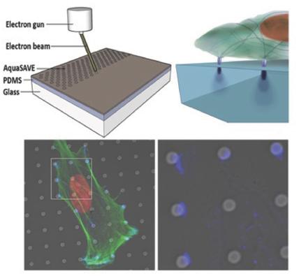
Cells directly probe and respond to the physicomechanical properties of their extracellular environment, a dynamic process which has been shown to play a key role in regulating both cellular adhesive processes and differential cellular function. Recent studies indicate that stem cells show lineage‐specific differentiation when cultured on substrates approximating the stiffness profiles of specific tissues. Although tissues are associated with a range of Young's modulus values for bulk rigidity, at the subcellular level, tissues are comprised of heterogeneous distributions of rigidity. Lithographic processes have been widely explored in cell biology for the generation of analytical substrates to probe cellular physicomechanical responses. In this work, it is shown for the first time that that direct‐write e‐beam exposure can significantly alter the rigidity of elastomeric poly(dimethylsiloxane) substrates and a new class of 2D elastomeric substrates with controlled patterned rigidity ranging from the micrometer to the nanoscale is described. The mechanoresponse of human mesenchymal stem cells to e‐beam patterned substrates was subsequently probed in vitro and significant modulation of focal adhesion formation and osteochondral lineage commitment was observed as a function of both feature diameter and rigidity, establishing the groundwork for a new generation of biomimetic material interfaces.
中文翻译:

间充质干细胞对电子束图案化弹性体表面的功能反应,呈现微米级到纳米级的异质刚性。
细胞直接探测并响应细胞外环境的物理力学特性,这是一个动态过程,已被证明在调节细胞粘附过程和差异细胞功能方面发挥着关键作用。最近的研究表明,当干细胞在接近特定组织硬度的基质上培养时,会表现出谱系特异性分化。尽管组织与一系列体积刚性的杨氏模量值相关,但在亚细胞水平上,组织由不均匀的刚性分布组成。光刻工艺在细胞生物学中已被广泛探索,用于生成分析基板以探测细胞物理力学反应。在这项工作中,首次表明直写电子束曝光可以显着改变弹性体聚(二甲基硅氧烷)基材的刚性和一类新型二维弹性体基材,其受控图案刚性范围从微米到微米。描述了纳米级。随后在体外探测了人类间充质干细胞对电子束图案基底的机械反应,并观察到粘着斑形成和骨软骨谱系定向的显着调节作为特征直径和刚性的函数,为新一代仿生学奠定了基础材料界面。
更新日期:2017-09-01
中文翻译:
间充质干细胞对电子束图案化弹性体表面的功能反应,呈现微米级到纳米级的异质刚性。
细胞直接探测并响应细胞外环境的物理力学特性,这是一个动态过程,已被证明在调节细胞粘附过程和差异细胞功能方面发挥着关键作用。最近的研究表明,当干细胞在接近特定组织硬度的基质上培养时,会表现出谱系特异性分化。尽管组织与一系列体积刚性的杨氏模量值相关,但在亚细胞水平上,组织由不均匀的刚性分布组成。光刻工艺在细胞生物学中已被广泛探索,用于生成分析基板以探测细胞物理力学反应。在这项工作中,首次表明直写电子束曝光可以显着改变弹性体聚(二甲基硅氧烷)基材的刚性和一类新型二维弹性体基材,其受控图案刚性范围从微米到微米。描述了纳米级。随后在体外探测了人类间充质干细胞对电子束图案基底的机械反应,并观察到粘着斑形成和骨软骨谱系定向的显着调节作为特征直径和刚性的函数,为新一代仿生学奠定了基础材料界面。


 京公网安备 11010802027423号