当前位置:
X-MOL 学术
›
J. Am. Coll. Cardiol.
›
论文详情
Our official English website, www.x-mol.net, welcomes your
feedback! (Note: you will need to create a separate account there.)
Effect of Losartan on Mitral Valve Changes After Myocardial Infarction
Journal of the American College of Cardiology ( IF 21.7 ) Pub Date : 2017-09-01 , DOI: 10.1016/j.jacc.2017.07.734 Philipp E Bartko 1 , Jacob P Dal-Bianco 1 , J Luis Guerrero 2 , Jonathan Beaudoin 3 , Catherine Szymanski 4 , Dae-Hee Kim 5 , Margo M Seybolt 2 , Mark D Handschumacher 1 , Suzanne Sullivan 2 , Michael L Garcia 2 , James S Titus 2 , Jill Wylie-Sears 6 , Whitney S Irvin 7 , Emmanuel Messas 8 , Albert A Hagège 8 , Alain Carpentier 8 , Elena Aikawa 7 , Joyce Bischoff 6 , Robert A Levine 4 ,
Journal of the American College of Cardiology ( IF 21.7 ) Pub Date : 2017-09-01 , DOI: 10.1016/j.jacc.2017.07.734 Philipp E Bartko 1 , Jacob P Dal-Bianco 1 , J Luis Guerrero 2 , Jonathan Beaudoin 3 , Catherine Szymanski 4 , Dae-Hee Kim 5 , Margo M Seybolt 2 , Mark D Handschumacher 1 , Suzanne Sullivan 2 , Michael L Garcia 2 , James S Titus 2 , Jill Wylie-Sears 6 , Whitney S Irvin 7 , Emmanuel Messas 8 , Albert A Hagège 8 , Alain Carpentier 8 , Elena Aikawa 7 , Joyce Bischoff 6 , Robert A Levine 4 ,
Affiliation
|
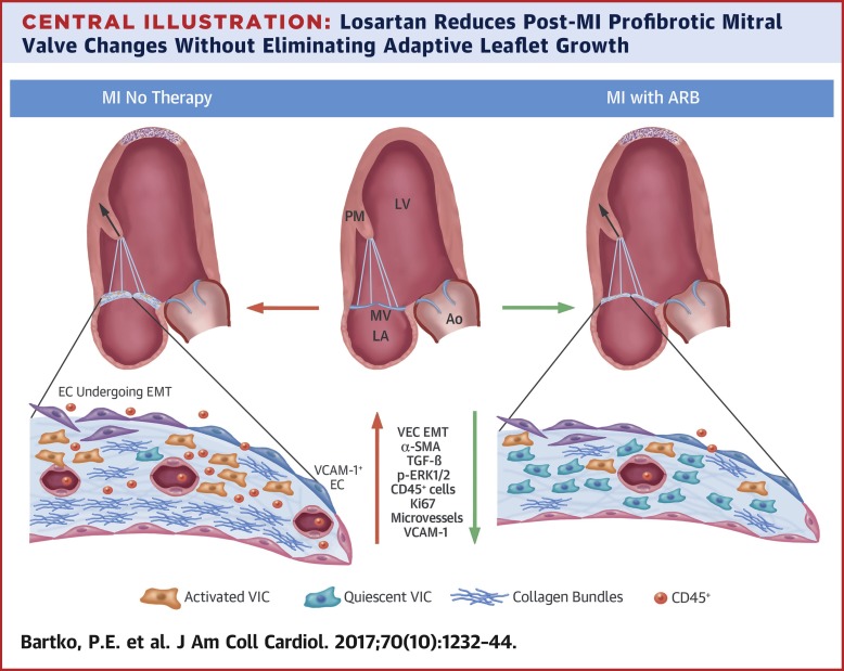
BACKGROUND
After myocardial infarction (MI), mitral valve (MV) tethering stimulates adaptive leaflet growth, but counterproductive leaflet thickening and fibrosis augment mitral regurgitation (MR), doubling heart failure and mortality. MV fibrosis post-MI is associated with excessive endothelial-to-mesenchymal transition (EMT), driven by transforming growth factor (TGF)-β overexpression. In vitro, losartan-mediated TGF-β inhibition reduces EMT of MV endothelial cells. OBJECTIVES
This study tested the hypothesis that profibrotic MV changes post-MI are therapeutically accessible, specifically by losartan-mediated TGF-β inhibition. METHODS
The study assessed 17 sheep, including 6 sham-operated control animals and 11 with apical MI and papillary muscle retraction short of producing MR; 6 of the 11 were treated with daily losartan, and 5 were untreated, with flexible epicardial mesh comparably limiting left ventricular (LV) remodeling. LV volumes, tethering, and MV area were quantified by using three-dimensional echocardiography at baseline and at 60 ± 6 days, and excised leaflets were analyzed by histopathology and flow cytometry. RESULTS
Post-MI LV dilation and tethering were comparable in the losartan-treated and untreated LV constraint sheep. Telemetered sensors (n = 6) showed no significant losartan-induced changes in arterial pressure. Losartan strongly reduced leaflet thickness (0.9 ± 0.2 mm vs. 1.6 ± 0.2 mm; p < 0.05; 0.4 ± 0.1 mm sham animals), TGF-β, and downstream phosphorylated extracellular-signal-regulated kinase and EMT (27.2 ± 12.0% vs. 51.6 ± 11.7% α-smooth muscle actin-positive endothelial cells, p < 0.05; 7.2 ± 3.5% sham animals), cellular proliferation, collagen deposition, endothelial cell activation (vascular cell adhesion molecule-1 expression), neovascularization, and cells positive for cluster of differentiation (CD) 45, a hematopoietic marker associated with post-MI valve fibrosis. Leaflet area increased comparably (17%) in constrained and losartan-treated sheep. CONCLUSIONS
Profibrotic changes of tethered MV leaflets post-MI can be modulated by losartan without eliminating adaptive growth. Understanding the cellular and molecular mechanisms could provide new opportunities to reduce ischemic MR.
中文翻译:

氯沙坦对心肌梗死后二尖瓣改变的影响
背景心肌梗死 (MI) 后,二尖瓣 (MV) 束缚刺激适应性小叶生长,但适得其反的小叶增厚和纤维化会增加二尖瓣关闭不全 (MR),使心力衰竭和死亡率加倍。MI 后 MV 纤维化与过度的内皮间质转化 (EMT) 相关,由转化生长因子 (TGF)-β 过度表达驱动。在体外,氯沙坦介导的 TGF-β 抑制降低了 MV 内皮细胞的 EMT。目的 本研究检验了 MI 后促纤维化 MV 变化在治疗上可及的假设,特别是通过氯沙坦介导的 TGF-β 抑制。方法 该研究评估了 17 只绵羊,包括 6 只假手术对照动物和 11 只患有心尖心肌梗塞和乳头肌收缩但未产生 MR 的绵羊;11 人中有 6 人每天服用氯沙坦,5 人未治疗,使用柔性心外膜网可比较限制左心室 (LV) 重构。通过在基线和 60 ± 6 天时使用三维超声心动图量化 LV 体积、束缚和 MV 面积,并通过组织病理学和流式细胞术分析切除的传单。结果 在氯沙坦治疗和未治疗的 LV 约束绵羊中,MI 后 LV 扩张和束缚是可比的。遥测传感器 (n = 6) 显示没有明显的氯沙坦引起的动脉压变化。氯沙坦显着降低小叶厚度(0.9 ± 0.2 毫米与 1.6 ± 0.2 毫米;p < 0.05;0.4 ± 0.1 毫米假动物)、TGF-β 和下游磷酸化细胞外信号调节激酶和 EMT(27.2 ± 12.0% 与. 51.6 ± 11.7% α-平滑肌肌动蛋白阳性内皮细胞,p < 0.05;7.2 ± 3.5% 假动物),细胞增殖,胶原沉积、内皮细胞活化(血管细胞粘附分子 1 表达)、新血管形成和分化簇 (CD) 45 阳性细胞,CD 是与 MI 后瓣膜纤维化相关的造血标记。在限制和氯沙坦处理的绵羊中,小叶面积增加了(17%)。结论 氯沙坦可以在不消除适应性生长的情况下调节 MI 后栓系 MV 小叶的促纤维化变化。了解细胞和分子机制可以为减少缺血性 MR 提供新的机会。在限制和氯沙坦处理的绵羊中,小叶面积增加了(17%)。结论 氯沙坦可以在不消除适应性生长的情况下调节 MI 后栓系 MV 小叶的促纤维化变化。了解细胞和分子机制可以为减少缺血性 MR 提供新的机会。在限制和氯沙坦处理的绵羊中,小叶面积增加了(17%)。结论 氯沙坦可以在不消除适应性生长的情况下调节 MI 后栓系 MV 小叶的促纤维化变化。了解细胞和分子机制可以为减少缺血性 MR 提供新的机会。
更新日期:2017-09-01
中文翻译:
氯沙坦对心肌梗死后二尖瓣改变的影响
背景心肌梗死 (MI) 后,二尖瓣 (MV) 束缚刺激适应性小叶生长,但适得其反的小叶增厚和纤维化会增加二尖瓣关闭不全 (MR),使心力衰竭和死亡率加倍。MI 后 MV 纤维化与过度的内皮间质转化 (EMT) 相关,由转化生长因子 (TGF)-β 过度表达驱动。在体外,氯沙坦介导的 TGF-β 抑制降低了 MV 内皮细胞的 EMT。目的 本研究检验了 MI 后促纤维化 MV 变化在治疗上可及的假设,特别是通过氯沙坦介导的 TGF-β 抑制。方法 该研究评估了 17 只绵羊,包括 6 只假手术对照动物和 11 只患有心尖心肌梗塞和乳头肌收缩但未产生 MR 的绵羊;11 人中有 6 人每天服用氯沙坦,5 人未治疗,使用柔性心外膜网可比较限制左心室 (LV) 重构。通过在基线和 60 ± 6 天时使用三维超声心动图量化 LV 体积、束缚和 MV 面积,并通过组织病理学和流式细胞术分析切除的传单。结果 在氯沙坦治疗和未治疗的 LV 约束绵羊中,MI 后 LV 扩张和束缚是可比的。遥测传感器 (n = 6) 显示没有明显的氯沙坦引起的动脉压变化。氯沙坦显着降低小叶厚度(0.9 ± 0.2 毫米与 1.6 ± 0.2 毫米;p < 0.05;0.4 ± 0.1 毫米假动物)、TGF-β 和下游磷酸化细胞外信号调节激酶和 EMT(27.2 ± 12.0% 与. 51.6 ± 11.7% α-平滑肌肌动蛋白阳性内皮细胞,p < 0.05;7.2 ± 3.5% 假动物),细胞增殖,胶原沉积、内皮细胞活化(血管细胞粘附分子 1 表达)、新血管形成和分化簇 (CD) 45 阳性细胞,CD 是与 MI 后瓣膜纤维化相关的造血标记。在限制和氯沙坦处理的绵羊中,小叶面积增加了(17%)。结论 氯沙坦可以在不消除适应性生长的情况下调节 MI 后栓系 MV 小叶的促纤维化变化。了解细胞和分子机制可以为减少缺血性 MR 提供新的机会。在限制和氯沙坦处理的绵羊中,小叶面积增加了(17%)。结论 氯沙坦可以在不消除适应性生长的情况下调节 MI 后栓系 MV 小叶的促纤维化变化。了解细胞和分子机制可以为减少缺血性 MR 提供新的机会。在限制和氯沙坦处理的绵羊中,小叶面积增加了(17%)。结论 氯沙坦可以在不消除适应性生长的情况下调节 MI 后栓系 MV 小叶的促纤维化变化。了解细胞和分子机制可以为减少缺血性 MR 提供新的机会。


 京公网安备 11010802027423号