当前位置:
X-MOL 学术
›
Biomater. Sci.
›
论文详情
Our official English website, www.x-mol.net, welcomes your
feedback! (Note: you will need to create a separate account there.)
Bilayered nanofibrous 3D hierarchy as skin rudiment by emulsion electrospinning for burn wound management†
Biomaterials Science ( IF 5.8 ) Pub Date : 2017-06-13 00:00:00 , DOI: 10.1039/c7bm00174f Pallabi Pal 1, 2, 3, 4, 5 , Prabhash Dadhich 1, 2, 3, 4, 5 , Pavan Kumar Srivas 1, 2, 3, 4, 5 , Bodhisatwa Das 1, 2, 3, 4, 5 , Dhrubajyoti Maulik 6, 7, 8, 9 , Santanu Dhara 1, 2, 3, 4, 5
Biomaterials Science ( IF 5.8 ) Pub Date : 2017-06-13 00:00:00 , DOI: 10.1039/c7bm00174f Pallabi Pal 1, 2, 3, 4, 5 , Prabhash Dadhich 1, 2, 3, 4, 5 , Pavan Kumar Srivas 1, 2, 3, 4, 5 , Bodhisatwa Das 1, 2, 3, 4, 5 , Dhrubajyoti Maulik 6, 7, 8, 9 , Santanu Dhara 1, 2, 3, 4, 5
Affiliation
|
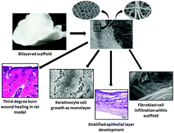
Mimicking skin extracellular matrix hierarchy, the present work aims to develop a bilayer skin graft comprising a porous cotton-wool-like 3D layer with membranous structure of PCL–chitosan nanofibers. Emulsion electrospinning with differential stirring periods of PCL–chitosan emulsion results in development of a bilayer 3D structure with varied morphology. The electrospun membrane has fiber diameter ∼274 nm and pore size ∼1.16 μm while fluffy 3D layer has fiber diameter ∼1.62 μm and pore size ∼62 μm. The 3D layer was further coated with collagen I isolated from Cirrhinus cirrhosus fish scales to improve biofunctionality. Surface coating with collagen I resulted in bundling the fibers together, thereby increasing their average diameter to 2.80 μm and decreasing pore size to ∼45 μm. The architecture and composition of the scaffold promotes efficient cellular activity where interconnected porosity with ECM resembling collagen I coating assists cellular adhesion, infiltration, and proliferation from initial days of fibroblast seeding, while keratinocytes migrate on the surface only without infiltrating in the membranous nanofiber layer. Anatomy of the scaffold arising due to variation in pore size distribution at different layers thereby facilitates compartmentalization and prevents initial cellular transmigration. The scaffold also assists in extracellular matrix protein synthesis and keratinocyte stratification in vitro. Further, the scaffold effectively integrates and attaches with third-degree burn wound margins created in rat models and accelerates healing in comparison to standard Tegaderm dressing™. The bilayer scaffold is thus a promising, readily available, cost-effective, off-the-shelf matrix as a skin substitute.
中文翻译:

双层纳米纤维3D层次结构通过乳剂静电纺丝作为皮肤底层,用于烧伤创面管理†
本研究模仿皮肤细胞外基质的层次结构,旨在开发一种双层皮肤移植物,该移植物包括具有PCL–壳聚糖纳米纤维膜结构的多孔棉样3D层。乳液静电纺丝在PCL-壳聚糖乳液的不同搅拌时间下产生了具有不同形态的双层3D结构。电纺膜的纤维直径约为274 nm,孔径约为1.16μm,而蓬松的3D层的纤维直径约为1.62μm,孔径约为62μm。3D层进一步涂有从Cirrhinus cirrhosus分离的胶原蛋白I鱼鳞可改善生物功能。用胶原蛋白I进行的表面涂层将纤维捆在一起,从而使它们的平均直径增加到2.80μm,并将孔径减小到〜45μm。支架的结构和组成可促进有效的细胞活性,其中孔隙率与类似于胶原蛋白I涂层的ECM相互联系,有助于细胞粘附,浸润和从成纤维细胞播种初期开始的增殖,而角质形成细胞仅在表面迁移而不渗入膜状纳米纤维层。由于在不同层上的孔径分布变化而产生的支架的解剖结构,从而促进了区室化并防止了最初的细胞迁移。支架还有助于细胞外基质蛋白的合成和角质形成细胞的分层体外。此外,与标准的Tegaderm敷料相比,该支架可有效地整合并附着在大鼠模型中产生的三度烧伤创口边缘,并加速愈合。因此,双层支架是有希望的,容易获得的,成本有效的,作为皮肤替代品的现成的基质。
更新日期:2017-06-13
中文翻译:
双层纳米纤维3D层次结构通过乳剂静电纺丝作为皮肤底层,用于烧伤创面管理†
本研究模仿皮肤细胞外基质的层次结构,旨在开发一种双层皮肤移植物,该移植物包括具有PCL–壳聚糖纳米纤维膜结构的多孔棉样3D层。乳液静电纺丝在PCL-壳聚糖乳液的不同搅拌时间下产生了具有不同形态的双层3D结构。电纺膜的纤维直径约为274 nm,孔径约为1.16μm,而蓬松的3D层的纤维直径约为1.62μm,孔径约为62μm。3D层进一步涂有从Cirrhinus cirrhosus分离的胶原蛋白I鱼鳞可改善生物功能。用胶原蛋白I进行的表面涂层将纤维捆在一起,从而使它们的平均直径增加到2.80μm,并将孔径减小到〜45μm。支架的结构和组成可促进有效的细胞活性,其中孔隙率与类似于胶原蛋白I涂层的ECM相互联系,有助于细胞粘附,浸润和从成纤维细胞播种初期开始的增殖,而角质形成细胞仅在表面迁移而不渗入膜状纳米纤维层。由于在不同层上的孔径分布变化而产生的支架的解剖结构,从而促进了区室化并防止了最初的细胞迁移。支架还有助于细胞外基质蛋白的合成和角质形成细胞的分层体外。此外,与标准的Tegaderm敷料相比,该支架可有效地整合并附着在大鼠模型中产生的三度烧伤创口边缘,并加速愈合。因此,双层支架是有希望的,容易获得的,成本有效的,作为皮肤替代品的现成的基质。


 京公网安备 11010802027423号