Our official English website, www.x-mol.net, welcomes your
feedback! (Note: you will need to create a separate account there.)
Biocompatible 3D printed polymers via fused deposition modelling direct C2C12 cellular phenotype in vitro
Lab on a Chip ( IF 6.1 ) Pub Date : 2017-08-01 00:00:00 , DOI: 10.1039/c7lc00577f Rowan P. Rimington 1, 2, 3, 4 , Andrew J. Capel 1, 2, 3, 4, 5 , Steven D. R. Christie 2, 3, 4, 5, 6 , Mark P. Lewis 1, 2, 3, 4
Lab on a Chip ( IF 6.1 ) Pub Date : 2017-08-01 00:00:00 , DOI: 10.1039/c7lc00577f Rowan P. Rimington 1, 2, 3, 4 , Andrew J. Capel 1, 2, 3, 4, 5 , Steven D. R. Christie 2, 3, 4, 5, 6 , Mark P. Lewis 1, 2, 3, 4
Affiliation

|
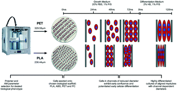
The capability to 3D print bespoke biologically receptive parts within short time periods has driven the growing prevalence of additive manufacture (AM) technology within biological settings, however limited research concerning cellular interaction with 3D printed polymers has been undertaken. In this work, we used skeletal muscle C2C12 cell line in order to ascertain critical evidence of cellular behaviour in response to multiple bio-receptive candidate polymers; polylactic acid (PLA), acrylonitrile butadiene styrene (ABS), polyethylene terephthalate (PET) and polycarbonate (PC) 3D printed via fused deposition modelling (FDM). The extrusion based nature of FDM elicited polymer specific topographies, within which C2C12 cells exhibited reduced metabolic activity when compared to optimised surfaces of tissue culture plastic, however assay viability readings remained high across polymers outlining viable phenotypes. C2C12 cells exhibited consistently high levels of morphological alignment across polymers, however differential myotube widths and levels of transcriptional myogenin expression appeared to demonstrate response specific thresholds at which varying polymer selection potentiates cellular differentiation, elicits pre-mature early myotube formation and directs subsequent morphological phenotype. Here we observed biocompatible AM polymers manufactured via FDM, which also appear to hold the potential to simultaneously manipulate the desired biological phenotype and enhance the biomimicry of skeletal muscle cells in vitro via AM polymer choice and careful selection of machine processing parameters. When considered in combination with the associated design freedom of AM, this may provide the opportunity to not only enhance the efficiency of creating biomimetic models, but also to precisely control the biological output within such scaffolds.
中文翻译:

通过融合沉积建模的生物相容性3D打印聚合物在体外可直接显示C 2 C 12细胞表型
在短时间内进行3D打印定制的生物可接受部件的能力推动了生物环境中增材制造(AM)技术的普及,但是有关细胞与3D打印聚合物相互作用的研究有限。在这项工作中,我们使用骨骼肌C 2 C 12细胞系来确定细胞对多种生物受体候选聚合物的反应的关键证据。通过熔融沉积建模(FDM)3D打印的聚乳酸(PLA),丙烯腈丁二烯苯乙烯(ABS),聚对苯二甲酸乙二醇酯(PET)和聚碳酸酯(PC )。FDM的基于挤出的性质引发了聚合物特定的形貌,其中C 2 C 12与组织培养塑料的优化表面相比,细胞显示出降低的代谢活性,但是,在概述了可行表型的聚合物中,测定活力的读数仍然很高。C 2 C 12细胞在聚合物中始终表现出高水平的形态学比对,但是,不同的肌管宽度和转录肌生成素表达水平似乎表明了反应特异性阈值,在该阈值下,不同的聚合物选择可增强细胞分化,引起过早的早期肌管形成并指导随后的形态表型。在这里,我们观察到通过FDM似乎还具有通过AM聚合物选择和仔细选择机器加工参数在体外同时操纵所需生物学表型和增强骨骼肌细胞仿生的潜力。当考虑与AM的相关设计自由度结合使用时,这可能提供机会,不仅可以提高创建仿生模型的效率,而且可以精确地控制此类支架内的生物输出。
更新日期:2017-08-22
中文翻译:

通过融合沉积建模的生物相容性3D打印聚合物在体外可直接显示C 2 C 12细胞表型
在短时间内进行3D打印定制的生物可接受部件的能力推动了生物环境中增材制造(AM)技术的普及,但是有关细胞与3D打印聚合物相互作用的研究有限。在这项工作中,我们使用骨骼肌C 2 C 12细胞系来确定细胞对多种生物受体候选聚合物的反应的关键证据。通过熔融沉积建模(FDM)3D打印的聚乳酸(PLA),丙烯腈丁二烯苯乙烯(ABS),聚对苯二甲酸乙二醇酯(PET)和聚碳酸酯(PC )。FDM的基于挤出的性质引发了聚合物特定的形貌,其中C 2 C 12与组织培养塑料的优化表面相比,细胞显示出降低的代谢活性,但是,在概述了可行表型的聚合物中,测定活力的读数仍然很高。C 2 C 12细胞在聚合物中始终表现出高水平的形态学比对,但是,不同的肌管宽度和转录肌生成素表达水平似乎表明了反应特异性阈值,在该阈值下,不同的聚合物选择可增强细胞分化,引起过早的早期肌管形成并指导随后的形态表型。在这里,我们观察到通过FDM似乎还具有通过AM聚合物选择和仔细选择机器加工参数在体外同时操纵所需生物学表型和增强骨骼肌细胞仿生的潜力。当考虑与AM的相关设计自由度结合使用时,这可能提供机会,不仅可以提高创建仿生模型的效率,而且可以精确地控制此类支架内的生物输出。


 京公网安备 11010802027423号