Our official English website, www.x-mol.net, welcomes your
feedback! (Note: you will need to create a separate account there.)
In situ generation of human brain organoids on a micropillar array
Lab on a Chip ( IF 6.1 ) Pub Date : 2017-07-20 00:00:00 , DOI: 10.1039/c7lc00682a Yujuan Zhu 1, 2, 3, 4, 5 , Li Wang 1, 2, 3, 4 , Hao Yu 1, 2, 3, 4 , Fangchao Yin 1, 2, 3, 4, 5 , Yaqing Wang 1, 2, 3, 4, 5 , Haitao Liu 1, 2, 3, 4, 5 , Lei Jiang 1, 2, 3, 4 , Jianhua Qin 1, 2, 3, 4, 6
Lab on a Chip ( IF 6.1 ) Pub Date : 2017-07-20 00:00:00 , DOI: 10.1039/c7lc00682a Yujuan Zhu 1, 2, 3, 4, 5 , Li Wang 1, 2, 3, 4 , Hao Yu 1, 2, 3, 4 , Fangchao Yin 1, 2, 3, 4, 5 , Yaqing Wang 1, 2, 3, 4, 5 , Haitao Liu 1, 2, 3, 4, 5 , Lei Jiang 1, 2, 3, 4 , Jianhua Qin 1, 2, 3, 4, 6
Affiliation

|
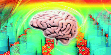
Brain organoids derived from human induced pluripotent stem cells can recapitulate the early stages of brain development, representing a powerful in vitro system for modeling brain development and diseases. However, the existing methods for brain organoid formation often require time-consuming procedures, including the initial formation of embryoid bodies (EBs) from hiPSCs, and subsequent neural induction and differentiation companied by multi-steps of cell transfer and encapsulation in a 3D matrix. Herein, we propose a simple strategy to enable in situ formation of massive brain organoids from hiPSCs on a micropillar array without tedious manual procedures. The optimized micropillar configurations allow for controlled EB formation, neural induction and differentiation, and generation of functional human brain organoids in 3D culture on a single device. The generated brain organoids were examined to imitate brain organogenesis in vivo at early stages of gestation with specific features of neuronal differentiation, brain regionalization, and cortical organization. By combining microfabrication techniques with stem cells and developmental biology principles, the proposed method can greatly simplify brain organoid formation protocols as compared to conventional methods, overcoming the potential limitations of cell contamination, lower throughput and variance of organoid morphology. It can also provide a useful platform for the engineering of stem cell organoids with improved functions and extending their applications in developmental biology, drug testing and disease modeling.
中文翻译:

在微柱阵列上原位生成人脑类器官
源自人类诱导的多能干细胞的脑类器官可以概括大脑发育的早期阶段,代表了一个强大的体外系统,可以为大脑发育和疾病建模。但是,现有的脑类器官形成方法通常需要耗时的过程,包括从hiPSC初步形成类胚体(EB),以及随后的神经诱导和分化,并伴随着多步细胞转移和封装在3D矩阵中。在此,我们提出了一种简单的策略来实现原位无需繁琐的人工操作,即可在微柱阵列上由hiPSC形成大量的脑器官。优化的微柱配置可在单个设备上的3D培养中控制EB的形成,神经诱导和分化,以及生成功能性人脑类器官。检查生成的脑类器官以模仿体内的脑器官发生在妊娠早期具有神经元分化,脑区域化和皮层组织的特定特征。通过将微细加工技术与干细胞和发育生物学原理相结合,与传统方法相比,该方法可以大大简化脑类器官的形成方案,克服了细胞污染,生产能力低和类器官形态变异的潜在局限性。它还可以为工程改造具有改进功能的干细胞类器官提供有用的平台,并扩展其在发育生物学,药物测试和疾病建模中的应用。
更新日期:2017-08-22
中文翻译:

在微柱阵列上原位生成人脑类器官
源自人类诱导的多能干细胞的脑类器官可以概括大脑发育的早期阶段,代表了一个强大的体外系统,可以为大脑发育和疾病建模。但是,现有的脑类器官形成方法通常需要耗时的过程,包括从hiPSC初步形成类胚体(EB),以及随后的神经诱导和分化,并伴随着多步细胞转移和封装在3D矩阵中。在此,我们提出了一种简单的策略来实现原位无需繁琐的人工操作,即可在微柱阵列上由hiPSC形成大量的脑器官。优化的微柱配置可在单个设备上的3D培养中控制EB的形成,神经诱导和分化,以及生成功能性人脑类器官。检查生成的脑类器官以模仿体内的脑器官发生在妊娠早期具有神经元分化,脑区域化和皮层组织的特定特征。通过将微细加工技术与干细胞和发育生物学原理相结合,与传统方法相比,该方法可以大大简化脑类器官的形成方案,克服了细胞污染,生产能力低和类器官形态变异的潜在局限性。它还可以为工程改造具有改进功能的干细胞类器官提供有用的平台,并扩展其在发育生物学,药物测试和疾病建模中的应用。


 京公网安备 11010802027423号