Our official English website, www.x-mol.net, welcomes your
feedback! (Note: you will need to create a separate account there.)
Micro-engineered perfusable 3D vasculatures for cardiovascular diseases
Lab on a Chip ( IF 6.1 ) Pub Date : 2017-07-18 00:00:00 , DOI: 10.1039/c7lc00607a Nishanth Venugopal Menon 1, 2, 3 , Hui Min Tay 2, 4, 5 , Soon Nan Wee 2, 6, 7 , King Ho Holden Li 1, 2, 3 , Han Wei Hou 2, 4, 5
Lab on a Chip ( IF 6.1 ) Pub Date : 2017-07-18 00:00:00 , DOI: 10.1039/c7lc00607a Nishanth Venugopal Menon 1, 2, 3 , Hui Min Tay 2, 4, 5 , Soon Nan Wee 2, 6, 7 , King Ho Holden Li 1, 2, 3 , Han Wei Hou 2, 4, 5
Affiliation
|
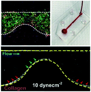
Vessel geometries in microengineered in vitro vascular models are important to recapitulate a pathophysiological microenvironment for the study of flow-induced endothelial dysfunction and inflammation in cardiovascular diseases. Herein, we present a simple and novel extracellular matrix (ECM) hydrogel patterning method to create perfusable vascularized microchannels of different geometries based on the concept of capillary burst valve (CBV). No surface modification is necessary and the method is suitable for different ECM types including collagen, matrigel and fibrin. We first created collagen-patterned, endothelialized microchannels to study barrier permeability and neutrophil transendothelial migration, followed by the development of a biomimetic 3D endothelial–smooth muscle cell (EC–SMC) vascular model. We observed a significant decrease in barrier permeability in the co-culture model during inflammation, which indicates the importance of perivascular cells in ECM remodeling. Finally, we engineered collagen-patterned constricted vascular microchannels to mimic stenosis in atherosclerosis. Whole blood was perfused (1–10 dyne cm−2) into the microdevices and distinct platelet and leukocyte adherence patterns were observed due to increased shear stresses at the constriction, and an additional convective flow through the collagen. Taken together, the developed hydrogel patterning technique enables the formation of unique pathophysiological architectures in organ-on-chip microsystems for real-time study of hemodynamics and cellular interactions in cardiovascular diseases.
中文翻译:

用于心血管疾病的微工程可灌注3D脉管系统
微工程体外血管几何血管模型对于概述病理生理微环境对于研究血流诱发的内皮功能障碍和心血管疾病中的炎症反应非常重要。在这里,我们提出了一种基于毛细管破裂阀(CBV)概念的新颖新颖的细胞外基质(ECM)水凝胶图案化方法,以创建具有不同几何形状的可灌注血管化微通道。无需表面修饰,该方法适用于不同的ECM类型,包括胶原蛋白,基质胶和纤维蛋白。我们首先创建了胶原蛋白模式的内皮化微通道,以研究屏障通透性和中性粒细胞跨内皮迁移,然后开发了仿生的3D内皮-平滑肌细胞(EC-SMC)血管模型。我们观察到炎症期间共培养模型的屏障通透性显着降低,这表明血管周细胞在ECM重塑中的重要性。最后,我们设计了胶原蛋白模式的收缩血管微通道,以模拟动脉粥样硬化中的狭窄。全血灌注(1–10达因厘米-2)进入微装置,并且由于在狭窄处的剪切应力增加以及胶原蛋白的额外对流流动,观察到了明显的血小板和白细胞粘附模式。综上所述,开发的水凝胶图案化技术能够在片上器官微系统中形成独特的病理生理结构,以实时研究心血管疾病中的血流动力学和细胞相互作用。
更新日期:2017-08-22
中文翻译:
用于心血管疾病的微工程可灌注3D脉管系统
微工程体外血管几何血管模型对于概述病理生理微环境对于研究血流诱发的内皮功能障碍和心血管疾病中的炎症反应非常重要。在这里,我们提出了一种基于毛细管破裂阀(CBV)概念的新颖新颖的细胞外基质(ECM)水凝胶图案化方法,以创建具有不同几何形状的可灌注血管化微通道。无需表面修饰,该方法适用于不同的ECM类型,包括胶原蛋白,基质胶和纤维蛋白。我们首先创建了胶原蛋白模式的内皮化微通道,以研究屏障通透性和中性粒细胞跨内皮迁移,然后开发了仿生的3D内皮-平滑肌细胞(EC-SMC)血管模型。我们观察到炎症期间共培养模型的屏障通透性显着降低,这表明血管周细胞在ECM重塑中的重要性。最后,我们设计了胶原蛋白模式的收缩血管微通道,以模拟动脉粥样硬化中的狭窄。全血灌注(1–10达因厘米-2)进入微装置,并且由于在狭窄处的剪切应力增加以及胶原蛋白的额外对流流动,观察到了明显的血小板和白细胞粘附模式。综上所述,开发的水凝胶图案化技术能够在片上器官微系统中形成独特的病理生理结构,以实时研究心血管疾病中的血流动力学和细胞相互作用。


 京公网安备 11010802027423号