当前位置:
X-MOL 学术
›
J. Am. Chem. Soc.
›
论文详情
Our official English website, www.x-mol.net, welcomes your
feedback! (Note: you will need to create a separate account there.)
In Situ Multimodal 3D Chemical Imaging of a Hierarchically Structured Core@Shell Catalyst
Journal of the American Chemical Society ( IF 14.4 ) Pub Date : 2017-05-12 00:00:00 , DOI: 10.1021/jacs.7b02177 Thomas L. Sheppard 1 , Stephen W. T. Price 2 , Federico Benzi 1 , Sina Baier 1 , Michael Klumpp 3 , Roland Dittmeyer , Wilhelm Schwieger 3 , Jan-Dierk Grunwaldt 1
Journal of the American Chemical Society ( IF 14.4 ) Pub Date : 2017-05-12 00:00:00 , DOI: 10.1021/jacs.7b02177 Thomas L. Sheppard 1 , Stephen W. T. Price 2 , Federico Benzi 1 , Sina Baier 1 , Michael Klumpp 3 , Roland Dittmeyer , Wilhelm Schwieger 3 , Jan-Dierk Grunwaldt 1
Affiliation

|
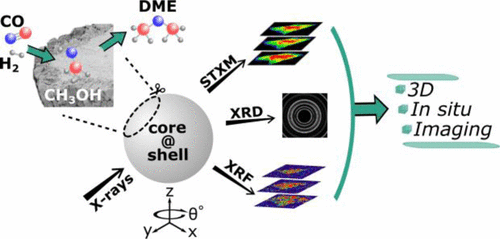
A Cu/ZnO/Al2O3@ZSM-5 core@shell catalyst active for one-step conversion of synthesis gas to dimethyl ether (DME) was imaged simultaneously and in situ using synchrotron-based micro X-ray fluorescence (μ-XRF), X-ray diffraction (μ-XRD), and scanning transmission X-ray microscopy (STXM) computed tomography (CT) with micrometer spatial resolution. An identical sample volume was imaged stepwise, first under oxidizing and reducing atmospheres (imitating calcination and activation processes), and then under model reaction conditions for DME synthesis (H2:CO:CO2 ratio of 16:8:1, up to 250 °C). The multimodal imaging methods offered insights into the active metal structure and speciation within the catalyst, and allowed imaging of both the catalyst core and zeolite shell in a single acquisition. Dispersion of nanosized Cu species was observed in the catalyst core during reduction, with formation of a metastable Cu+ phase at the core–shell interface. Under DME reaction conditions at 1 bar, the coexistence of Cu0 in the active catalyst core together with partially oxidized Cu species was unraveled. The zeolite shell and core–shell interface remained stable under all conditions, preserving the bifunctional nature of the catalyst. These observations are inaccessible using standard bulk techniques like X-ray absorption spectroscopy (XAS) and XRD, demonstrating the potential of multimodal in situ X-ray CT for characterization of hierarchically designed materials, which stand to benefit tremendously from such 3D spatially resolved measurements.
中文翻译:

分层结构核@壳催化剂的原位多峰3D化学成像
铜/氧化锌/铝2 ö 3 @ ZSM-5芯@壳催化剂活性为合成气的单步转化为二甲醚(DME)中的溶液同时成像和原位使用基于同步加速器的微X射线荧光(μ- XRF),X射线衍射(μ-XRD)和具有微米级空间分辨率的扫描透射X射线显微镜(STXM)计算机断层扫描(CT)。首先在氧化和还原气氛下(模仿煅烧和活化过程),然后在用于DME合成的模型反应条件下(H 2:CO:CO 2),逐步对相同的样品体积成像。比率为16:8:1,最高可达250°C)。多峰成像方法提供了对催化剂中活性金属结构和形态的深入了解,并允许在一次采集中对催化剂核和沸石壳进行成像。在还原过程中,在催化剂核心中观察到了纳米级Cu物种的分散,在核-壳界面处形成了亚稳态的Cu +相。在1 bar的DME反应条件下,Cu 0共存分解出活性催化剂核中的铜和部分氧化的铜。沸石壳和核-壳界面在所有条件下均保持稳定,从而保留了催化剂的双功能性质。使用标准的批量技术(例如X射线吸收光谱(XAS)和XRD)无法获得这些观察结果,这证明了多峰原位X射线CT表征分层设计材料的潜力,这些材料将从此类3D空间分辨测量中受益匪浅。
更新日期:2017-05-27
中文翻译:

分层结构核@壳催化剂的原位多峰3D化学成像
铜/氧化锌/铝2 ö 3 @ ZSM-5芯@壳催化剂活性为合成气的单步转化为二甲醚(DME)中的溶液同时成像和原位使用基于同步加速器的微X射线荧光(μ- XRF),X射线衍射(μ-XRD)和具有微米级空间分辨率的扫描透射X射线显微镜(STXM)计算机断层扫描(CT)。首先在氧化和还原气氛下(模仿煅烧和活化过程),然后在用于DME合成的模型反应条件下(H 2:CO:CO 2),逐步对相同的样品体积成像。比率为16:8:1,最高可达250°C)。多峰成像方法提供了对催化剂中活性金属结构和形态的深入了解,并允许在一次采集中对催化剂核和沸石壳进行成像。在还原过程中,在催化剂核心中观察到了纳米级Cu物种的分散,在核-壳界面处形成了亚稳态的Cu +相。在1 bar的DME反应条件下,Cu 0共存分解出活性催化剂核中的铜和部分氧化的铜。沸石壳和核-壳界面在所有条件下均保持稳定,从而保留了催化剂的双功能性质。使用标准的批量技术(例如X射线吸收光谱(XAS)和XRD)无法获得这些观察结果,这证明了多峰原位X射线CT表征分层设计材料的潜力,这些材料将从此类3D空间分辨测量中受益匪浅。


 京公网安备 11010802027423号