当前位置:
X-MOL 学术
›
Chem. Rev.
›
论文详情
Our official English website, www.x-mol.net, welcomes your
feedback! (Note: you will need to create a separate account there.)
Stimulated Emission Depletion Microscopy
Chemical Reviews ( IF 51.4 ) Pub Date : 2017-03-06 00:00:00 , DOI: 10.1021/acs.chemrev.6b00653 Hans Blom 1 , Jerker Widengren 2
Chemical Reviews ( IF 51.4 ) Pub Date : 2017-03-06 00:00:00 , DOI: 10.1021/acs.chemrev.6b00653 Hans Blom 1 , Jerker Widengren 2
Affiliation
|
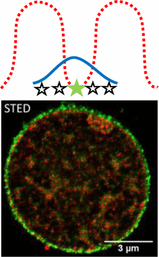
Despite its short history, diffraction-unlimited fluorescence microscopy techniques have already made a substantial imprint in the biological sciences. In this review, we describe how stimulated emission depletion (STED) imaging originally evolved, how it compares to other optical super-resolution imaging techniques, and what advantages it provides compared to previous golden-standards for biological microscopy, such as diffraction-limited optical microscopy and electron microscopy. We outline the prerequisites for successful STED imaging experiments, emphasizing the equally critical roles of instrumentation, sample preparation, and photophysics, and describe major evolving strategies for how to push the borders of STED imaging even further in life science. Finally, we provide examples of how STED nanoscopy can be applied, within three different fields with particular potential for STED imaging experiments: neuroscience, plasma membrane biophysics, and subcellular clinical diagnostics. In these areas, and in many more, STED imaging can be expected to play an increasingly important role in the future.
中文翻译:

受激发射耗竭显微镜
尽管历史不长,但无限制衍射荧光显微镜技术已在生物科学中留下了重要的烙印。在这篇综述中,我们描述了受激发射损耗(STED)成像最初是如何演变的,如何与其他光学超分辨率成像技术进行比较,以及与以前的生物显微镜金标准(例如衍射极限光学)相比具有什么优势?显微镜和电子显微镜。我们概述了成功进行STED成像实验的先决条件,强调了仪器,样品制备和光物理学的同等重要作用,并描述了如何在生命科学领域进一步拓展STED成像边界的主要发展策略。最后,我们提供了如何应用STED纳米显微镜的示例,在三个不同领域具有STED成像实验的特殊潜力:神经科学,质膜生物物理学和亚细胞临床诊断。在这些领域以及更多领域,预计STED成像在未来将扮演越来越重要的角色。
更新日期:2017-03-06
中文翻译:
受激发射耗竭显微镜
尽管历史不长,但无限制衍射荧光显微镜技术已在生物科学中留下了重要的烙印。在这篇综述中,我们描述了受激发射损耗(STED)成像最初是如何演变的,如何与其他光学超分辨率成像技术进行比较,以及与以前的生物显微镜金标准(例如衍射极限光学)相比具有什么优势?显微镜和电子显微镜。我们概述了成功进行STED成像实验的先决条件,强调了仪器,样品制备和光物理学的同等重要作用,并描述了如何在生命科学领域进一步拓展STED成像边界的主要发展策略。最后,我们提供了如何应用STED纳米显微镜的示例,在三个不同领域具有STED成像实验的特殊潜力:神经科学,质膜生物物理学和亚细胞临床诊断。在这些领域以及更多领域,预计STED成像在未来将扮演越来越重要的角色。


 京公网安备 11010802027423号