研究领域
 查看导师最新文章
(温馨提示:请注意重名现象,建议点开原文通过作者单位确认)
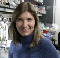
Professor Gradinaru’s research has focused on developing technologies for neuroscience and using them to probe circuits underlying locomotion, reward, and sleep. Together with collaborators, mentors, and trainees, Gradinaru has developed and applied neuronal activity readout and control tools such as optogenetics (Gradinaru et al, Cell 2010) and tissue clearing by CLARITY (Yang et al, Cell 2014; Gradinaru et al, Annual Review of Biophysics 2018) to dissect circuitry underlying movement, mood, and sleep disorders (Gradinaru et al, Science, 2009; Xiao et al, Neuron, 2016; Cho et al, Neuron, 2017). To extract functional information from cleared tissue the group also reported on methods for multi-color, multi-RNA imaging in deep tissue. In most recent work, to facilitate delivery of genetic controllers and sensors they have developed viral vector screening methods, with first efforts resulting in capsids capable of crossing the Blood-Brain-Barrier for non-invasive brain-wide transduction in adults after systemic delivery (Deverman et al, Nat.Biotech.2016) and a method for sparse stochastic Golgi-like genetic labeling for morphology assessment (Chan et al, NatureNeurosc. 2017); ongoing work in this area focuses on improving viral vectors for cell-type specificity and larger packaging ability (Bedbrook et al, Annual Review of Neuroscience 2018)
Prof. Gradinaru has received the NIH Director’s New Innovator Award and a Presidential Early Career Award for Scientists and Engineers and has been honored as a World Economic Forum Young Scientist and as one of Cell’s 40 under 40. Gradinaru is also a Sloan Fellow, Pew Scholar, Moore Inventor, Vallee Scholar, and Allen Brain Institute NGL Council Member, and received the inaugural Peter Gruss Young Investigator Award by the Max Planck Florida Institute for Neuroscience. In 2017 she was the Early-Career Scientist Winner in the Innovators in Science Award in Neuroscience (Takeda and the New York Academy of Sciences) and in 2018 she received a Gill Transformative award.
Viviana Gradinaru has also been very active in teaching and service, participating with lab members in regular technology training workshops at Caltech and for summer courses at Cold Spring Harbor Laboratory as well as running the CLOVER Center (Beckman Institute for CLARITY, Optogenetics and Vector Engineering), which provides training and access to the group's reagents and methods for the broader research community.
(1) Methods for precise neuromodulation (Optogenetics): Optogenetics uses light together with genetically encoded, light-sensitive proteins to modulate or monitor the function of specific cell types within living heterogeneous tissue. Optogenetics provided neuroscientists with means to activate or inhibit the firing of a defined population of neurons at physiological, millisecond timescales and study the effects on living, freely moving organisms. By targeting expression of the opsin(s) to a defined class of neuron within a specific brain structure, it is possible to understand the function of those neurons with a precision not possible with electrical or chemical methods. Through optogenetic experiments, neuroscientists are rapidly unraveling how individual neurons and circuits work together to control mood, learning, memory, desire and sensory and motor function as well as how these circuits are altered in disease states. Although now a mature field, in the early days starting with 2005 there were significant challenges that we have solved: many opsins, especially pumps, were not well tolerated by mammalian cells and therefore could not be used in vivo. We have worked out subcellular and transcellular trafficking strategies that resulted in potent and safe optogenetic tools, which includes inhibitors that span the visible spectrum, and generalizable strategies for targeting cells based not on genetic identity, but on morphology or tissue topology, to allow versatile targeting when promoters are not known or in genetically intractable organisms. The same principles used to better traffic opsins outside of the endoplasmic reticulum and into the membrane work for an array of newly described opsins for neuroscience and are likely to help with tolerability in mammalian cells of microbial opsins yet to be discovered or engineered.
a.Gradinaru V, Zhang F, Ramakrishnan C, Mattis J, Prakash R, Diester I, Goshen I, Thompson KR, Deisseroth K. Molecular and cellular approaches for diversifying and extending optogenetics. Cell. 2010 Apr 2;141(1):154-65. PMCID: PMC4160532. F1000 must read. Download
b.Bedbrook CN, Rice AJ, Yang KK, Ding X, Chen S, LeProust EM, Gradinaru V, Arnold FH. Structure-guided SCHEMA recombination generates diverse chimeric channelrhodopsins. PNAS 2017 Mar 10. PMID:28283661 Download
c.Herwig L, Rice AJ, Bedbrook CN, Zhang RK, Lignell A, Cahn JK, Renata H, Dodani SC, Cho I, Cai L, Gradinaru V, Arnold FH. Directed Evolution of a Bright Near-Infrared Fluorescent Rhodopsin Using a Synthetic Chromophore. Cell Chem Biol. 2017 Feb 20. PMID:28262559 Download
(2) Mechanisms of Deep Brain Stimulation by optogenetic deconstruction of diseased brain circuitry: Deep brain stimulation (DBS) is a powerful therapeutic option for intractable movement and affective disorders. The benefits of DBS are immediate and dramatic, manifested as instantaneous improvements in motor function in the case of PD patients. However, due to the nonspecificity of electrical stimulation, the mechanisms behind the effects of DBS are still highly controversial. In order to understand the role of specific cell types underlying effective DBS treatment, we have successfully: (1) developed and optimized optogenetic technologies (molecular and hardware) for safe and effective use in behaving mammals, including readout methods such as the optrode; and (2) employed the above developed optogenetic toolkit to deconstructing diseased brain circuitry, with focus on Parkinson's disease. This work challenged the traditional perception that deep brain stimulation acts mainly by inhibiting local cell bodies at the stimulation site by showing that controlling axons in the stimulation area was sufficient to restore motor behavior in parkinsonian animals. The framework and tools developed are generalizable across many brain circuits to study intact and disordered brain processes and were indeed used subsequently by many projects within our group and outside.
a.Gradinaru V, Mogri M, Thompson KR, Henderson JM, Deisseroth K. Optical deconstruction of parkinsonian neural circuitry. Science. 2009 Apr 17;324(5925):354-9. PubMed PMID: 19299587. Science News Focus. F1000 exceptional. Download.
b.Xiao C, Cho JR, Zhou C, Treweek BJ, Chan K, McKinney SL, Yang B, and Gradinaru V. Cholinergic Mesopontine Signals Govern Locomotion and Reward Through Dissociable Midbrain Pathways. Neuron 2016 Apr 20;90(2)33-47. PMCID: PMC4840478. Download.
c.Cho JR, Treweek JB, Robinson JE, Xiao C, Bremner LR, Greenbaum A, Gradinaru V. Dorsal Raphe Dopamine Neurons Modulate Arousal and Promote Waking by Salient Stimuli. Neuron 2017 Jun PMID:28602690. Download
(3) Methods for anatomical mapping of intact circuits (Tissue Clearing, CLARITY): Even with the power of optogenetics for control and readout of brain networks, a standing challenge is knowing which circuits to modulate for an intended therapeutic effect: we do not have detailed maps of connectivity across large brain volumes. This can be a serious problem, as our DBS optogenetic study above showed. That electrical DBS might act fundamentally through white matter away from the electrode site highlights the need for better brain maps. It is however difficult to create such maps for phenotypically distinct fine axons that run in bundles throughout the brain when the traditional method involves sectioning the tissue in paper-thin slices, imaging each slice, and putting it all back together with imaging software: it is slow, tedious, costly, and error prone. We invested in tissue clearing instead to remove the lipids, which obstruct the view, and created a new method known as CLARITY (Nature, 2013), which renders the tissue transparent for easy visualization and identification of cellular components and their molecular identity without slicing. Tissue clearing complements optogenetics, in that it can reveal, with ease, circuit-wide effects of optogenetic manipulations and also aid in mapping novel circuits that need tuning in disease. In an attempt to perfect the execution of CLARITY, our group has recently reported (Cell, 2014) the first case of whole-body clearing – transparent rodents that can be used to obtain detailed maps of both central and peripheral nerves at their target organs throughout the body. Such nerves could then be modulated with optogenetics in animal models of disease to understand what needs tuning to improve symptoms and the resulting knowledge could facilitate better therapies that rely on, for example, electrically stimulating nerves for better organ function or for decreasing chronic pain.
a.Chung K, Wallace J, Kim SY, Kalyanasundaram S, Andalman AS, Davidson TJ, Mirzabekov JJ, Zalocusky KA, Mattis J, Denisin AK, Pak S, Bernstein H, Ramakrishnan C, Grosenick L, Gradinaru V, Deisseroth K. Structural and molecular interrogation of intact biological systems. Nature. 2013 May 16;497(7449):332-7. PMID: PMC4092167. Download
b.Yang B, Treweek JB, Kulkarni RP, Deverman BE, Chen CK, Lubeck E, Shah S, Cai L, Gradinaru V. Single-cell phenotyping within transparent intact tissue through whole-body clearing. Cell. 2014 Aug 14;158(4):945-58. PMCID: PMC4153367. Highlighted by NIH, Nature, Science, F1000. Scientific American 10 World Changing Ideas 2014. Nature Biotechnology News and Views. Download
c.Treweek, J.B.; Chan, K.Y.; Flytzanis, N.C.; Yang, B.; Deverman, B.E.; Greenbaum, A.; Lignell, A.; Xiao, C.; Cai, L.; Ladinsky, M.S.; Bjorkman, P.J.; Fowlkes, C.C.; Gradinaru, V. Whole-Body Tissue Stabilization and Selective Extractions via Tissue-Hydrogel Hybrids for High Resolution Intact Circuit Mapping and Phenotyping. Nature Protocols. 2015 Nov;10(11):1860-96. PMID: 26492141. Download
d.Greenbaum A, Chan K, Dobreva T, Brown D, Balani DH, Boyce R, Kronenberg HM, McBride HJ and Gradinaru V, Bone CLARITY: clearing, imaging, and computational analysis of osteoprogenitors within intact bone marrow Science Translational Medicine 2017 PMID:28446689 Download
(4) Methods for optical monitoring of neuronal activity and RNA detection at depth: In addition to optogenetic control of neuronal activity we need feedback on how exactly the tissue is responding to light modulation, and tune it up or down accordingly – for example to stop seizures. I have worked on two related topics: Optical Voltage Sensors and Imaging of Single Molecule RNA in Cleared Tissue. In collaboration with Prof. Frances Arnold at Caltech I have worked on voltage sensing with microbial opsins for all-optical control and readout of neuronal networks. Opsins can be engineered for diverse properties, including increased opsin radiance whose level tracks the membrane voltage changes with high temporal precision in defined cell types. My group used directed evolution of opsins to make them better at reporting action potentials. Changes in RNA transcripts can also report on activity history of brain circuits. Preserving spatial relationships while accessing the transcriptome of selected cells is a crucial feature for advancing many biological areas, from developmental biology to neuroscience. Collaborating with Profs. Cai and Pierce at Caltech we recently reported on methods for multi-color, multi-RNA, imaging in deep tissues. By using single-molecule hybridization chain reaction (smHCR), PACT tissue hydrogel embedding and clearing and light-sheet microscopy we detected single-molecule mRNAs in ~mm-thick tissue samples (reference 4 below).
a.Flytzanis NC, Bedbrook CN, Chiu H, Engqvist MK, Xiao C, Chan KY, Sternberg PW, Arnold FH, Gradinaru V. Archaerhodopsin variants with enhanced voltage-sensitive fluorescence in mammalian and Caenorhabditis elegans neurons. Nat Commun. 2014 Sep 15;5:4894. PMCID: PMC4166526. Selected by Nature Methods for “Methods to Watch.”
Download
b.Shah S, Lubeck E, Schwarzkopf M, He TF, Greenbaum A, Sohn CH, Lignell A, Choi HM, Gradinaru V* (co-corresponding), Pierce NA*, Cai L*. “Single-molecule RNA detection at depth via hybridization chain reaction and tissue hydrogel embedding and clearing.” Development. 2016 Jun 24. pii: dev.138560. [Epub ahead of print] PMID: 27342713
c.DePas, W.H., Starwart-Lee, R., Van Sambeek, L., Kumar, S.R., Gradinaru, V* (co-corresponding), Newman, D.K.*, Exposing the three-dimensional biogeography and metabolic states of pathogens in cystic fibrosis sputum via hydrogel embedding, clearing, and rRNA labeling. mBio 2016; PMID:27677788
(5) Viral-based approaches to non-invasive whole-brain cargo delivery: Genetically-encoded tools that can be used to visualize, monitor, and modulate mammalian neurons are revolutionizing neuroscience. These tools are particularly powerful in rodents and invertebrate models where intersectional transgenic strategies are available to restrict their expression to defined cell populations. However, use of genetic tools in non-transgenic animals is often hindered by the lack of vectors capable of safe, efficient, and specific delivery to the desired cellular targets. To begin to address these challenges, we have developed an in vivo Cre-based selection platform (CREATE) for identifying adeno-associated viruses (AAVs) that more efficiently transduce genetically defined cell populations. Our platform's novelty and power arises from the additional selective pressure imparted by a recovery step that amplifies only those capsid variants that have functionally transduced a genetically-defined, Cre-expressing target cell population. The Cre-dependent capsid recovery works within heterogeneous tissue samples without the need for additional steps such as selective capsid recovery approaches that require cell sorting or secondary adenovirus infection. As a first test of the CREATE platform, we selected for viruses that transduced the brain after intravascular delivery and found a novel vector, AAV-PHP.B, that is 40- to 90-fold more efficient at transducing the brain than the current standard, AAV9. AAV-PHP.B transduces most neuronal types and glia across the brain. We also demonstrate here how whole-body tissue clearing can facilitate transduction maps of systemically delivered genes. Since CNS disorders are notoriously challenging due to the restrictive nature of the blood brain barrier our discovery that recombinant vectors can be engineered to overcome this barrier is enabling for the whole field. With the exciting advances in gene editing via the CRISPR-Cas, RNA interference and gene replacement strategies, the availability of potent gene delivery methods provided by vectors such as our reported AAV-PHP.B is key to advancing the field of genome engineering.
a.Deverman BE, Pravdo P, Simpson B, Banerjee A, Kumar, S.R., Chan K, Wu WL, Yang B, Gradinaru V. Cre-Dependent Capsid Selection Yields AAVs for Global Gene Transfer to the Adult Brain. Nature Biotechnol. 2016 Feb 34(2):204-9. PMID: 26829320 PMCID in Process. F1000 Special Significance. Scientific American in Advances, Neuroscience. (published 2016). Download
b.K Chan, M Jang, B Yoo, A Greenbaum , N Ravi , W Wu , L Sánchez-Guardado, C Lois, S Mazmanian, B Deverman, V Gradinaru, Engineered adeno-associated viruses for efficient and noninvasive gene delivery throughout the central and peripheral nervous systems NatureNeuro 2017Jun PMID: 28671695 Download
c.Allen WE, Kauvar IV, Chen MZ, Richman EB, Yang SJ, Chan K, Gradinaru V, Deverman BE, Luo L, Deisseroth K. Global representations of goal-directed behavior in distinct cell types of mouse neocortex. Neuron 2017 7;94(4):891-907.e6. PMID: 28521139 Download
d.RC Challis, SR Kumar, KY Chan, C Challis, MJ Jang, PS Rajendran, JD Tompkins, K Shivkumar, BE Deverman, V Gradinaru* Widespread and targeted gene expression by systemic AAV vectors: Production, purification, and administration. BioRxiv doi: https://doi.org/10.1101/246405
Biochemistry, Structural, and Molecular Cell Biology; Biological Engineering; Neuroscience; Systems Biology; Evolutionary and Organismal Biology


 京公网安备 11010802027423号