Article

Genetic Diversity of Trypanosomes Infesting Cattle from Savannah District in North of Côte d’Ivoire Using Conserved Genomic Signatures: rRNA, ITS1 and gGAPDH

by
Jean-Yves Ekra
1,2,3,*,
Eliakunda Michael Mafie
1,*,
Edouard K. N’Goran
4,
Dramane Kaba
5,
Biégo Guillaume Gragnon
6 and
Jagan Srinivasan
3,*
1
Department of Veterinary Microbiology, Parasitology and Biotechnology, Sokoine University of Agriculture, Morogoro 67125, Tanzania
2
SACIDS Africa Centre of Excellence for Infectious Diseases, SACIDS Foundation for One Health, Sokoine University of Agriculture, Morogoro 67125, Tanzania
3
Department of Biology and Biotechnology, Worcester Polytechnic Institute, 100 Institute Rd., Worcester, MA 01609, USA
4
Unité de Formation et de Recherche (UFR) des Sciences Biologiques, Département de Biochimie-Génétique, Université Peleforo Gon Coulibaly, Korhogo BP1328, Côte d’Ivoire
5
Unité de Recherche «Trypanosomoses», Institut Pierre Richet 01, Bouaké BP1500, Côte d’Ivoire
6
Laboratoire National d’Appui au Développement Agricole (LANADA), Korhogo BP1328, Côte d’Ivoire
*
Authors to whom correspondence should be addressed.
Pathogens 2024, 13(3), 262; https://doi.org/10.3390/pathogens13030262
Submission received: 6 February 2024 / Revised: 14 March 2024 / Accepted: 15 March 2024 / Published: 19 March 2024

Abstract

:
The potential danger to livestock from African animal trypanosomiasis is well known. However, the trypanosome species circulating in cattle and their genetics are poorly understood. After different alignments according to three regions (ITS1, gGAPDH and rRNA gene) of the trypanosome genome, phylogenetic analyses were used to show the genetic diversity of the different species that were circulating in the cattle in three regions (Bagoue, Poro and Tchologo) of Côte d’Ivoire. These analyses were performed by alignment of ITS1; by alignment of partial 18S, ITS1, 5.8S, ITS2 and partial 28S rRNA genes; and by alignment of gGAPDH gene with sequences of Trypanosomes found in GenBank. Three species were identified (T. vivax, T. theileri and T. congolense) in the cattle in the three northern regions of Côte d’Ivoire. T. vivax and T. theileri were the most abundant species in the present study. Contrary to the other primers used in this study, the ITS1 primers were not able to amplify T. theileri. We observed mixed infections between T. theileri and the other two species identified (T. vivax and T. congolense). As far as primers are concerned, in some cases, rRNA was able to identify the same species of trypanosomes that the ITS1 and gGAPDH primers were able to identify. Two main distinct groups of T. theileri complex were identified. The T. congolense and T. vivax strains were close to African strains, such as those from Kenya, Nigeria and Cameroon, unlike the T. theileri strain. Three trypanosome species (T. vivax, T. theileri and T. congolense) circulate in cattle in the Savannah district of Côte d’Ivoire. The genetic diversity of the trypanosome species encountered in this study cannot be classified as intraspecies according to geographical area and breed of cattle they infect.

1. Introduction

Trypanosomes are protozoan parasites that cause disease in humans (sleeping sickness) and cattle (Nagana) in sub-Saharan Africa [1]. In socio-economic terms, Trypanosoma brucei, T. congolense and T. vivax are the most important trypanosome species in sub-Saharan Africa. They are transmitted by tsetse flies (Glossina sp.) as a key biological vector [2]. The geographical distribution of tsetse flies is limited to sub-Saharan Africa [3].
Some species of trypanosomes can be transmitted by other vectors, such as Trypanosoma vivax, which can also be mechanically transmitted by Tabanidae and Stomoxys [4]. Several techniques can be used to diagnose them, but the most widely used is microscopy. Parasites are detected by optical microscopy of wet blood films or thick and thin fixed blood films stained with Giemsa [5]. Greater sensitivity can be achieved by centrifuging hematocrit to concentrate the parasite load [6]. However, these techniques do not allow the differentiation of intraspecific Trypanosoma spp. and are not sensitive enough to detect low levels of parasitemia [7]. Antibody detection methods, such as indirect ELISA, have been widely used in epidemiological studies.
However, these methods do not prove the existence of an ongoing infection and cannot differentiate between Trypanosoma spp. infections [7,8,9]. Molecular methods, notably the polymerase chain reaction (PCR), have been used to establish species-specific and interspecies detection and can detect intraspecific diversity [10]. But primers using microsatellites have shown their limitations [5]. Species-specific primers only amplify the target species and will not amplify unidentified or diverse trypanosomes that do not carry the target sequence. Other types of primers that amplify regions conserved in all trypanosome species have been developed. These include PCRs targeting the internal transcribed space (ITS) region of ribosomal genes [10,11,12]; PCRs targeting partial 18S, ITS1, 5.8S, ITS2 and partial 28S rRNA genes; and gGAPDH genes [13].
This method has been widely used due to its high sensitivity, which is attributed to a high copy number, and the feasibility of interspecies length variation allowing visual discrimination by gel electrophoresis. Amplicon sequencing can further increase sensitivity and enable intraspecific identification. These primers have been used to obtain a higher prevalence of T. vivax in Tanzanian tsetse populations than those based on satellite DNA sequences [13]. Thanks to these primers, new trypanosomes have been discovered in recent years. The identification of T. simiae tsavo was made possible by the failure of trypanosome hybridization with existing DNA probes [14]. Similarly, T. godfreyi was described when isoenzyme and DNA analysis indicated a trypanosome that differed from the already known species found in Glossina morsitans submorsitans in Gambia. Surveys of tsetse populations in Tanzania revealed a parasite that did not amplify with existing PCR primers. This led to the designation of “T. godfreyilike” [13] and “T. brucei-like” [15] parasites in tsetse flies.
In Côte d’Ivoire, trypanosome diagnostic studies using the PCR technique have been exclusively carried out with microsatellite primers [15,16,17,18]. The primers used are specific to the T. vivax, T brucei and T. congolense savannah and forest species. It is therefore difficult to know whether other trypanosome species exist in the natural environment of Côte d’Ivoire. Information on the genetic diversity of trypanosomes in Côte d’Ivoire is poorly known and limited. This constitutes a major weakness in the development of an effective trypanosome control strategy in Côte d’Ivoire. The lack of knowledge of this diversity constitutes a health risk for both humans and animals. In the context of One Health, it is therefore necessary to investigate the diversity of these parasites in Côte d’Ivoire.
The objective of this work was to investigate trypanosome species diversity and their genetic diversity circulating in cattle in three regions of northern Côte d’Ivoire.

2. Materials and Methods

2.1. Sample Collection in the Field

The criteria for selecting farms and animals for blood sampling were the same as those described in our previous study [16]. Blood samples were collected from cattle in the field in a period from August to November 2021. The animals were chosen after a visual evaluation of vital indicators such as mucosal condition, respiratory rate, heart rate, pulse, body temperature, lymph nodes and animal behavior (standing position). Blood samples were prioritized for animals with low vital signs. After appropriate immobilization, a sample was taken from the jugular vein using EDTA tubes, which were kept in cold storage until the PCR analysis was performed. In addition to the blood samples, farm, and individual cattle data, such as age, breed according to the breeder, sex and body condition score, were recorded on a sample card for each herd.

2.2. DNA Extraction, PCR Amplification and Sequencing

DNA from blood samples was extracted using a commercially available kit (Quick-DNA™ Miniprep Plus Kit) according to the instructions of the manufacturer.
To better understand the diversity of trypanosomes, several genome regions were selected, notably ITS1, rRNA and gGAPDH. The first time, the ITS1 region of the ribosomal RNA gene (rDNA) was amplified with ITS1 primers. The trypanosomes were identified according to their band size, as described by [11]. The reaction mixture constituted of 2 μL of template, 1X of one taq buffer, each dNTP at 200 μM, each primer at 1 μM and 0.5 U of one taq DNA polymerase (NEW ENGLAND BiolabsRinc, Ipswich, MA, USA), adjusted at 25 μL with nuclease-free water. The cycling conditions were 94 °C for 5 min, with 35 cycles: 94 °C for 40 s, 58 °C for 40 s, 72 °C for 90 s, 72 °C for 5 min.
Secondly, the regions of the genome grouping the partial 18S, ITS1, 5.8S, ITS2 and the partial 28S were amplified by nested PCR with two pairs of ITS primers (ITS1, ITS2, ITS3 and ITS4) following the protocol described by [19]. This PCR was also performed in a 25 μL total reaction. The reaction mixtures in the first run constituted of 1X of one taq buffer, each dNTP at 200 μM, each primer (ITS1 and ITS2) at 0.4 μM, 1.25 U of one taq DNA polymerase (NEW ENGLAND BiolabsRinc) and 1 μL of DNA as template. For the second run, the first PCR product (2 μL) was added to 23 μL of a new mix composed of the same components as the first with the primers replaced by ITS3 and ITS4. The cycling conditions of both rounds were 95 °C for 7 min, with 35 cycles: 94 °C for 1 min, 55 °C for 1 min, 72 °C for 120 s, 72 °C for 5 min.
Finally, the glycosomal glyceraldehyde-3-phosphate dehydrogenase gene (gGAPDH) was amplified by nested PCR, as described by Hamilton [20], using degenerate primers (G3, G5, G4a, G1, G4b and Gs). The program was 95 °C for 3 min, with 35 cycles: 95 °C for 1 min, 55 °C for 30 s, 72 °C for 1 min and 72 °C for 10 min for both rounds. PCR products were electrophoresed in 1.5% agarose (BioTools Inc., Chuo-ku, Tokyo, Japan) in TBE buffer and stained using safe view (4 μL for 100 mL of TBE) before being visualized under UV light. The expected fragment sizes and primers sequences are shown in Supplementary Table S1.
For the sequencing of the targeted genes, the PCR products and the primers were submitted to ETON Company for sequencing by Sanger method. Afterwards, the obtained fragments were edited and aligned using CLUSTAL W program in BioEdit software to detect any conserved polymorphism between the strains. The nucleotide sequences reported in the present study are available in the DDBJ/EMBL/GenBank databases under the accession numbers presented in Table 1.

2.3. Analysis of Sequences

Sequence cleaning, inspection and editing were performed in BioEdit. IDs were assigned to each sequence. The IDs of the different sequences were assigned according to the following formula: The sequence ID consist of the species name, the locality abbreviation, the breed abbreviation of the infected animal, the animal number and the target genome region (Table 1). The trypanosome species corresponding to the sequences were identified by BLAST in NCBI Blastn by local alignment. Based on the three genome regions previously amplified by PCR, the alignment and comparison were only performed on the parts of the genome which were available in GenBank. Using the CLUSTAL W multiple alignment program [21] in BioEdit software, the trypanosome genome region of the ITS1 gene, gGAPDH gene and the composite region of partial 18S, ITS1, 5.8S, ITS2 and partial 28S rRNA gene nucleotide sequences were aligned with those of related trypanosomes, which were available in DDBJ/EMBL/GenBank databases. According to each region of the trypanosome genome targeted in this study, three alignments were created with trypanosome species frequently found in Africa.
For phylogenetic analysis, rooted trees were performed. Before that, the sequences were compared at two levels: (i) between sequences obtained in the current study and available reference sequences, and (ii) between our own sequences identified as belonging to the same species or group.
According to each region which was targeted by PCR, three neighbor-joining trees were constructed in MEGA7 [22]. The evolutionary history was inferred using maximum likelihood (ML) analysis based on the Kimura 2-parameter model (for ITS1 and gGAPDH gene) and on the Tamura 3-parameter model [23] for regions of partial 18S, ITS1, 5.8S, ITS2 and partial 28S rRNA genes. Pairwise distance estimation was performed using the Maximum Composite Likelihood (MCL) approach. The probability of inferred branches was assessed by the approximate likelihood ratio test (aLRT), an alternative to the non-parametric bootstrap estimation of branch support [24,25].

3. Results

3.1. Sequence Identification

The present study generated 68 new sequences (Table 1). They included 4 internal transcribed spacer 1 (ITS1) sequences, 44 ribosomal DNA (18S, ITS1, 5.8S, ITS2 and 28S) sequences and 20 new sequences of the glycosomal glyceraldehyde-3-phosphate dehydrogenase gene (gGAPDH). The ITS1 sizes obtained were between 158 and 253 bp for the three sequences which shared 94.31–100% of their identity with T. vivax sequences found in NCBI. The 581 bp sequence was the only one that shared similarity (84%) with Trypanosoma congolense sequences in the NCBI database.
For the 18S, ITS1, 5.8S, ITS2 and 28S regions, the sizes were between 550 and 1458 bp. Three and two of the 44 sequences, respectively, had a high degree of shared identity with the T. congolense (81.17–85.64%) and T. vivax (84.88–96.72%) species. The other sequences were very close to the T. theileri species, with shared identities of between 83.02 and 99.43%.
For the gGAPDH gene, we had eight sequences that were close to the T. vivax species with 84.55–90.83% similarity in identity, ranging in size from 300 to 752 bp. Eleven (11) sequences were identified as very close to T. theileri, with identity percentages between 91.23 and 97.98%. Of the twenty sequences obtained based on this region of the trypanosome genome, only one sequence was identified as being very close to the T. congolense species, with an identity similarity of 90.08%, with the accession number AJ620289.1 as reference.

3.2. Mixed Infection and Mixed Identification by Primers

In this study, we obtained mixed infections from animals and species infection cases that were identified simultaneously by at least two of the types of primers used. This can be seen in Table 1, so the IDs with the same letter in the superscripts labeled in red are cases of mixed infection, and the IDs with the same number in the superscripts written in blue are cases of species identified by at least two of the primer types in this study.
For mixed infections, we had two cases of T. theileri/T. vivax couplings and one case of a T. theileri/T. congolense coupling. As examples, we list the following IDS: T. theileri_Fer_M_362_RNA (OR973760) and T. vivax_Ferk_M_362 (PP210927), as well as T. theileri_Mben_M_272_RNA (PP188050) and T. congo_Mben_M_272_RNA (PP188052).
In the case of trypanosome species identified by more than one primer, we observed two possibilities in this study: ITS1/rRNA coupling or GAPDH/rRNA coupling. We obtained one case with an ITS1/rRNA coupling (T. congo_Mben_M_271 (PP210925) and T. congo_Mben_M_271_RNA (PP188049)), which enabled us to identify the same strain of T. congolense. The GAPDH/rRNA pairing enabled us to identify seven T. vivax strains (T. vivax_Diko_N_225_RNA (PP188048) and T. vivax_Diko_N_225_GAPDH (OR966685)) and six T. theileri strains (Table 1).

3.3. Sequence Comparison between the Different Trypanosome Species Identified

Of the ITS1 nucleotide sequences of T. vivax (Supplementary Figure S1), the different individuals identified had 245 nucleotides in common, corresponding to a similarity of 90.40%. We found 16 insertions/deletions (Indel) and 10 nucleotide substitutions. As for the T. vivax GAPDH gene sequence, there was a 30% similarity (90 matches) between the sequences, 130 nucleotide substitutions and 136 insertions/deletions (Indel) when all sequences were considered simultaneously.
Nucleotide sequences covering the partial 18S, ITS1, 5.8S, ITS2 and partial 28S rRNA gene regions of T. theileri shared a similarity of 27.43% (203 nucleotides), with 309 insertions/deletions (Indel) and 381 nucleotide substitutions between all these individuals. For the T. theileri GAPDH gene sequences, there was 67.50% similarity, which corresponds to 162 nucleotide matches between all these sequences, 84 nucleotide substitutions and 6 insertions/deletions (Indel), when comparing all sequences simultaneously (Supplementary Figure S2).
Comparing the T. congolense sequences with reference to the partial 18S, ITS1, 5.8S, ITS2 and partial 28S rRNA genes, we observed 36 insertions/deletions (Indel), 90 nucleotide substitution and 239 identical nucleotides; this represents 68.480% similarity between the three T. congolense genotypes identified (Supplementary Figure S3).

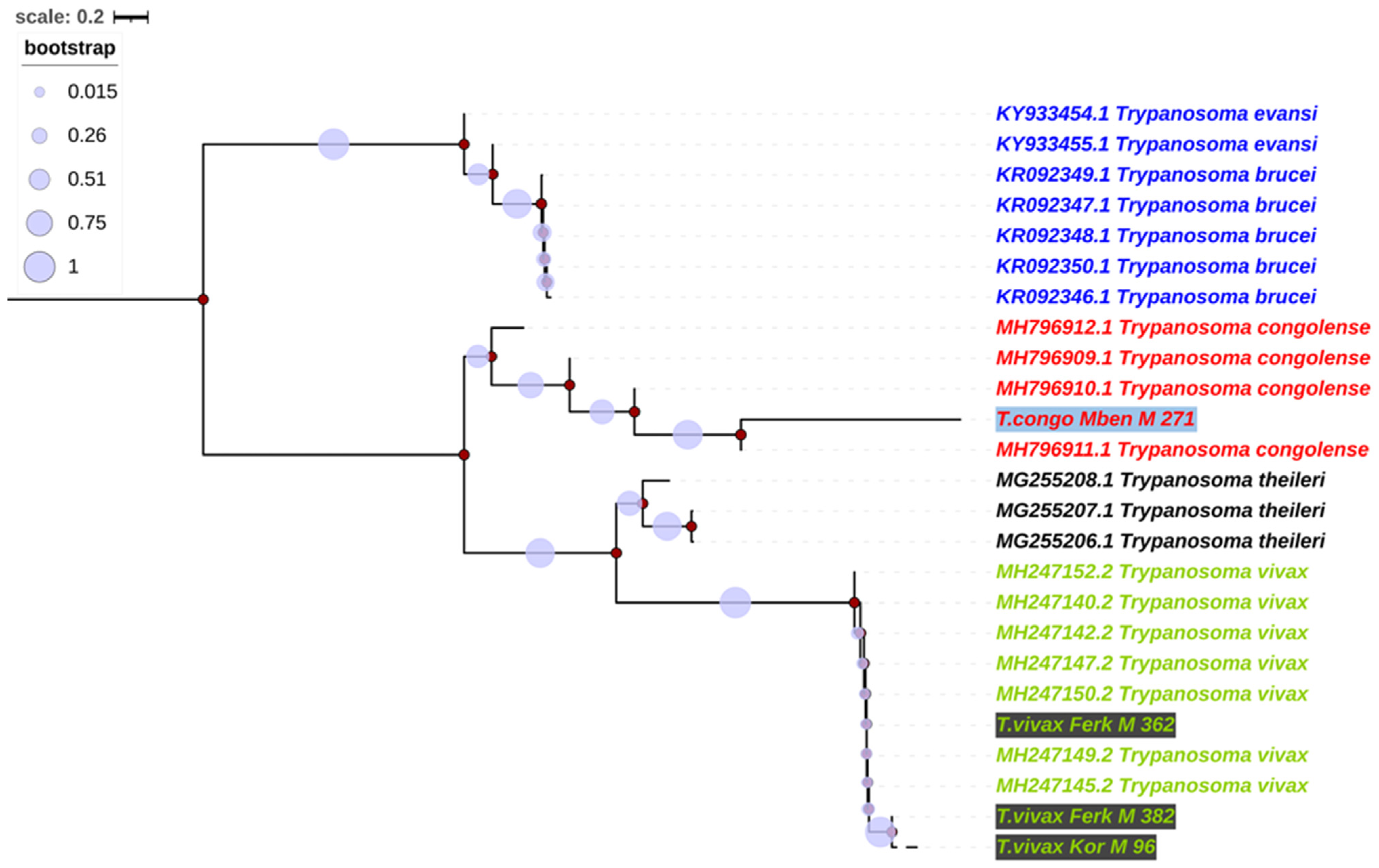
3.4. Alignment and Phylogenetics of Trypanosome Species According to Internal Transcribed Spacer 1 (ITS1)

The internal transcribed spacer 1 (ITS1) alignment was used to construct phylogenetic trees using neighbor joining, a maximum likelihood method based on the Kimura two-parameter model [25]. The ITS1 sequences of this study clustered with different reference sequences (Figure 1), and three sequences formed strong groups with T. vivax sequences from the NCBI database. This group formed a separate clade (bootstrap value: 90%) with a clade formed by T. congolense, T. theileri, T. brucei and T. evansi. One ITS1 sequence of this study was identified as a T. congolense sequence. This sequence is part of the clade of T. congolense with a bootstrap of 60% (Figure 1).

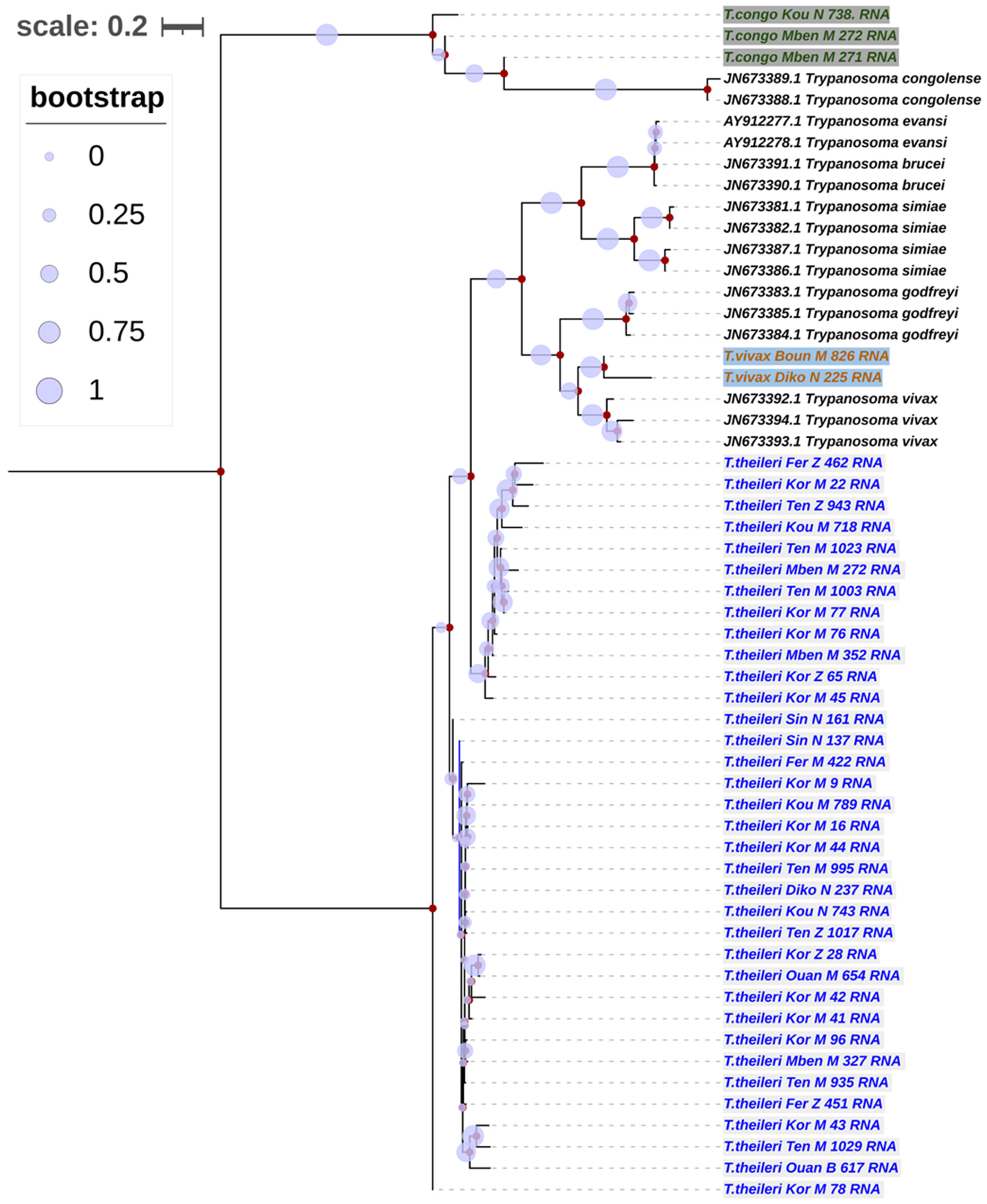
3.5. Phylogenetic Trees of Trypanosomes Species According to Partial 18S, ITS1, 5.8S, ITS2 and Partial 28S rRNA Genes

Based on the alignment of the partial 18S, ITS1, 5.8S, ITS2 and partial 28S rRNA genes for the sequences generated in this study with those sequences from the NCIBI, a phylogenetic tree was constructed. The distances between the different sequences were estimated using the Maximum Composite Likelihood (MCL).
The sequences of T. vivax (2 sequences), T. congolense (3 sequences) and T. theileri (39 sequences) species were identified as close to the sequences obtained in the current study. The alignment from the partial 18S, ITS1, 5.8S, ITS2 and partial 28S rRNA genes for the sequence generated showed a phylogenetic tree with two main groups of T. theileri (Figure 2). The first group comprised 12/39 sequences, and the second group had 29/39 sequences. The two large, distinct T. theileri groups formed by our sequences had a bootstrap value of 57% (Figure 2). The two sequences of T. vivax formed a strong clade with those from the NCBI database. This T. vivax clade was part of a big clade constituted of clades formed by T. godfreyi, T. simiae, T. brucei and T. evansi, which are more closely related to the T. theileri taxon 1, with a bootstrap value of 79% (Figure 2).

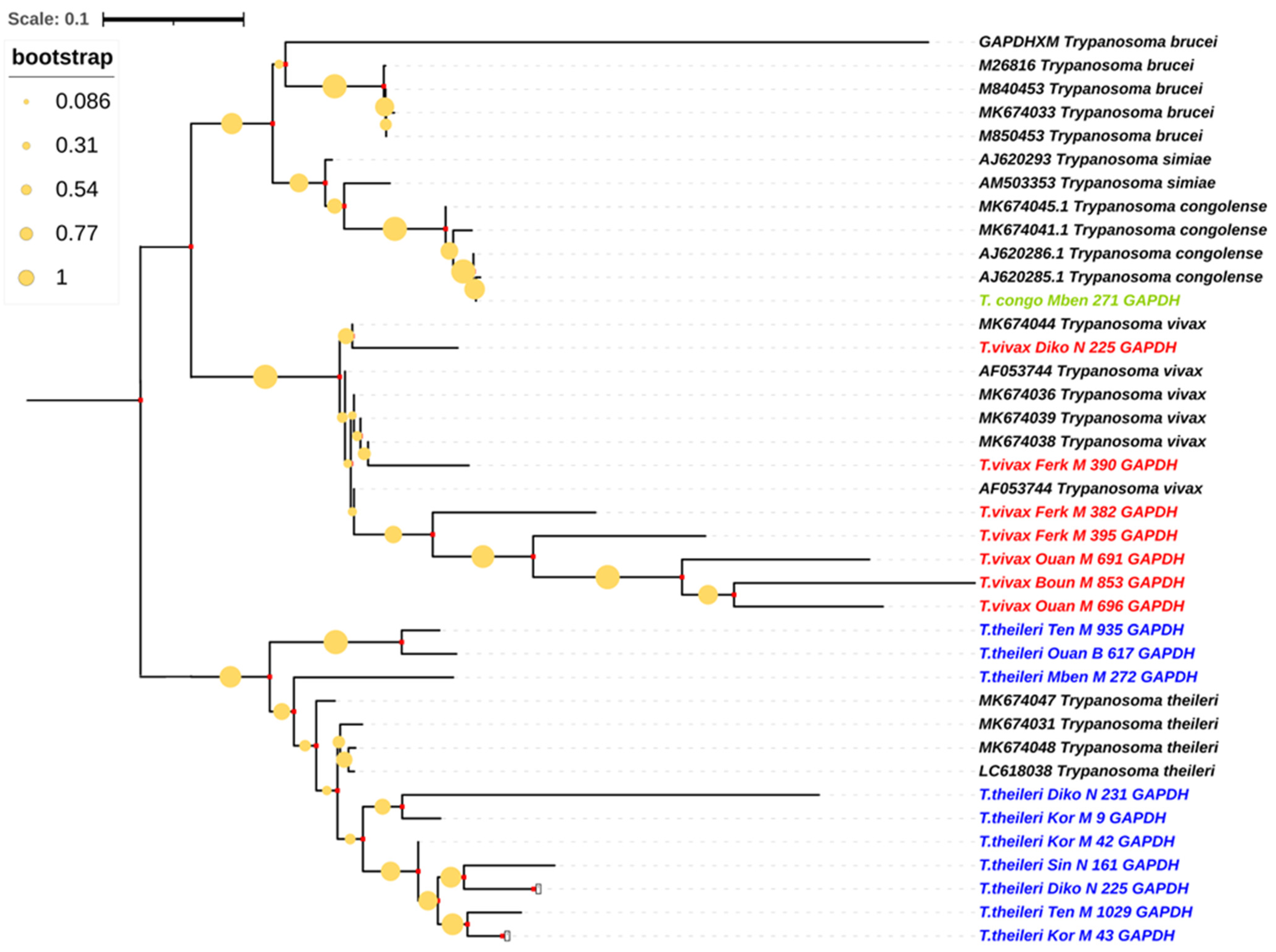
3.6. Phylogenetic Trees of Trypanosome Species According to Glycosomal Glyceraldehyde-3-Phosphate Dehydrogenase Gene (gGAPDH)

The Maximum Composite Likelihood (MCL) approach allowed us to estimate the distance between the sequences of this study and those from the NCIBI GenBank.
Sequences identified as T. vivax, T. theileri and T. congolense had strongly constituted clades according to their reference’s sequences. Here, two clades formed in the generated sequences, the T. vivax clade and the big clade formed by the T. theileri clade, the T. brucei clade and the T. congolense clade. The T. vivax clade formed a single clade which was very distinct, with a 100% bootstrap value. The T. congolense sequence was closer to the T. theileri clade than to the one of the T. vivax clade (Figure 3).

4. Discussion

Analysis of PCR product sequences according to three different regions of the trypanosome genome obtained from the blood of cattle collected in the Bagoue, Tchologo and Poro regions has enabled us to identify three trypanosome species: T. vivax, T. congolense and T. theileri.
The ITS1 primers were unable to identify the T. theileri species. The ability of ITS1 primers to detect more T. vivax and T. congolense species and fewer T. theileri was demonstrated by Njiru et al. [11]. Indeed, the ITS1 primer was designed to improve the detection of pathogenic trypanosome species by minimizing its homology with non-pathogenic species, notably T. theileri, which is non-pathogenic [5]. This could be because during primer design, it may not have been considered due to its non-pathogenic nature.
Primers amplifying the gGAPDH region and the one amplifying the partial 18S, ITS1, 5.8S, ITS2 and partial 28S rRNA genes were used to identify the three species encountered in this study. In contrast, the prevalence of T. congolense and T. theileri species obtained with primers amplifying the partial 18S, ITS1, 5.8S, ITS2 and partial 28S rRNA genes was high compared to their prevalence with gGAPDH. This could be explained by the presence of more copies of partial 18S, ITS1, 5.8S, ITS2 and partial 28S rRNA genes than copies of the gGAPDH gene. It is important to note that the situation was different for the T. vivax species, whose high prevalence was revealed by the gGAPDH. This low prevalence of T. vivax according to partial 18S, ITS1, 5.8S, ITS2 and partial 28S rRNA genes was also observed by Auty et al. [29], who used a different primer for T. vivax amplification. This could be explained by a problem specific to this region of the T. vivax species.
Here, we have a sensitivity and specificity of primers that would relatively depend on the target region and the trypanosome species in question.
In this study, we noted more T. vivax sequences than T. congolense sequences, which was also noted by other researchers [16,17,30]. T vivax and T. theileri were present in mechanical vectors, notably Tabanidae. The present study area, in this case the savannah district, is a predilection zone for Tabanidae [31]. Several researchers [29,30,31] have demonstrated the role of Tabanidae in the transmission of T. vivax. The work of [31] showed the dispersion and abundance of Tabanidae in the different ecological facies of this study area. Twelve (12) species were identified, with Tabanus laverani being the most encountered species with an ADP of 906 individuals/trap/day. The Tabanidae species are also potential vectors of the T. theileri species [4,32,33]. Kostygov et al. [34] demonstrated the life cycle of two different species of the Trypanosoma theileri complex in Tabanidae. The strong presence of T. vivax and T. theileri species are therefore closely linked to the strong presence of the Tabanidae vectors of these two species, whose population is high in this area [29,32].
Comparison between the sequences of individuals gave us 68.48%, 27.43% and 90.40% similarity between the nucleotide sequences of T. congolense, T. theileri and T. vivax species, respectively, considering the portions including the ITS1 part. The 27.43% similarity between T. theileri sequences was low. This may be due to the high number of sequences compared at the T. theileri species level. According to keita [20,35], sequence comparison is a function of sequence number and this has an impact on the identity rate. When the datasets are small enough, very precise alignments may be calculated. Alignments of huge datasets that have developed with numerous indels will most likely have high error rates, and gene trees constructed from these alignments will similarly have significant errors [36].
We a high diversity of the T. theileri complex. The blast results showed that the strains were close to certain sources from horizons that are geographically far from the present study area, such as North America, South America, Asia and Europe. This could be explained by the fact that the gGAPDH and rRNA regions of the T. theileri species are so conserved that it is impossible to distinguish between those from Africa and other continents. On the other hand, it may be due to the low availability of sequences from African strains of this part of the T. theileri genome in the NCBI database. Given the low pathogenicity of this species, it has been neglected by African researchers. In fact, they have made greater use of specific primers and have been more focused on three species: T. brucei, T. congolense and T. vivax [37]. By observing the phylogenetic trees according to the rRNA gene we were able to classify the T. theileri complex into two distinct main groups. These observations are in line with those of Kostygov [34], who was able to show that two different species of the T. theileri complex, for which we had no prior information on the morphology of their blood forms, belong to two different clades of the complex, named TthI and TthII. However, these species have not been distinguished by morphology, morphometry, ultrastructure or development in the vector. Similarly, this distinction of the T. theileri species into two different clades is not possible when considering the geographical areas and the hosts of the T. theileri species to which they are genetically close according to phylogenetic trees.
The T. congolense species observed in this study are closely related to strains from the greater West African zone, more specifically Nigeria and Cameroon. The phylogenetic proximity of the T. congolense populations observed in this study can be broken down into two subspecies. The strain obtained in the Kouto department is genetically close to the savannah-type T. congolense subspecies. The strains obtained in the M’Bengue area are genetically close to the forest-type T. congolense subspecies. In fact, T. congolense species belong to the Nannomonas genus, which has been the subject of much morphological and genetic research. Currently, the recognized genotypes of the nannomonas genus are savannah-type T. congolense, West African forest/river-type T. congolense and Kilifi-type T. congolense [13,35,38].
According to the gGAPDH gene, we found 30% similarity between T. vivax individuals and 67.50% at the T. theileri level despite the high number of individuals compared at T. theileri level. This observation demonstrates the high diversity found within the trypanosomes of the T. vivax species. This high diversity of the T. vivax species could be linked to the large number of vectors it has, notably mechanical vectors (Tabanidae, Stomoxes) and tsetse flies [39]. In fact, this species’ ability to spend a stage of its cycle in the digestive tract of different vectors may play a role in the perpetual modification of its genetic material. According to the present phylogenetic tree, the Trypanosoma vivax species encountered in this study are very close to the Kenyan strains, based on the ITS1 part of the genome. On the other hand, based on the gGAPDH gene, the T. vivax species are close to those from Cameroon (Central Africa). According to Adams [40], it would be better to classify T. vivax variants into types A, B and C on the basis of genetic variants. So, in the present case, based on the ITS1 region, the strains are types A or B, but they are type C according to the gGAPDH gene. It is important to note that the geographical nomenclature used in previous works to name trypanosomes can be misleading. Clearly, taking genetic data into account in historical taxonomic classifications is not an easy task. The nomenclature suggested by Adams [40] for naming groups A, B and C should be further developed, taking into account variations on the part of the T. vivax genome by comparing more strains from different parts of Africa based on different parts of the genome, like the small subunit ribosomal RNA gene (SSU rDNA), the mitochondrial DNA or the kinetoplast DNA (minicircles and maxicircles).

5. Conclusions

Analysis of the ITS1, rRNA and gGAPDH regions of trypanosomes circulating in cattle in the Savannah district of Côte d’Ivoire identified three trypanosome species, including a species that had been neglected (T. theileri) in terms of study in Côte d’Ivoire as well as certain species that are important as livestock pathogens (T. vivax and T. congolense). The observed variability in these three strains cannot be classified according to the geographical area and breed of cattle they infect. In addition, due to the fact that livestock farming has been recognized for many years as being under pressure from pathogens originating from a variety of vectors, the addition of phylogenetic information raises many questions about livestock trypanosomes, particularly regarding transmission, host sharing and pathogenicity. This is important, as livestock morbidity and mortality due to T. congolense and T. vivax infections continues to cause heavy losses to livestock farmers. These strains should be studied in other domestic animals (sheep, goats, etc.), as well as in wild animals. Further phylogenetic analysis will probably be needed to explore these complex relationships.

Supplementary Materials

The following supporting information can be downloaded at: https://www.mdpi.com/article/10.3390/pathogens13030262/s1, Table S1: Trypanosomes band sizes expected and primers sequences; Figure S1: Cluster W alignment of T. vivax strain based on Internal Transcribed Spacer 1 (ITS1); Figure S2. Cluster W alignment of T. theileri strain according to the Glycosomal glyceraldehyde-3-phosphate dehydrogenase gene (gGAPDH); Figure S3. Cluster W alignment of T. congolense strain according to partial 18S, ITS1, 5.8S, ITS2 and partial 28S rRNA genes.

Author Contributions

Conceptualization, J.-Y.E.; data curation, J.-Y.E. and E.M.M.; formal analysis, J.-Y.E.; funding acquisition, J.-Y.E., E.M.M. and E.K.N.; investigation, J.-Y.E.; methodology, J.-Y.E. and E.M.M.; project administration, E.M.M.; resources, D.K., B.G.G. and J.S.; software, J.-Y.E.; supervision, E.M.M., E.K.N. and J.S.; validation, E.M.M., E.K.N., D.K., B.G.G. and J.S.; visualization, E.K.N., D.K., B.G.G. and J.S.; writing—original draft, J.-Y.E.; writing—review and editing, J.-Y.E., E.M.M., E.K.N. and B.G.G. All authors have read and agreed to the published version of the manuscript.

Funding

The Partnership for Skills in Applied Sciences, Engineering and Technology (PASET) funded this research through the Regional Scholarship and Innovation Fund (RSIF), which was awarded to Jean-Yves Ekra to pursue doctoral studies at the SACIDS Africa Centre of Excellence for Infectious Diseases, SACIDS Foundation for One Health, Sokoine University of Agriculture, Morogoro, Tanzania. The APC was funded by PASET-RSIF, a sustainable pan-African science fund supporting high quality doctoral training and research in African universities and other institutions of higher learning. The funders had no say in the study’s design, data collection and analysis, the decision to publish or the manuscript preparation. The study’s results and conclusions are those of the authors and do not necessarily reflect the views of the sponsors.

Institutional Review Board Statement

Following approval from the Ministry of Animal Resources Halieutics (MIRAH), sample collection was carried out as part of epidemiological surveillance operations overseen by the National Laboratory for Support to Agricultural Development (LANADA). For epidemiological monitoring activities, local authorities do not require an ethical declaration. With the approval of the owner, any veterinarian may collect blood from domestic animals as part of a prophylactic or diagnostic program. No laboratory animals were used in this work. Outside of those necessary for regular screening and diagnostic procedures, no samples were taken. Following an explanation and clarification of the study’s objectives, the farmers consented to have blood drawn from their animals. Every animal sampled received a free deworming medicine. The wounds on the animals were cleansed and disinfected.

Informed Consent Statement

Informed consent was obtained from all subjects involved in the study.

Data Availability Statement

All sequences generated in the present study are available in the GenBank database under the accession numbers in Table 1.

Acknowledgments

We thank the Côte d’Ivoire Ministry of Animal Resources and Halieutics (MIRAH) for assisting in field data collection. We appreciate the Ferkéssédougou Breeders’ Professional Organisation (O.P.E.F). We acknowledge the assistance of the staff of UFR (Unité de Formation et de Recherche) of Biology Sciences of Péléforo Gon Coulibaly University of Korhogo (Côte d’Ivoire) in collaboration with the National Laboratory for Support to Agricultural Development (LANADA) for their scientific support and the conservation of samples after sampling. We acknowledge the Pierre Richet of Bouake institute (Côte d’Ivoire) and the Worcester Polytechnic Institute (USA), where the molecular work was conducted. We acknowledge the assistance of the staff of Professor Jagan’s lab at Worcester Polytechnic Institute for their assistance with DNA sequencing. We acknowledge Rao Reeta, the head of the Department of Biology and Biotechnology of Worcester Polytechnic Institute, for her help in accessing the institution and all the connections we were able to benefit from. We acknowledge the assistance of Derrick S. Casey, the Assistant Director of Research Operations at Worcester Polytechnic Institute, for his help with administrative and financial procedures.

Conflicts of Interest

The authors declare no conflicts of interest in this study.

References

  1. Semayat, O.; Maireg, H. Review on Prevalence of Bovine Trypanosomosis in Ethiopia. Afr. J. Agric. Res. 2018, 13, 1–6. [Google Scholar] [CrossRef]
  2. Solano, P.; Ravel, S.; de Meeûs, T. How Can Tsetse Population Genetics Contribute to African Trypanosomiasis Control. Trends Parasitol. 2010, 26, 255–263. [Google Scholar] [CrossRef] [PubMed]
  3. Rogers, D.J.; Williams, B.G. Tsetse Distribution in Africa: Seeing the Wood and the Trees. In Large Scale Ecology and Conservation Biology: The 35th Symposium of Conservation Biology, University of Southampton, 1993; Wiley–Blackwell: Hoboken, NJ, USA, 1994; pp. 247–271. [Google Scholar]
  4. Mulandane, F.C.; Snyman, L.P.; Brito, D.R.A.; Bouyer, J.; Fafetine, J.; Van Den Abbeele, J.; Oosthuizen, M.; Delespaux, V.; Neves, L. Evaluation of the Relative Roles of the Tabanidae and Glossinidae in the Transmission of Trypanosomosis in Drug Resistance Hotspots in Mozambique. Parasites Vectors 2020, 13, 219. [Google Scholar] [CrossRef] [PubMed]
  5. Desquesnes, M.; Bengaly, Z.; Berthier, D.; Bossard, G.; Cene, B.; Ilboudo, H. Compendium of Diagnostic Protocols of the OIE Reference Laboratory for Animal Trypanosomoses of African Origin; OIE: Montpellier, France, 2017. [Google Scholar]
  6. Paris, J.; Murray, M.; McOdimba, F. A Comparative Evaluation of the Parasitological Techniques Currently Available for the Diagnosis of African Trypanosomiasis in Cattle. Acta Trop. 1982, 39, 307–316. [Google Scholar] [PubMed]
  7. Desquesnes, M.; Gonzatti, M.; Sazmand, A.; Thévenon, S.; Bossard, G.; Boulangé, A.; Gimonneau, G.; Truc, P.; Herder, S.; Ravel, S.; et al. A Review on the Diagnosis of Animal Trypanosomoses. Parasites Vectors 2022, 15, 64. [Google Scholar] [CrossRef]
  8. Ouzzani, M.; Hammady, H.; Fedorowicz, Z.; Elmagarmid, A. Rayyan-a Web and Mobile App for Systematic Reviews. Syst. Rev. 2016, 5, 210. [Google Scholar] [CrossRef]
  9. Food and Agriculture Organization of the United Nations. A Field Giude for Diadnosis, Treatment and Preventio of Affrican Animal Trypanosomisis; Food and Agriculture Organization of The United Nations: Rome, Italy, 1998; pp. 99–100. [Google Scholar]
  10. Solano, P.; Michel, J.F.; Lefrançois, T.; De La Rocque, S.; Sidibé, I.; Zoungrana, A.; Cuisance, D. Polymerase Chain Reaction as a Diagnosis Tool for Detecting Trypanosomes in Naturally Infected Cattle in Burkina Faso. Vet. Parasitol. 1999, 86, 95–103. [Google Scholar] [CrossRef]
  11. Njiru, Z.K.; Constantine, C.C.; Guya, S.; Crowther, J.; Kiragu, J.M.; Thompson, R.C.A.; Dávila, A.M.R. The Use of ITS1 rDNA PCR in Detecting Pathogenic African Trypanosomes. Parasitol. Res. 2005, 95, 186–192. [Google Scholar] [CrossRef]
  12. Njiru, Z.K.; Mikosza, A.S.J.; Matovu, E.; Enyaru, J.C.K.; Ouma, J.O.; Kibona, S.N.; Thompson, R.C.A.; Ndung’u, J.M. African Trypanosomiasis: Sensitive and Rapid Detection of the Sub-Genus Trypanozoon by Loop-Mediated Isothermal Am-plification (LAMP) of Parasite DNA. Int. J. Parasitol. 2008, 38, 589–599. [Google Scholar] [CrossRef] [PubMed]
  13. Malele, I.; Craske, L.; Knight, C.; Ferris, V.; Njiru, Z.; Hamilton, P.; Lehane, S.; Lehane, M.; Gibson, W. The Use of Specific and Generic Primers to Identify Trypanosome Infections of Wild Tsetse Flies in Tanzania by PCR. Infect. Genet. Evol. J. Mol. Epi-demiol. Evol. Genet. Infect. Dis. 2003, 3, 271–279. [Google Scholar] [CrossRef] [PubMed]
  14. Majiwa, P.A.; Maina, M.; Waitumbi, J.N.; Mihok, S.; Zweygarth, E. Trypanosoma (Nannomonas) Congolense: Molecular Characterization of a New Genotype from Tsavo, Kenya. Parasitology 1993, 106, 151–162. [Google Scholar] [CrossRef]
  15. Hamilton, P.B.; Adams, E.R.; Malele, I.I.; Gibson, W.C. A Novel, High-Throughput Technique for Species Identification Reveals a New Species of Tsetse-Transmitted Trypanosome Related to the Trypanosoma Brucei Subgenus, Trypanozoon. Infect. Genet. Evol. J. Mol. Epidemiol. Evol. Genet. Infect. Dis. 2008, 8, 26–33. [Google Scholar] [CrossRef]
  16. Ekra, J.Y.; N’goran, E.K.; Mboera, L.E.G.; Gragnon, B.G.; Assovié, K.R.N.; Mafie, E.M. Molecular Epidemiological Survey of Pathogenic Trypanosomes in Naturally Infected Cattle in Northern Côte d’ivoire. Parasites Hosts Dis. 2023, 61, 127–137. [Google Scholar] [CrossRef] [PubMed]
  17. Yao, L.S.; Gragnon, B.G.; Dri, K.L.N.; Coulibaly, B.; Komono, D. Prévalence Trypanosomienne Dans Le Bassin Cotonnier En Zone Soudanaise de Côte d’Ivoire. IAV Hassan II 2020, 8, 417–424. [Google Scholar]
  18. N’Djetchi, M.K.; Ilboudo, H.; Koffi, M.; Kaboré, J.; Kaboré, J.W.; Kaba, D.; Courtin, F.; Coulibaly, B.; Fauret, P.; Kouakou, L.; et al. The Study of Trypanosome Species Cir-culating in Domestic Animals in Two Human African Trypanosomiasis Foci of Côte d’Ivoire Identifies Pigs and Cattle as Potential Reservoirs of Trypanosoma Brucei Gambiense. PLoS Negl. Trop. Dis. 2017, 11, e0005993. [Google Scholar] [CrossRef]
  19. Cox, A.; Tilley, A.; Mcodimba, F.; Fyfe, J.; Eisler, M.; Hide, G.; Welburn, S. A PCR Based Assay for Detection and Differentiation of African Trypanosome Species in Blood. Exp. Parasitol. 2005, 111, 24–29. [Google Scholar] [CrossRef] [PubMed]
  20. Hamilton, P.B.; Stevens, J.R.; Gaunt, M.W.; Gidley, J.; Gibson, W.C. Trypanosomes Are Monophyletic: Evidence from Genes for Glyceraldehyde Phosphate Dehydrogenase and Small Subunit Ribosomal RNA. Int. J. Parasitol. 2004, 34, 1393–1404. [Google Scholar] [CrossRef] [PubMed]
  21. Thompson, J.D.; Higgins, D.G.; Gibson, T.J. CLUSTAL W: Improving the Sensitivity of Progressive Multiple Sequence Alignment through Sequence Weighting, Position-Specific Gap Penalties and Weight Matrix Choice. Nucleic Acids Res. 1994, 22, 4673–4680. [Google Scholar] [CrossRef] [PubMed]
  22. Kumar, S.; Stecher, G.; Tamura, K. MEGA7: Molecular Evolutionary Genetics Analysis Version 7.0 for Bigger Datasets. Mol. Biol. Evol. 2016, 33, 1870–1874. [Google Scholar] [CrossRef]
  23. Tamura, K. Estimation of the Number of Nucleotide Substitutions When There Are Strong Transition-Transversion and G+C-Content Biases. Mol. Biol. Evol. 1992, 9, 678–687. [Google Scholar] [CrossRef] [PubMed]
  24. Anisimova, M.; Gascuel, O. Approximate Likelihood-Ratio Test for Branches: A Fast, Accurate, and Powerful Alternative. Syst. Biol. 2006, 55, 539–552. [Google Scholar] [CrossRef] [PubMed]
  25. Kimura, M. A Simple Method for Estimating Evolutionary Rates of Base Substitutions through Comparative Studies of Nu-cleotide Sequences. J. Mol. Evol. 1980, 16, 111–120. [Google Scholar] [CrossRef]
  26. Saitou, N.; Nei, M. The Neighbor-Joining Method: A New Method for Reconstructing Phylogenetic Trees. Mol. Biol. Evol. 1987, 4, 406–425. [Google Scholar] [CrossRef] [PubMed]
  27. Felsenstein, J. Confidence Limits on Phylogenies: An Approach Using the Bootstrap. Evolution 1985, 39, 783–791. [Google Scholar] [CrossRef]
  28. Tamura, K.; Nei, M.; Kumar, S. Prospects for Inferring Very Large Phylogenies by Using the Neighbor-Joining Method. Proc. Natl. Acad. Sci. USA 2004, 101, 11030–11035. [Google Scholar] [CrossRef] [PubMed]
  29. Auty, H.; Anderson, N.E.; Picozzi, K.; Lembo, T.; Mubanga, J.; Hoare, R.; Fyumagwa, R.D.; Mable, B.; Hamill, L.; Cleaveland, S.; et al. Trypanosome Diversity in Wildlife Species from the Serengeti and Luangwa Valley Ecosystems. PLoS Negl. Trop. Dis. 2012, 6, e1828. [Google Scholar] [CrossRef] [PubMed]
  30. Djohan, V.; Kaba, D.; Rayaissé, J.B.; Dayo, G.K.; Coulibaly, B.; Salou, E.; Dofini, F.; Kouadio, A.D.M.K.; Menan, H.; Solano, P. Detection and Identification of Pathogenic Trypanosome Species in Tsetse Flies along the Comoé River in Côte d’Ivoire. Parasite 2015, 22, 18. [Google Scholar] [CrossRef] [PubMed]
  31. Acapovi-Yao, G.; Mavoungou, J.; Kohagne, L.; Ta, D. Dynamique et Dispersion Spatiale Des Tabanidae Dans Différents Faciès Écologiques de Korhogo En Côte d’Ivoire. Entomol. Faun.—Faun. Entomol. 2017, 70, 13–22. [Google Scholar]
  32. Desquesnes, M.; Dia, M.L. Mechanical Transmission of Trypanosoma Congolense in Cattle by the African Tabanid Atylotus Agrestis. Exp. Parasitol. 2003, 105, 226–231. [Google Scholar] [CrossRef]
  33. Abdi, R.D. Epidemiology of Tsetse Flies and Trypanosomes with a Case Study in Ethiopia. Afr. Focus 2016, 29, 109–116. [Google Scholar] [CrossRef]
  34. Kostygov, A.Y.; Frolov, A.O.; Malysheva, M.N.; Ganyukova, A.I.; Drachko, D.; Yurchenko, V.; Agasoi, V.V. Development of Two Species of the Trypanosoma Theileri Complex in Tabanids. Parasites Vectors 2022, 15, 95. [Google Scholar] [CrossRef] [PubMed]
  35. Keita, M.L.; Medkour, H.; Sambou, M.; Dahmana, H.; Mediannikov, O. Tabanids as Possible Pathogen Vectors in Senegal (West Africa). Parasites Vectors 2020, 13, 500. [Google Scholar] [CrossRef] [PubMed]
  36. Liu, K.; Linder, C.R.; Warnow, T. Multiple Sequence Alignment: A Major Challenge to Large-Scale Phylogenetics. PLoS Curr. 2010, 2, RRN1198. [Google Scholar] [CrossRef] [PubMed]
  37. Ekra, J.Y.; N’goran, E.K.; Mboera, L.E.G.; Mafie, E.M. Prevalence of Bovine Trypanosomiasis in Côte d’Ivoire: Systematic Review and Meta-Analysis. Onderstepoort J. Vet. Res. 2023, 90, e1–e8. [Google Scholar] [CrossRef]
  38. Majiwa, P.A.O.; Hamers, R.; Van Meirvenne, N.; Matthyssens, G. Evidence for Genetic Diversity in Trypanosoma (Nan-nomonas) Congolense. Parasitology 1986, 93, 291–304. [Google Scholar] [CrossRef]
  39. Desquesnes, M.; Dia, M.L. Trypanosoma Vivax: Mechanical Transmission in Cattle by One of the Most Common African Tabanids, Atylotus Agrestis. Exp. Parasitol. 2003, 103, 35–43. [Google Scholar] [CrossRef]
  40. Adams, E.R.; Hamilton, P.B.; Rodrigues, A.C.; Malele, I.I.; Delespaux, V.; Teixeira, M.M.G.; Gibson, W. New Trypanosoma (Duttonella) Vivax Genotypes from Tsetse Flies in East Africa. Parasitology 2010, 137, 641–650. [Google Scholar] [CrossRef] [PubMed]
Figure 1. Phylogenetic analysis based on internal transcribed spacer 1 (ITS1). The nucleotide sequences of T. vivax described in this study are written in green with a gray background, and the nucleotide sequences of T. congolense are written in red with a blue background. Evolutionary history was inferred using the neighbor-joining method [26]. The percentages of replicate trees in which the associated taxa clustered together in the bootstrap test (1000 replicates) are shown next to the branches [27]. The tree is drawn to scale, with branch lengths in the same units as those of the evolutionary distances used to infer the phylogenetic tree. The evolutionary distances were computed using the Maximum Composite Likelihood method [28] and are in the units of the number of base substitutions per site. Evolutionary analyses were conducted in MEGA7 [22] and iTOL (https://itol.embl.de/itol.cgi; accessed on 23 February 2024).
Pathogens 13 00262 g001
Figure 2. Phylogenetic analysis based on partial 18S, ITS1, 5.8S, ITS2 and partial 28S rRNA genes. The nucleotide sequences described in this study are written in green, orange and blue, respectively, with a background for T. vivax, T. congolense and T. theileri species. Evolutionary history was inferred using the neighbor-joining method [26]. The percentages of replicate trees in which the associated taxa clustered together in the bootstrap test (1000 replicates) are shown next to the branches [27]. The evolutionary distances were computed using the Maximum Composite Likelihood method [28] and are in the units of the number of base substitutions per site. The rate of variation among sites was modeled with a gamma distribution (shape parameter = 3). All ambiguous positions were removed for each sequence pair. Evolutionary analyses were conducted in MEGA7 [22] and iTOL (https://itol.embl.de/itol.cgi; accessed on 23 February 2024).
Pathogens 13 00262 g002
Figure 3. Phylogenetic analysis based on glycosomal glyceraldehyde-3-phosphate dehydrogenase gene (gGAPDH). The nucleotide sequences described in this study are written in green, red and blue, respectively, for T. vivax, T. congolense and T. theileri species. Evolutionary history was inferred using the neighbor-joining method [26]. The percentage of replicate trees in which the associated taxa clustered together in the bootstrap test (1000 replicates) are shown next to the branches [27]. The evolutionary distances were computed using the Maximum Composite Likelihood method [28] and are in the units of the number of base substitutions per site. All ambiguous positions were removed for each sequence pair. Evolutionary analyses were conducted in MEGA7 [22] and iTOL (https://itol.embl.de/itol.cgi; accessed on 25 February 2024).
Pathogens 13 00262 g003
Table 1. Sequences of trypanosomes identified in this study.
Sequence IDAccession NumberBovine Race and Location Length of Sequence (bp) Similarity (%) Reference Sequence AccessionOrigin Country of Reference
ITS1
T.vivax_Kor_M_96 aPP210924Méré/Korhogo 209 97.56 MH247152.2 T. vivax Kenya
T.congo_Mben_M_271 1PP210925Méré/Mbengue 666 80.57 MK756202.1 T. congolense Nigeria
T.vivax_Ferk_M_382PP210926Méré/Ferkéssédougou 250 94.31 MN213748.1 T. vivax Sub-Saharan Africa
T.vivax_Ferk_M_362 bPP210927Méré/Ferkéssédougou 250 100 MN213748.1 T. vivax Sub-Saharan Africa
18S, ITS1, 5.8S, ITS2, 28S
T.theileri_Kor_M_9_RNA  2PP188043Méré/Korhogo 932 92.60 LC440407.1 T. theileri Mongolia
T.theileri_Kor_M_16_RNAPP188044Méré/Korhogo 939 99.43 OQ341207.1 T. theileri Ecuador
T.theileri_Kor_M_22_RNAOR973744Méré/Korhogo 707 84.18 LC440405.1 T. theileri Mongolia
T.theileri_Kor_Z_28_RNAOR973745Zébu/Korhogo 872 94.13 OQ341206.1 T. theileri Ecuador
T.theileri_Kor_M_41_RNAPP188045Méré/Korhogo 930 93.87 LC440408.1 T. theileri Mongolia
T.theileri_Kor_M_42_RNA  5OR973746Méré/Korhogo 776 92.22 AB569248.1 T. theileri Japan
T.theileri_Kor_M_43_RNA  6OR973747Méré/Korhogo 957 90.27 KY412803.1 T. theileri Austria
T.theileri_Kor_M_44_RNAOR973748Méré/Korhogo 912 99.01 OQ341206.1 T. theileri Ecuador
T.theileri_Kor_M_45_RNAPP188046Méré/Korhogo 939 98.73 LC440405.1 T. theileri Mongolia
T.theileri_Kor_Z_52_RNAOR973749Zébu/Korhogo 930 99.45 OQ341205.1 T. theileri Ecuador
T.theileri_Kor_Z_65_RNAOR973750Zébu/Korhogo 744 90.87 OQ341215.1 T. theileri Ecuador
T.theileri_Kor_M_76_RNAOR973751Méré/Korhogo 764 94.89 AB569249.1 T. theileri Japan
T.theileri_Kor_M_77_RNAOR973752Méré/Korhogo 939 97.23 AB569249.1 T. theileri Japan
T.theileri_Kor_M_78_RNAOR973753Méré/Korhogo 687 88.35 AB569248.1 T. theileri Japan
T.theileri_Kor_M_96_RNA aOR973754Méré/Korhogo 914 98.57 KY412803.1 T. theileri Austria
T.theileri_Sin_N_119_RNAOR973755N’Dama/Sinématiali 617 88.77 OQ341215.1 T. theileri Ecuador
T.theileri_Sin_N_137_RNA OR973756 N’Dama/Sinématiali 716 99.16 LC440408.1 T. theileri Mongolia
T.theileri_Sin_N_161_RNAPP188047N’Dama/Sinématiali 931 98.83 OQ341205.1 T. theileri Ecuador
T.vivax_Diko_N_225_RNA  3PP188048N’Dama/Dikodougou 575 84.88 KM391836.1 T. vivax Nigeria
T.theileri_Diko_N_237_RNA OR973757 N’Dama/Dikodougou 925 98.26 OQ341205.1 T. theileri Ecuador
T.congo_Mben_M_271_RNA  1PP188049Méré/M’Bengué 815 85.40 U22319.1 T. congolense Ecuador
T.theileri_Mben_M_272_RNA  cPP188050Méré/M’Bengué 918 93.69 OQ341215.1 T. theileri Ecuador
T.congo_Mben_M_272_RNA  c  9PP188052Méré/M’Bengué 710 85.76 U22319.1 T. congolense Kenya
T.theileri_Mben_M_327_RNA OR973758 Méré/M’Bengué 819 97.68 LC440408.1 T. theileri Ecuador
T.theileri_Mben_M_352_RNAOR973759Méré/M’Bengué 750 96.55 AB569249.1 T. theileri Japan
T.theileri_Fer_M_362_RNA  bOR973760Méré/Ferkéssédougou 708 95.28 AB569249.1 T. theileri Japan
T.theileri_Fer_M_422_RNA OR973761 Méré/Ferkéssédougou 757 97.74 OQ341209.1 T. theileri Ecuador
T.theileri_Fer_Z_451_RNAOR973762Zébu/Ferkéssédougou 904 96.50 KY412803.1 T. theileri Austria
T.theileri_Fer_Z_462_RNA OR973763 Zébu/Ferkéssédougou 763 85.87 LC440405.1 T. theileri Mongolia
T.theileri_Ouan_B_617_RNA  4 OR973764 Baoulé/Ouangolodougou 700 87.43 JX178185.1 T. theileri USA
T.theileri_Ouan_M_654_RNA OR973765 Méré/Ouangolodougou 880 95.72 OQ341206.1 T. theileri Ecuador
T.theileri_Kou_M_718_RNA OR973766 Méré/Kouto 931 89.04 AB569249.1 T. theileri Japan
T.congo_Kou_N_738_RNAPP188051N’Dama/Kouto 1457 85.64 U22319.1 T. congolense Ecuador
T.theileri_Kou_N_743_RNAOR973767N’Dama/Kouto 934 98.93 OQ341206.1 T. theileri Ecuador
T.theileri_Kou_M_789_RNAOR973768Méré/Kouto 924 99.14 KY412803.1 T. theileriAustria
T.vivax_Boun_M_826_RNAPP188053Méré/Boundiali 549 96.72 KC196660.1 T. vivaxGambia
T.vivax_Boun_M_854_RNAOR973769Méré/Boundiali 740 83.02 AB569249.1 T. theileri Japan
T.theileri_Ten_M_935_RNA OR973770 Méré/Tengréla 922 99.23 KY412803.1 T. theileri Austria
T.theileri_Ten_Z_943_RNA OR973771 Zébu/Tengréla 888 89.19 OQ341215.1 T. theileri Ecuador
T.theileri_Ten_M_995_RNA OR973772 Méré/Tengréla 937 98.79 OQ341205.1 T. theileri Ecuador
T.theileri_Ten_M_1003_RNA OR973773 Méré/Tengréla 925 96.95 AB569249.1 T. theileri Japan
T.theileri_Ten_Z_1017_RNA OR973774 Zébu/Tengréla 1048 97.97 OQ341206.1 T. theileri Ecuador
T.theileri_Ten_M_1023_RNA OR973775 Méré/Tengréla 940 97.12 AB569249.1 T. theileri Japan
T.theileri_Ten_M_1029_RNA 8 OR973776 Méré/Tengréla 1168 91.27 KY412803.1 T. theileri Austria
gGAPDH
T.theileri_Kor_M_9_GAPDH  2OR966684Méré/Korhogo 865 97.38 XM_029023637.1 T. theileri United Kingdom
T.vivax_Diko_N_225_GAPDH  3OR966685N’Dama/Dikodougou 752 90.83 MK674038.1 T. vivax Cameroon
T.vivax_Ferk_M_390_GAPDHOR966686Méré/Ferkéssédougou 424 91.23 MK674038.1 T. vivax Cameroon
T.vivax_Ferk_M_395_GAPDHOR966687Méré/Ferkéssédougou 391 89.88 MK674038.1 T. vivax Cameroon
T.theileri_Ouan_B_617_GAPDH  4OR966688Baoulé/Ouangolodougou 342 93.08 XM_029023637.1 T. theileri United Kingdom
T.vivax_Ouan_M_691_GAPDHOR966689Méré/Ouangolodougou 331 84.72 MK674007.1 T. vivax Cameroon
T.vivax_Ouan_M_696_GAPDHOR966690Méré/Ouangolodougou 339 84.55 MK674007.1 T. vivax Cameroon
T.vivax_Boun_M_853_GAPDHOR966691Méré/Boundiali 317 85.96 MK674007.1 T. vivax Cameroon
T.theileri_Ten_M_935_GAPDH 7OR966692Méré/Tengréla 349 95.13 XM_029023637.1 T. theileri United Kingdom
T.theileri_Kor_M_42_GAPDH  5OR966693Méré/Korhogo 504 97.35 XM_029023637.1 T. theileri United Kingdom
T.theileri_Kor_M_43_GAPDH  6OR966694Méré/Korhogo 294 97.29 XM_029023637.1 T. theileri United Kingdom
T.theileri_Sin_N_161_GAPDHOR966695N’Dama/Sinématiali 323 89.30 LC618038.1 T. theileri Japan
T.theileri_Diko_N_231_GAPDHOR966696N’Dama/Dikodougou 245 88.36 HQ664784.1 T. theileri Brazil
T.congolense_Mben_M_271_GAPDHOR966697Méré/M’Bengué 270 90.08 AJ620289.1 T. congolense Cameroon
T.theileri_Mben_M_272_GAPDHOR966698Méré/M’Bengué 303 96.08 AJ620282.1 T. theileri Cameroon
T.vivax_Ferk_M_382_GAPDHOR966699Méré/Ferkéssédougou 300 91.04 MK674044.1 T. vivax Cameroon
T.theileri_Ouan_B_617_GAPDHOR966700Baoulé/Ouangolodougou 337 93.31 XM_029023637.1 T. theileri United Kingdom
T.vivax_Boun_N_816_GAPDHOR966701N’Dama/Boundiali 481 88.12 MK674044.1 T. vivax Cameroon
T.theileri_Ten_M_935_GAPDH  7OR966702Méré/Tengréla 338 97.98 LC618038.1 T. theileri Japan
T.theileri_Ten_M_1029_GAPDH  8OR966703Méré/Tengréla 328 92.76 XM_029023637.1 T. theileri United Kingdom
ITS1: internal transcribed spacer 1, ITS2: internal transcribed spacer 2, bp: base pair, T: Trypanosoma, gGAPDH: glycosomal glyceraldehyde-3-phosphate dehydrogenase gene, ID: identifying; IDs with same letter superscripts labeled in red are cases of mixed infection; IDs with same number superscripts written in blue are cases of species identified by at least two of the primer types in this study.
Disclaimer/Publisher’s Note: The statements, opinions and data contained in all publications are solely those of the individual author(s) and contributor(s) and not of MDPI and/or the editor(s). MDPI and/or the editor(s) disclaim responsibility for any injury to people or property resulting from any ideas, methods, instructions or products referred to in the content.

