


No CrossRef data available.
Article contents
A Pattern Processing Method to Map Nanoscale Phases by EBSD
Published online by Cambridge University Press: 08 April 2022
Abstract
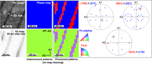
The crystallographic analysis of nanoscale phases with dimensions well below the spatial probing volume of electron backscatter diffraction (EBSD) traditionally rely on electron microscopy in transmission (either in SEM or TEM), because EBSD patterns are invariably dominated by the matrix phase contribution and present seemingly no trace from such nanoscale phases. Yet, this study shows that such nanoscale features generate a very faint but valuable secondary diffraction signal which can be retrieved. A diffraction pattern postprocessing method is presented which focuses on the detection of such secondary signal emitted by nanoscale minority phases in overlapped patterns dominated by a dominant matrix signal. The predominant, majority phase contribution in EBSD patterns is removed by a close-neighbor pattern subtraction routine, after which both the conventional Hough indexing method as well as pattern matching methods can be used to reveal the crystallography, spatial distribution, morphology, and orientation of nanoscale minority phases initially absent from EBSD maps. Nanolamellar pearlitic steel, which has long been out of reach for EBSD, has been chosen as an application example.
Keywords
- Type
- Software and Instrumentation
- Information
- Copyright
- Copyright © The Author(s), 2022. Published by Cambridge University Press on behalf of the Microscopy Society of America
References
Bhadeshia, H (2018). Solution to the Bagaryatskii and Isaichev ferrite–cementite orientation relationship problem. Mater Sci Technol 34, 1666–1668.CrossRefGoogle Scholar
Brodu, E & Bouzy, E (2017). Depth resolution dependence on sample thickness and incident energy in on-axis transmission Kikuchi diffraction in scanning electron microscope (SEM). Microsc Microanal 23, 1096–1106.CrossRefGoogle Scholar
Durgaprasad, A, Giri, S, Lenka, S, Kundu, S, Mishra, S, Chandra, S, Doherty, RD & Samajdar, I (2017). Defining a relationship between pearlite morphology and ferrite crystallographic orientation. Acta Mater 129, 278–289.CrossRefGoogle Scholar
Eggeman, A, Krakow, R & Midgley, A (2015). Scanning precession electron tomography for three-dimensional nanoscale orientation imaging and crystallographic analysis. Nat Commun 6, 7267.CrossRefGoogle ScholarPubMed
Fullwood, DT, Sanderson, S, Baird, S, Christensen, J, Homer, ER & Johnson, OK (2021). Determining grain boundary position and geometry from EBSD data: Limits of accuracy. Microsc Microanal 28, 1–13.Google Scholar
Guo, N & Liu, Q (2012). Back-scattered electron imaging combined with EBSD technique for characterization of pearlitic steels. J Microsc 246, 221–228.CrossRefGoogle ScholarPubMed
Kobler, A & Kubel, C (2018). Towards 3D crystal orientation reconstruction using automated crystal orientation mapping transmission electron microscopy (ACOM-TEM). Beilstein J Nanotechnol 9, 602–660.CrossRefGoogle Scholar
Lenthe, WC, Germain, L, Chini, MR, Gey, N & De Graef, M (2020). Spherical indexing of overlap EBSD patterns for orientation-related phases – Application to titanium. Acta Mater 188, 579–590.CrossRefGoogle Scholar
Liu, HH, Schmidt, S, Poulsen, HF, Godfrey, A, Liu, ZQ, Sharon, JA & Huang, X (2011). Three-dimensional orientation mapping in the transmission electron microscope. Science 332, 833–834.CrossRefGoogle ScholarPubMed
Masoumi, M, Batista Delima, N, Tressia, G, Sinatora, A & Goldenstein, H (2019). Microstructure and crystallographic orientation evolutions below the superficial white layer of a used pearlitic rail. Journal of Materials Research and Technology 8, 6275–6288.CrossRefGoogle Scholar
Ram, F & De Graef, M (2018). Phase differentiation by electron backscatter diffraction using the dictionary indexing approach. Acta Mater 144, 352–364.CrossRefGoogle Scholar
Shi, Q, Zhou, Y, Zhong, H, Loisnard, D, Dan, C, Zhang, F, Chen, Z, Wang, H & Roux, S (2021). Indexation of electron diffraction patterns at grain boundaries. Mater Charact 182, 111553.CrossRefGoogle Scholar
Singh, S, Guo, Y, Winiarski, B, Burnett, TL, Withers, PJ & De Garef, M (2018). High resolution low kV EBSD of heavily deformed and nanocrystalline aluminium by dictionary-based indexing. Nat Sci Rep 8, 10991.CrossRefGoogle ScholarPubMed
Sitzman, SD, Nolze, G & Nowell, MM (2010). EBSD pattern quality and its use in evaluating sample surface condition. Microsc Microanal 16(Suppl 2), 698–699.CrossRefGoogle Scholar
Sneddon, GC, Trimby, PW & Cairney, JM (2016). Transmission Kikuchi diffraction in a scanning electron microscope: A review. Mater Sci Eng R 110, 1–12.CrossRefGoogle Scholar
Takahashi, T, Ponge, D & Raabe, D (2007). Investigation of orientation gradients in pearlite in hypoeutectoid steel by use of orientation imaging microscopy. Steel Res Int 78, 38–44.CrossRefGoogle Scholar
Valery, A, Rauch, EF, Clement, L & Lorut, F (2017). Retrieving overlapping crystals information from TEM nano-beam electron diffraction patterns. J Microsc 268, 208–218.10.1111/jmi.12599CrossRefGoogle ScholarPubMed
Winkelmann, A, Jablon, BM, Tong, VS, Trager-Cowan, C & Mingard, KP (2020). Improving EBSD precision by orientation refinement with full pattern matching. J Microsc 277, 79–92.CrossRefGoogle ScholarPubMed
Winkelmann, A & Nolze, G (2010). Analysis of Kikuchi band contrast reversal in electron backscatter diffraction patterns of silicon. Ultramicroscopy 110, 190–194.CrossRefGoogle ScholarPubMed
Winkelmann, A, Nolze, G, Cios, G & Tokarski, T (2018). Mapping of local lattice parameter ratios by projective Kikuchi pattern matching. Phys Rev Mater 2, 123803.CrossRefGoogle Scholar
Zhang, YD, Esling, C, Calcagnotto, M, Zhao, X & Zuo, L (2007). New insights into crystallographic correlations between ferrite and cementite in lamellar eutectoid structures, obtained by SEM–FEG/EBSD and an indirect two-trace method. J Appl Crystallogr 40, 849–856.CrossRefGoogle Scholar
Zhong, N, Wang, X, Guo, Z & Rong, Y (2011). Orientation relationships between ferrite and cementite by edge-to-edge matching principle. J Mater Sci Technol 27, 475–480.CrossRefGoogle Scholar