


No CrossRef data available.
Article contents
Calcium, Ca2+-ATPase, Calmodulin, and Calbindin D-28KD Localization in Testis of Leptodactylus chaquensis (Anura: Leptodactylidae)
Part of:
Micrographia Collection
Published online by Cambridge University Press: 17 March 2022
Abstract
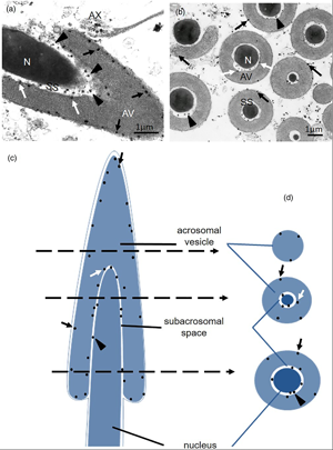
The intracellular localization of Ca2+, Ca2+-ATPase, Calmodulin, and Calbindin D-28KD have been studied in testes of the toad Leptodactylus chaquensis, using ultracytochemical and immunohistochemical techniques. The Ca2+ presences in the nucleus and into the mitochondria of the germ cells, together with the activity of Ca2+-ATPase detected in the nuclear envelope and mitochondrial crests, suggest the participation of this transporter in the storage of Ca2+. In Sertoli cells, Ca2+ deposits were also found in vesicles and lamellar bodies. Calmodulin and Calbindin D-28KD were revealed in the cytoplasm of both cell types. At the spermatozoon level, the cation deposits were located in the subacrosomal space and in the acrosomal vesicle. Ca2+-ATPase activity was observed in the acrosomal and plasma membranes of the gamete that suggests the existence of a transport system responsible for maintaining low cytoplasmic Ca2+ levels. The activity of Ca2+-ATPase and the location of Ca2+ deposits in gamete tail would be related to flagellar movement. The colocalization of Ca2+ deposits and their binding proteins in efferent duct cells would probably be associated with secretory activity. Considering that intracellular Ca2+ is present in different gonadal cells, this work would provide a better understanding of the cation importance in the testicular functions of this species.
- Type
- Micrographia
- Information
- Copyright
- Copyright © The Author(s), 2022. Published by Cambridge University Press on behalf of the Microscopy Society of America
References
Alavi, SMH & Cosson, J (2006). Sperm motility in fishes. (II) Effects of ions and osmolality: A review. Cell Biol Int 30, 1–14. doi:10.1016/j.cellbi.2005.06.004CrossRefGoogle ScholarPubMed
Ando, T, Fujimoto, K, Mayahara, H, Miyajima, H & Ogawa, K (1981). A new one-step method for the histochimestry and cytochimestry of Ca2+-ATPase activity. Acta Histochem Cytochem 14, 705–726.CrossRefGoogle Scholar
Andreuccetti, P, Denis-Donini, S, Burrini, AG & Campanella, C (1984). Calcium ultrastructural localization in Xenopus laevis eggs following activation by pricking or by calcium ionophore A23187. J Exp Zool 229, 295–308.CrossRefGoogle ScholarPubMed
Arenas Ríos, E, Cambrón Ruiz, A & Ambríz García, D (2010). Bases fisiológicas de la capacitación y de la reacción acrosomal del espermatozoide. ContactoS 78, 5–11.Google Scholar
Berridge, MJ, Bootman, MD & Roderick, HL (2003). Calcium signalling: Dynamics, homeostasis and remodelling. Nat Rev Mol Cell Biol 4(7), 517–529. doi:10.1038/nrm1155CrossRefGoogle ScholarPubMed
Borke, JL, Caride, A, Verma, AK, Penniston, JT & Kumar, R (1989). Plasma membrane calcium pump and 28-kDa calcium binding protein in cells of rat kidney distal tubules. Am J Physiol 257(5 Pt 2), F842–F849. doi:10.1152/ajprenal.1989.257.5.F842Google ScholarPubMed
Boschek, CB (2008). Different conformational switches underlie the calmodulin-dependent modulation of calcium pumps and channels. Biochemistry 47(6), 1640–1651. doi:10.1021/bi701987nCrossRefGoogle ScholarPubMed
Carafoli, E (1982). The transport of calcium across the inner membrane of mitochondria. In Membrane Transport of Calcium, Carafoli, E (Ed.), pp. 109–1399. London: Academic Press.Google Scholar
Carafoli, E, Santella, L, Branca, D & Brini, M (2001). Generation, control and processing of cellular calcium signals. Crit Rev Biochem Mol Biol 36, 107–260. doi:10.1080/20014091074183CrossRefGoogle ScholarPubMed
Crespo, CA, Medina, MF, Ramos, I & Fernández, SN (2014). Homeostasis and secretion of calcium in the oviductal mucosa of toad Rhinella arenarum. J Exp Zool A Ecol Genet Physiol 321(8), 432–441. doi:10.1002/jez.1874CrossRefGoogle ScholarPubMed
Crespo, CA, Ramos, I, Medina, MF & Fernández, SN (2009). Analysis of Bufo arenarum oviductal secretion during the sexual cycle. Zygote 18(1), 60–80. doi:10.1017/S0967199409005462Google Scholar
Darszon, A, Beltrán, C, Félix, R, Nishigaki, T & Treviño, CL (2001). Ion transport in sperm signaling. Dev Biol 240, 1–14. doi:10.1006/dbio.2001.0387CrossRefGoogle ScholarPubMed
Darszon, A, Nishigaki, T, Beltrán, C & Treviño, CL (2011). Calcium channel in the development, maturation, and function of spermatozoa. Physiol Rev 91, 1305–1355. doi:10.1152/physrev.00028.2010CrossRefGoogle ScholarPubMed
Félix, R (2005). Molecular physiology and pathology of Ca2+-conducting channels in the plasma membrane of mammalian sperm. Reproduction 129(3), 251–262. doi:10.1530/rep.1.00478CrossRefGoogle ScholarPubMed
Feng, HL, Hershlag, A, Han, YB & Zheng, LJ (2006). Localizations of intracellular calcium and Ca2+-ATPase in hamster spermatogenic cells and spermatozoa. Micros Res Tech 69, 618–623. doi:10.1002/jemt.20329CrossRefGoogle ScholarPubMed
Golpour, A, Pšenička, M & Niksirat, H (2016 a). Subcellular localization of calcium deposits during zebrafish (Danio rerio) oogenesis. Micron 80, 6–13. doi:10.1016/j.micron.2015.09.004CrossRefGoogle ScholarPubMed
Golpour, A, Pšenička, M & Niksirat, H (2016 b). Ultrastructural localization of intracellular calcium during spermatogenesis of sterlet (Acipenser ruthenus). Micros Microanal 22, 1155–1161. doi:10.1017/S1431927616011958CrossRefGoogle Scholar
Golpour, A, Pšenička, M & Niksirat, H (2017). Subcellular distribution of calcium during spermatogenesis of zebrafish, Danio rerio. J Morphol 278, 1149–1159. doi:10.1002/jmor.20701CrossRefGoogle ScholarPubMed
Grasso, P & Reichert, LE Jr. (1989). Follicle-stimulating hormone receptor-mediated uptake of 45 Ca2+ by proteoliposomes and cultured rat Sertoli cells: Evidence for involvement of voltage-activated and voltage-independent calcium channels. Endocrinology 125, 3029–3036. doi:10.1210/endo-125-6-3029CrossRefGoogle Scholar
Hagiwara, Y & Dan, JC (1969). Effect of lack of calcium on the starfish acrosome. Dev Growth Differ 11(1), 29–39. doi:10.1111/j.1440-169x.1969.00029.xCrossRefGoogle ScholarPubMed
Herpetological Animal Care and Use Committee (HACC) (2004). Guidelines for the Use of Live Amphibians and Reptiles in Field and Laboratory Research, 2th ed. American Society of Ichthyologists and Herpetologists. Lawrence, Kansas, USA.Google Scholar
Herrera, E, Salas, K, Lagos, N, Benos, DJ & Reyes, JG (2001). Temperature dependence of intracellular Ca2+ homeostasis in rat meiotic and postmeiotic spermatogenic cells. Reproduction 122, 545–551. doi:10.1530/rep.0.1220545CrossRefGoogle ScholarPubMed
Hiroshi, N & Vacquier, VD (2003). Store-operated calcium channels trigger exocytosis of the sea urchin sperm acrosomal vesicle. Biochem Biophys Res Commun 304, 285–292. doi:10.1016/s0006-291x(03)00587-4CrossRefGoogle Scholar
Inpanbutr, N & Taylor, AN (1992). Expression of calbindin-D28k in developing and growing chick testes. Histochemistry 97(4), 335–339. doi:10.1007/BF00270035CrossRefGoogle ScholarPubMed
Iruzubieta Villagra, L, Ramos, I, Cisint, S, Crespo, CA & Fernández, SN (2018). Electron microscopy observations on testis and spermatozoa of Leptodactylus chaquensis (Anura, Leptodactylidae). Micron 105, 35–46. doi:10.1016/j.micron.2017.11.007CrossRefGoogle Scholar
Jones, KT (2007). Intracellular calcium in the fertilization and development of mammalian eggs. Clin Exp Pharmacol Physiol 34, 1084–1089. doi:10.1111/j.1440-1681.2007.04726.xCrossRefGoogle ScholarPubMed
Jülich, G, Haider, SG, Passia, D & Goslar, HG (1982). Jahreszeitliche Veränderungen der Adenosintriphosphatase-Aktivität in den Germinalcysten des Testis vom Grasfrosch Rana temporaria. Eine enzymhistochemische Studie [Seasonal changes in the adenosine triphosphatase activity in the germinal cysts of the testis of the common frog Rana temporaria. An enzyme histochemical study]. Acta Histochem 71(2), 191–200. In German.CrossRefGoogle Scholar
Klein, RL, Yen, SS & Thureson-Klein, A (1972). Critique on the K-pyroantimoniante method for semiquantitative stimation of cations on conjunction with electron microscopy. J Histochem Cytochem 20, 65–78. doi:10.1177/20.1.65CrossRefGoogle Scholar
Krapf, D, O'Brien, E, Maidagán, PM, Morales, ES, Visconti, PE & Arranz, SE (2014). Calcineurin regulates progressive motility activation of Rhinella (Bufo) arenarum sperm through dephosphorylation of PKC substrates. J. Cell. Physiol 229, 1378–1386. doi:10.1002/jcp.24571CrossRefGoogle ScholarPubMed
Mann, T & Lutwak-Mann, C (1981). Male Reproductive Function and Semen. Springer, London: Springer-Verlag. doi:10.1007/978-1-4471-1300-3.CrossRefGoogle Scholar
Means, AR, Dedman, JR, Tash, JS, Tindall, DJ, van Sickle, M & Welsh, MJ (1980). Regulation of the testis Sertoli cell by follicle stimulating hormone. Annu Rev Physiol 42, 59–70. doi:10.1146/annurev.ph.42.030180.000423CrossRefGoogle ScholarPubMed
Means, AR & Rasmussen, CD (1988). Calcium, calmodulin, and cell proliferation. Cell Calcium 9, 313–319. doi:10.1016/0143-4160(88)90012-7CrossRefGoogle ScholarPubMed
Medina, MF (2005). Componentes de la secreción oviductal involucrados en la fecundación. Tesis Doctoral. Universidad Nacional de Tucumán, Argentina.Google Scholar
Medina, MF, Crespo, CA, Ramos, I & Fernández, SN (2009). Role of cations as components of jelly coats in Bufo arenarum fertilization. Zygote 18, 69–80. doi:10.1017/S0967199409990037CrossRefGoogle ScholarPubMed
Medina, MF, Crespo, CA, Ramos, I & Fernández, SN (2012). Effect of oviductal secretion components on the fertilizing capacity of amphibian sperm: Biological and ultrastructural studies. Micron 43(2–3), 223–228. doi:10.1016/j.micron.2011.08.001CrossRefGoogle ScholarPubMed
Niksirat, H, Andersson, L, James, P, Kouba, A & Kozák, P (2014). Proteomic profiling of the signal crayfish Pacifastacus leniusculus egg and spermatophore. Anim Reprod Sci 149(3–4), 335–344. doi:10.1016/j.anireprosci.2014.07.024CrossRefGoogle ScholarPubMed
Niksirat, H, James, P, Andersson, L, Kouba, A & Kozák, P (2015). Label-free protein quantification in freshly ejaculated versus post-mating spermatophores of the noble crayfish Astacus astacus. J Proteomics 123, 70–77. doi:10.1016/j.jprot.2015.04.004CrossRefGoogle ScholarPubMed
Niksirat, H & Kouba, A (2016). Subcellular localization of calcium deposits in the noble crayfish Astacus astacus spermatophore: Implications for post-mating spermatophore hardening and spermatozoon maturation. J Morphol 277(4), 445–452. doi:10.1002/jmor.20509CrossRefGoogle ScholarPubMed
Ravindranath, N, Papadopoulos, V, Vornberger, W, Zitzmann, D & Dym, M (1994). Ultrastructural distribution of calcium in the rat testis. Biol Reprod 51, 50–62. doi:10.1095/biolreprod51.1.50CrossRefGoogle ScholarPubMed
Sakata, Y, Saegusa, H, Zong, S, Osanai, M, Murakoshi, T, Shimizu, Y, Noda, T, Aso, T & Tanabe, T (2002). Ca(v)2.3 (alpha1E) Ca2+ channel participates in the control of sperm function. FEBS Lett 516, 229–233. doi:10.1016/s0014-5793(02)02529-2CrossRefGoogle ScholarPubMed
Santi, CM, Darszon, A & Hernández-Cruz, A (1996). A dihydropyridine-sensitive T-type Ca2+ current is the main Ca2+ current carrier in mouse primary spermatocytes. Am J Physiol 271(5 Pt 1), C1583–C1593. doi:10.1152/ajpcell.1996.271.5.C1583CrossRefGoogle ScholarPubMed
Spicer, SS, Hardin, JH & Greene, WB (1968). Nuclear precipitates in pyroantimonate-osmium tetroxide-fixed tissues. J Cell Biol 39, 216–221. doi:10.1083/jcb.39.1.216CrossRefGoogle ScholarPubMed
Sullivan, MH & Cooke, BA (1986). The role of [Ca2+]i in steroidogenesis in Leydig cells. Stimulation of intracellular free Ca2+ by lutropin (LH), luliberin (LHRH agonist), and cyclic AMP. Biochem J 236, 45–51. doi:10.1042/bj2360045CrossRefGoogle Scholar
Suzuki, S & Sugi, H (1989). Evaluation of the pyroantimonate method for detecting intracellular calcium localization in smooth muscle fibers by the X-ray microanalysis of cryosections. Histochemistry 92(2), 95–101. doi:10.1007/BF00490226CrossRefGoogle ScholarPubMed
Tidow, H, Hein, KL, Baekgaard, L, Palmgren, MG & Nissen, P (2010). Expression, purification, crystallization and preliminary X-ray analysis of calmodulin in complex with the regulatory domain of the plasma-membrane Ca2+-ATPase ACA8. Acta Crystallogr Sect F Struct Biol Cryst Commun 66(Pt 3), 361–363. doi:10.1107/S1744309110003805CrossRefGoogle ScholarPubMed
Timmermans, JAH, Bindels, RJM & Van Os, CH (1995). Stimulations of plasma membrane Ca2+ pump by calbindina D-28K and calmodulin is additive in EGTA-free solutions. J Nutr 125, 1981–1986. doi:10.1093/jn/125.suppl_7.1981SCrossRefGoogle Scholar
Treviño, CL, Santi, CM, Beltrán, C, Hernández-Cruz, A, Darszon, A & Lomeli, H (1998). Localization of inositol trisphosphate and ryanodine receptors during mouse spermatogenesis: Possible functional implications. Zygote 6(2), 159–172. doi:10.1017/s0967199498000094CrossRefGoogle Scholar
Vacca, LL (1982). Duoble bridge techniques of immunocytochemistry. In Techniques in Immunocytochemistry, Bullock, GR & Petrusz, P (Eds.), pp. 154–182. New York: Academic Press.Google Scholar
Williams-Ashman, HG (1988). Perspectives in the male sexual physiology of eutherian mammals. In The Physiology of Reproduction, Knobil, E & Neill, J (Eds.), pp. 727–751. New York: Raven Press.Google Scholar
Yanagimachi, R (2011). Mammalian sperm acrosome reaction: Where does it begin before fertilization? Biol Reprod 85(1), 4–5. doi:10.1095/biolreprod.111.092601CrossRefGoogle ScholarPubMed
Yanagimachi, R & Usui, N (1974). Calcium dependence of the acrosome reaction and activation of guinea-pig spermatozoa. Exp Cell Res 89, 161–174. doi:10.1016/0014-4827(74)90199-2CrossRefGoogle ScholarPubMed
Yoshida, M & Yoshida, K (2011). Sperm chemotaxis and regulation of flagellar movement by Ca2+. Mol Hum Reprod 17(8), 457–465. doi:10.1093/molehr/gar041CrossRefGoogle ScholarPubMed