1. Introduction
In various bone and bone structure diseases and defects, the use of innovative biomaterials represents a new trend in the treatment of these pathological conditions. Surgery using these materials is often accompanied by inflammatory reactions at the site of intervention, and severe, longer-lasting inflammatory processes can lead to a change in the symmetry of the bones or joints. Early detection and assessment of the severity of the inflammatory pre-process using certain inflammatory blood biomarkers and serum enzymes can be of great help in early detection of the inflammatory process, initiating adequate therapy, improving prognosis and preventing severe changes in tissue structure and site symmetry.
Nowadays, articular cartilage research is even more important due to the increasing incidence of cases with osteoarthritis and rheumatoid arthritis. These diseases are caused by alterations in the structure and functionality of articular cartilage [
1]. Since articular cartilage has no direct blood supply and is of low cell density, which allows only extremely limited self-renewal, cartilage defects heal very poorly compared to other tissues in the body [
2]. Several surgical treatment options to enhance cartilage repair have been developed. Techniques involving the subchondral bone, for example, microfracture, as well as techniques that do not disturb the cement line, for example, autologous chondrocyte implantation or the use of seeding cells and scaffolds, are commonly employed [
3,
4]. Nowadays, regenerative medicine deals with the development of biomaterials that can mimic cartilage matrix and restore function at the defect site [
5,
6]. Currently used biomaterials must be biocompatible, noncarcinogenic, corrosion-resistant, and has low toxicity and wear [
7,
8]. The use of bone cement is one of the methods applicable for cartilage reconstruction. Calcium phosphate cement (CPC) is similar to the inorganic phase of bone, which allows its application in bone reconstruction [
9]. CPC is characterized by positive biological activities (biocompatibility, osteoconductivity, osteoinductivity), and lower exothermic reaction temperature. For these reasons, CPC has become one of the most promising bone repair materials for the maxillofacial area, dentistry and orthopedics [
10]. One of the ways to evaluate the response of the organism to the material used, the type of cartilage reconstruction and the efficacy of treatment is the assessment of the acute phase response through the measurement of acute phase reactants.
Acute phase proteins (APPs) represent a group of proteins that change in concentration in response to alterations in homeostasis, tissue injury, inflammation, infections, trauma or necrosis. In addition to their importance as an indicator of disease, their main diagnostic importance is that the concentrations of APPs are correlated with the degree of tissue damage, the severity of the disease, and the rate of recovery of impaired functions after treatment [
11]. Furthermore, they may be useful markers in assessing disease progression, monitoring and control of post-operative conditions, as well as in monitoring treatment efficacy [
12]. From this point of view, some APPs might be useful markers for the evaluation of post-operative complications also after cartilage reconstruction, and for assessing the extent of surgical trauma. Regenerative medicine is an intensively developing area of research, with many published results, but a general understanding of the inflammatory responses in pigs following the reconstruction of cartilage defects is still lacking in the published literature. Little is known about the response of the organism to calcium phosphate cement regarding cartilage repair, especially which combinations of inflammatory markers would be the best for providing useful information about the ongoing inflammatory process in the experimental animals. Therefore, the aim of this study was to evaluate the alterations in the values of five APPs and the activities of some enzymes in pigs within the first 30 days following the restoration of experimentally induced articular cartilage defects using the tetracalcium phosphate/nanomoneite cement powder with amino acids (CAL).
2. Materials and Methods
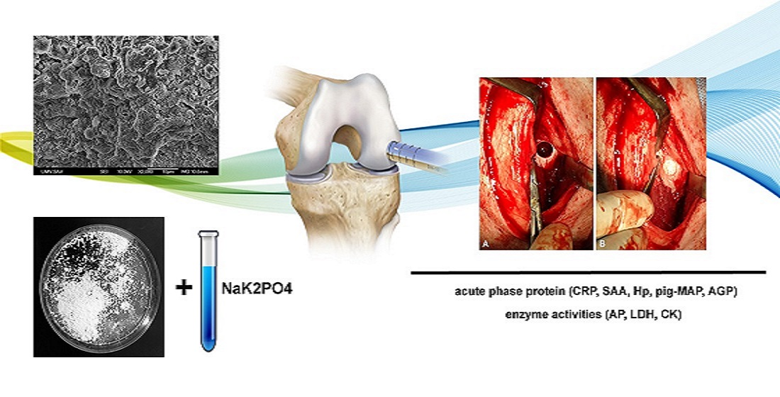
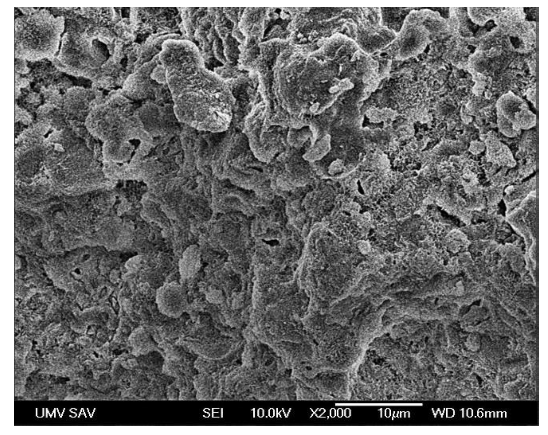
2.1. Preparation of Biomaterials
Tested tetracalcium phosphate cement (CAL) containing four amino acids (L-proline/glycine/4-hydroxyproline/L-lysine = 22:42:22:14—ratio of individual compounds) which have been added and dissolved in orthophosphoric acid solution. The content of amino acids in CAL cement per 1000 mg was as follows: L-proline 8.9 mg/glycine 17.8 mg/4-hydroxyproline 8.9 mg/L-lysine 4.4 mg. The cement powder has Ca/P mole ratio close to 1.67. The final CAL cement powder contained 4 wt% of amino acids. Tetracalcium phosphate (Ca
4(PO
4)
2O, TTCP) was prepared by the synthesis of an equimolar mixture consisting of calcium carbonate (CaCO
3, analytical grade, Merck KGaA, Darmstadt, Germany) and dicalcium phosphate anhydrous (DCPA) (CaHPO
4 (Ph.Eur.), Fluka, Buchs, Switzerland) using the temperature 1450 °C for 5 h. The planetary ball mill (Fritsch, 730 rpm, ZrO
2 balls and vessel) was used for 2 h regrading to preparation of the final product. Subsequently, the purity was analyzed using X-ray powder diffraction analysis (XRD, Philips X Pert Pro). The CAL injectable biocement scaffold was prepared by the mixture of the CAL cement powder with the hardening liquid (2% NaH
2PO
4 sterile solution;
Figure 1).
2.2. Animals
The blood samples and all procedures with animals were performed in accordance with the ethical standards and guidelines approved by the institutional Committee on protection of animals used for scientific purposes and complied with the institutional Code of Ethics for Scientists - Directive 74/2019/UVLF. The study was approved by the Ethics Committee of the State Veterinary and Food Administration of Slovakia under the No. 4650/17-221.
Seven clinically healthy Large White and Landrace female pigs from a breeding farm with good herd health management (PD Agro, Michalovce, Slovakia) were selected. The animals were 5 months old, and their body weight was 75.8 ± 1.9 kg at arrival to the Clinic of Swine of the University of Veterinary Medicine and Pharmacy in Kosice (Slovakia), where they were acclimatized 30 days before starting the experiment. At the clinic, two or three pigs were housed together in a solid floor pen with straw bedding. After surgery, they were housed in pens individually. Commercial feed mixture was provided according to the category of pigs, and they had free access to drinking water. Standard physical examination was performed before the inclusion of pigs into the study. This was oriented to the evaluation of the general health state of the animals, including feed intake, behavior, respiratory rates, as well as measuring of body temperature [
13].
The health state of the pigs was evaluated daily throughout the whole study period. In addition to the monitoring of general health state, local inflammatory signs in the surgical wound (heat, swelling, pain, discharge) were assessed after the surgical intervention. Furthermore, signs and grade of lameness were evaluated according to the lameness scoring system (from no detectable lameness, through minor to major lameness) described by Main et al. [
14].
2.3. Anaesthesia, Surgical Procedure and Postoperative Care
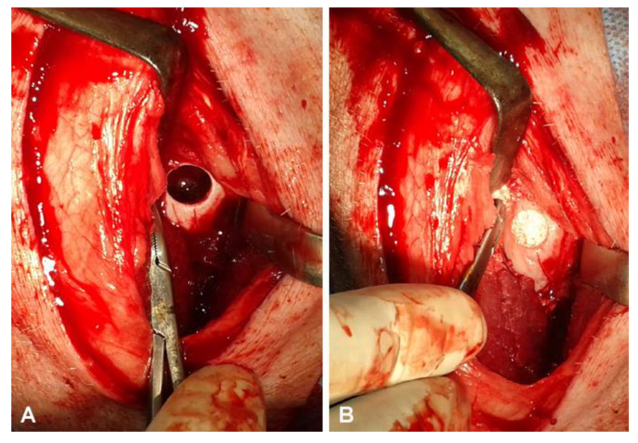
Anesthesia, the surgical procedure (creation of defect, filling of the defect with biocement,
Figure 2), and postoperative care were described by Tóthová et al. [
15].
2.4. Sample Collection
The effect of the surgical intervention on health state in the experimental animals was evaluated through the measurement of the concentrations of serum amyloid A (SAA, mg/L), haptoglobin (Hp, g/L), C-reactive protein (CRP, mg/L), α1-acid glycoprotein (AGP, g/L), pig major acute phase protein (pig-MAP, g/L), and for the enzyme activities of creatine kinase (CK, µkat/L), alkaline phosphatase (AP, µkat/L), and lactate dehydrogenase (LD, µkat/L) in blood samples. Blood was collected from v. jugularis externa one day before surgery. Further samples were obtained on days 7, 14 and 30 after surgery. Blood samples were taken into 4.4 mL serum gel separator tubes without additives and anticoagulants (Sarstedt, Nümbrecht, Germany). After allowing blood samples to coagulate at room temperature, sera were obtained by centrifugation at 4000 g for 15 min and then transferred to Eppendorf tubes for biochemical analyses. The remaining aliquots of sera were kept frozen at −20 °C for further laboratory analyses.
2.5. Laboratory Analyses
Commercial multispecies enzyme-linked immunosorbent assay (ELISA) kits (TP-802, Tridelta Development, Kildare, Ireland) were used for the quantification of SAA values. Porcine CRP was analyzed by solid-phase ELISA immunoassay using TA-901 kits (Tridelta Developmet, Kildare, Ireland). Haptoglobin was determined spectrophotometrically in microplates using colorimetric kits (TP-801, Tridelta Development, Kildare, Ireland). Porcine AGP was tested using commercially available Porcine A1AGP sandwich ELISA kit (MBS051651, MyBioSource, San Diego, CA, USA) and the concentrations of pig-MAP were measured by sandwich ELISA with monoclonal antibodies using commercial ACUVET pig-MAP ELISA kit (AC/PME 01, ACUVET BIOTECH, Zaragoza, Spain). For all analysis, serum samples were tested according to the manufacturer’s instructions. The absorbance was read on automatic microplate reader Opsys MR (The Dynex Technologies, Chantilly, VA, USA). The concentrations of the studied parameters were calculated from the calibration curve generated with the use of the computer software Revelation QuickLink version 4.25 (Dynex Technologies, Chantilly, VA, USA). The activities of serum CK, AP, and LD were determined by clinical chemistry assay kits (Randox, Crumlin, UK) using an automated biochemical analyzer Alizé (Lisabio, Poully en Auxois, France).
2.6. Statistical Analyses
The arithmetic means (x) and standard deviations (SD) were calculated using descriptive statistical procedures. Kolmogorov–Smirnov Test for normality was applied to evaluate the distribution of data. The normally distributed data with equal variance were subjected to repeated-measures one-way ANOVA to assess the changes during the perioperative period, the non-parametric Friedman test was used for non-normally distributed data. The significance of differences in values between the different sample collection times was evaluated by Tukey–Kramer and Dunn’s Multiple Comparisons post hoc test. Significance was considered at the 5% probability level. All calculations were performed by the GraphPad Prism V5.02 (GraphPad Software Inc., San Diego, CA, USA) computer program.
3. Results
No signs of inflammation of the surgical wound were detected by clinical examination, and no discharge from the wound was observed in any of the animals. Four of the seven pigs showed reduction of appetite and clinical signs of lameness without inflammatory processes in other organ systems. They had locomotor problems with failure to get up and walk normally, they had an abnormal posture and disturbed gait. In three animals, the basic clinical parameters (appetite, behavior, and rectal temperature) were not affected and showed improvement with no serious complications.
The average values of acute phase proteins recorded during the perioperative period are shown in
Table 1. The concentrations of SAA, Hp, CRP and pig-MAP showed statistically significant dynamics of changes during the perioperative period. The concentrations of SAA were very low before the surgical intervention and increased significantly (approximately 100-fold on average) 7 days after surgery. In the next postoperative period, its values decreased gradually until day 30 post surgery. In the concentrations of Hp, a significant increase (approximately 4-fold) was recorded 7 days after surgery in relation to the values recorded prior to surgery. Its values decreased only slightly 14 days after surgery; they were, however, still significantly higher than those before surgery. A significant decrease of values was obtained 30 days after surgery compared to the values 7 days post surgery. A similar trend of significant increase 7 days after the surgical intervention (approximately 3-fold) was also observed in the concentrations of CRP. From day 14 after the repair, a gradual non-significant decrease was observed until day 30 after surgery. Similarly, the concentrations of pigMAP increased significantly (almost 3-fold) 7 days post surgery. Its values showed a gradual decrease from day 14 post surgery, and the concentrations recorded 30 days after the surgical intervention were significantly lower compared to those obtained 7 days after cartilage repair. In the values of AGP, no significant changes were recorded during the evaluated perioperative period.
The average activities of the evaluated enzymes are presented in
Table 2. The changes observed in the activity of CK during the perioperative period were significant. A significant increase in the values was found on day 7 after the surgical intervention compared to those obtained prior to surgery. The activity of CK was non-significantly lower after this period, and at the end of the monitored period was still higher than before surgery. Similar, but non-significant dynamics of changes during the evaluated period was found in the activity of LD, with increased activity 7 days after the intervention and a subsequent gradual decline until day 30 after surgery. An opposite trend of changes was recorded in the activity of AP. Its values were non-significantly lower 7 days after surgery and then started, compared to the values 7 days after surgery, to gradually increase until the end of the postoperative period. The highest mean AP activity was obtained 30 days after surgery, which was significantly higher than the value recorded 7 days post surgery. The changes observed in AP activity during the studied perioperative period were significant.
4. Discussion
Injectable CAL cements represent acellular CPC biomaterial with amino acids, which allows osteochondral defect treatment regardless of the shape of the defect site. In our previous work, macroscopic, histological and radiological analyses of osteochondral defects in pigs clearly showed the excellent healing process after treatment with CAL. There was no evidence of inflammation three months after the implantation. The newly formed hyaline-like tissue had morphological and structural features comparable to the untreated cartilage tissue, with good integration to the adjacent tissue [
16]. Although CPC has a poor degradability due to the fact that CPC scaffolds without any modification are microporous, and such scaffolds have limited pore interconnections [
17]. Macroporosity within CPCs enables faster degradation of CPC scaffold by increasing of interaction with the biological adjacent tissue. Pore sizes smaller then 100 μm may result in ingrowth of unmineralized bone tissue or fibrous tissue [
18].
The pig as an experimental animal model in orthopedics has some advantages, such as joint size, joint loading mechanisms, weight, lack of spontaneous healing of any significant defects, bone trabecular thickness and the arrangement of the collagen network, which mimics a human joint [
19,
20]. In cartilage research, scientists are most interested in searching for markers that may give early information on whether cartilage reconstruction has been effective and the healing process is proceeding normally without complications. It has been shown that systemic inflammatory markers can be used to monitor postoperative period and early detection of postoperative complications after surgical treatment of cartilage damage. Acute phase proteins belong to the protein markers that could also be successfully used in regenerative and reconstruction medicine, where they might be important in the early diagnosis of complications after reconstructing defects, detecting uncontrolled inflammatory responses, and preventing long-term convalescence. It was shown by Aulin et al. [
21] in rabbits that the concentrations of SAA increased profoundly after the creation of full thickness osteochondral defect in the femorotibial joints and decreased to initial values four weeks postoperatively. A marked inflammatory response was also observed by Tóthová et al. [
15] in pigs following the reconstruction of experimentally created defects of articular cartilage using tetracalcium phosphate/nanomonetite cement powder with and without amino acids (CAK and C), which was characterized by the increase of SAA, Hp and CRP, while their values reached the preoperative concentrations 1 month after the surgical intervention suggesting normal postoperative healing. In the present study, the reconstruction of surgically induced defects using the cement powder CAL was accompanied by a significant increase of SAA, Hp, as well as CRP after the surgical intervention. Although their concentrations decreased gradually within one-month post surgery, the values recorded at the end of this period were higher than those obtained prior to surgery. Furthermore, the standard deviations recorded in pigs after the surgical intervention were very high, reflecting different reactivity of animals to the induction of cartilage defects and their repair, operative trauma, abnormal postoperative healing, possible continuation of inflammatory processes, or insufficient treatment response in some animals [
22]. Another reason for the ongoing inflammatory processes within one month after implantation might be the slow degradation rate and the presence of microporosity in the used CAL scaffolds. The impairment of health state was also observable clinically in four of seven animals and manifested with appetite disorders and clinical signs of more serious lameness. In these animals, higher values of SAA, Hp and CRP were mainly observed. In addition to the above-mentioned factors, the type and composition of the used biocement material might affect the postoperative inflammatory processes, and thus the concentrations of these acute phase proteins in the experimental animals. Therefore, in light of these results, further studies would be important for determining whether this CPC powder with amino acids is suitable for possible cartilage regeneration in human or veterinary medicine.
The evaluation of some other potential APPs in pigs, including α
1-acid glycoprotein (AGP) and pig major acute phase protein (pig-MAP), may be useful in practice, providing important information for the examination of their health state and diagnosis of some diseases [
23,
24]. The data presented by González-Ramón et al. [
25] suggest that pig-MAP show promise as a good marker of acute inflammatory processes and pathologies in pigs. Marked increases in pig-MAP concentrations were found in different acute bacterial and experimental viral infections, as well as in pigs with several diseases in field conditions or in animals with surgical trauma [
26,
27,
28]. Furthermore, pig-MAP was found to be an excellent marker of distress and to determine the influence of stressors on the animal health status [
29,
30]. The presented study showed significantly elevated concentrations of pig-MAP 7 days after the reconstruction of surgically created defects of the articular cartilage when compared to preoperative concentrations, and then gradually decreased until the end of the one-month postoperative period. Alterations in the pig-MAP concentrations were found in surgically castrated piglets in the suckling period, with significantly higher values in surgically castrated than in intact males on the day 1 after castration, and these differences disappeared by the day 10 after castration [
31]. Studies dealing with the possible use of pig-MAP in reconstruction medicine to monitor postoperative inflammatory processes and to evaluate the healing processes after the repair of cartilage damage are still lacking, and need to be further studied.
The data regarding the usefulness of AGP in the determination of health disorders in pigs are not so consistent. In general, AGP in most animal species is described clearly as a positive acute phase protein. Elevated concentrations have also been reported in pigs with some inflammatory diseases, especially respiratory infections caused by
Actinobacillus pleuropneumoniae and
Mycoplasma hyopneumoniae [
23]. Surprisingly, AGP in pigs was found by Heegaard et al. [
32] to act as a negative acute phase reactant in several infections under experimental conditions and aseptic inflammatory processes with a significant decline of values. Despite the ongoing inflammatory responses after the restoration of experimental articular cartilage defects, evidenced by the significant increase of the SAA, Hp, CRP and pig-MAP concentrations, the obtained data suggest no marked alteration in the concentrations of AGP during the evaluated perioperative period. Although the values tended to slightly decrease in the postoperative period, the changes were not statistically significant. These results suggest that AGP is not a suitable marker for the evaluation of health disorders after the restoration of cartilage defects.
In the activity of CK, a significant increase was obtained 7 days after the reconstruction of cartilage defects using the biocement powder CAL, with a subsequent gradual decrease. This pattern is comparable to the changes observed by Tóthová et al. [
21] in pigs after the repair of created cartilage defects using the biocement powder C, and may be caused by the muscle damage after muscle-cutting surgery. Very similar, but not so marked alterations were found in the serum activity of LD following the restoration of cartilage, which similarly to CK may be related to the damage of skeletal muscle and injury due to the surgical procedure [
33]. An opposite trend was found in the activity of AP, showing a more marked decline in concentrations 7 days after the cartilage repair and a subsequent gradual elevation to slightly higher values when compared to those obtained prior to surgery. In tissue engineering, marked expression of AP indicates the success of osteogenesis, as its specific isoform is highly expressed in bones, and thus is the most frequently used biomarker for the evaluation of active osteoblastic bone formation and osteogenic activity [
34,
35]. According to Miao and Scutt [
36], the AP expression was also found in cartilage tissues, and compared to other enzymes, the time-shifted gradually increasing values observed in our study after cartilage repair might reflect this function. Because of limited literature data available about the changes in the AP activity after the restoration of defects in cartilage tissues, further studies would be helpful. Furthermore, because of some limitations of the study, including, e.g., the number of animals used and the wider range of standard deviations (e.g., SAA, CRP), further investigations are needed to obtain more satisfactory results.
5. Conclusions
In conclusion, in the study presented, the data indicate significant changes in the concentrations of SAA, Hp, CRP, as well as pig-MAP following the reconstruction of surgically created defects of articular cartilage using CAL cement. The results showed a marked increase in values 7 days after surgery and a subsequent gradual decrease until the end of the one-month postoperative period. However, the values recorded at the end of this period were higher compared to those obtained prior to surgery. In the concentrations of AGP, no significant alterations were observed in pigs during the evaluated perioperative period, making it less suitable for the monitoring of the period after the repair of cartilage defects. Changes were also found in the activities of CK and LD, with an increase 7 days post surgery, and then decreased gradually. The activity of AP decreased after surgery and started to increase until the end of the evaluated period. The presented findings suggest that SAA, Hp, CRP and pig-MAP might be applicable biomarkers of acute phase response for the monitoring of postoperative period after the repair of articular cartilage defects using calcium phosphate biocement powders.


 ,
, 


