Article

Whole-Genome Sequencing of Corallococcus sp. Strain EGB Reveals the Genetic Determinants Linking Taxonomy and Predatory Behavior

1
Key Laboratory of Agricultural Environmental Microbiology, Ministry of Agriculture and Rural Affairs, College of Life Sciences, Nanjing Agricultural University, Nanjing 210095, China
2
Institute of Botany, Jiangsu Province and Chinese Academy of Sciences, Nanjing 210014, China
3
Key Laboratory of Plant Immunity, Nanjing Agricultural University, Nanjing 210095, China
*
Author to whom correspondence should be addressed.
Genes 2021, 12(9), 1421; https://doi.org/10.3390/genes12091421
Submission received: 24 August 2021 / Revised: 13 September 2021 / Accepted: 13 September 2021 / Published: 15 September 2021
(This article belongs to the Section Microbial Genetics and Genomics)

Abstract

:
Corallococcus sp. strain EGB is a Gram-negative myxobacteria isolated from saline soil, and has considerable potential for the biocontrol of phytopathogenic fungi. However, the detailed mechanisms related to development and predatory behavior are unclear. To obtain a comprehensive overview of genetic features, the genome of strain EGB was sequenced, annotated, and compared with 10 other Corallococcus species. The strain EGB genome was assembled as a single circular chromosome of 9.4 Mb with 7916 coding genes. Phylogenomics analysis showed that strain EGB was most closely related to Corallococcus interemptor AB047A, and it was inferred to be a novel species within the Corallococcus genus. Comparative genomic analysis revealed that the pan-genome of Corallococcus genus was large and open. Only a small proportion of genes were specific to strain EGB, and most of them were annotated as hypothetical proteins. Subsequent analyses showed that strain EGB produced abundant extracellular enzymes such as chitinases and β-(1,3)-glucanases, and proteases to degrade the cell-wall components of phytopathogenic fungi. In addition, 35 biosynthetic gene clusters potentially coding for antimicrobial compounds were identified in the strain EGB, and the majority of them were present in the dispensable pan-genome with unexplored metabolites. Other genes related to secretion and regulation were also explored for strain EGB. This study opens new perspectives in the greater understanding of the predatory behavior of strain EGB, and facilitates a potential application in the biocontrol of fungal plant diseases in the future.

1. Introduction

Myxobacteria are a type of Gram-negative deltaproteobacteria that are distributed all over the world [1]. To date, hundreds of isolates of myxobacteria have been cultured from various natural sources including soil, compost, or trees. Unlike other bacteria, Myxobacteria exhibit several complex social behaviors, and one distinct trait is that their cells move by swarming or gliding on surfaces for social communication [2]. Once nutrients are depleted, fruiting bodies are born naked, followed by differentiation into myxospores. Due to their complex lifecycle, Myxobacteria produce abundant proteins to participate in signal transduction pathways, so as to coordinate cell–cell communication and regulate social motility [3]. In addition, myxobacteria prey on other microorganisms including fungi and bacteria, thus placing myxobacteria at the top of the microbial food chain [4,5]. The effective predation ability of myxobacteria is thought to rely on macromolecule degradation enzymes and secondary metabolites [6]. Myxobacteria can produce abundant secondary metabolites for potential applications as ecological weapons against living microorganisms [7]. Therefore, predatory behavior could make some myxobacteria suitable for the biological control of plant diseases. Five genera have been described within myxobacteria, and Myxococcus and Corallococcus are the two dominant genera, which possess ten and twelve species as of 2020 [8]. Among these, Myxococcus xanthus is the best-studied myxobacterium, and is widely used as an exemplar for the investigation of myxobacterial biology [9]. In contrast, species within the Corallococcus genus are poorly studied.
In our previous research, Corallococcus sp. strain EGB, a myxobacteria strain isolated from the soil, exhibited efficient biological control of various phytopathogenic fungi such as Fusarium oxysporum [10]. Several aspects, including hydrolytic activity and secondary metabolite, have been investigated to help understand its developmental and antifungal behaviors. A β-(1,3)-glucanase (lamC) derived from Corallococcus sp. strain EGB, displays exo-mode activity toward β-(1,3)/(1,6)-linked glucan substrates and endo-mode activity on β-(1,4)-linked glucan and xylan substrates, and shows lytic and antifungal activity against the plant pathogen Magnaporthe oryzae [11,12]. Another chitin hydrolase CcCti1 is identified from Corallococcus sp. strain EGB, and exhibits efficient antifungal activity against rice blast fungus [13]. Moreover, 32 volatile organic compounds have been identified from Corallococcus sp. strain EGB, and several compounds, such as isooctanol, could significantly inhibit the mycelial growth of F. oxysporum f. sp. cucumerinum and Penicillium digitatum [14]. These studies reveal that Corallococcus sp. strain EGB has potential applications in the control of plant pathogenic fungi; however, the detailed antifungal mechanism is still poorly investigated.
With the boom in high-throughput sequencing, whole-genome sequencing has been increasingly used in daily research. The availability of genome sequences not only allows for the rapid and reliable taxonomic classification of new strains, but also provides deeper insights into organismal evolution and gene functions [8]. Currently, more than 50 draft genomes are available for Myxococcus and Corallococcus strains, and the relatively larger genome size is a feature of Myxobacter [8,15]. These genomes possess several thousand proteins, many of which are hypothetical proteins. Comparative genomics analyses of organisms within the Myxococcaceae have clearly demonstrated many novel species, and Corallococcus genomes could group into two major clades [8,15]. Both the Corallococcus pan-genome and Myxococcus/Pyxidicoccus pan-genome are large and open, and Myxococcus/Pyxidicoccus spp. genomes are more diverse than those of Corallococcus species [8]. In both genera, a high number of biosynthetic gene clusters (BGCs) have been identified, and they are enriched in accessory pan-genomes with potential selective advantages during predation [8,15].
Here, we describe a novel Corallococcus genome, which displays efficient biological control of various phytopathogenic fungi. We assembled the chromosome-level genome of Corallococcus sp. strain EGB, and performed a comparative genome analysis with another ten species in the genus Corallococcus. The phylogenomics approach was used to confirm the taxonomic classification of strain EGB. Genes related to secretion, regulation, extracellular enzymes, and biosynthetic gene clusters (BGCs) were further analyzed to expand the understanding of the development and predatory behavior of strain EGB.

2. Materials and Methods

2.1. Culturing and DNA Isolation of Corallococcus sp. Strain EGB

Corallococcus sp. strain EGB (CCTCC No. M2012528) was previously isolated from saline soil, and was cultivated in VY/4 medium (1% yeast cells and 1% CaCl2, PH 7.0, w/v). Genomic DNA of Corallococcus sp. strain EGB was extracted using the methods described previously with minor modifications [16].

2.2. Genome Sequencing, Assembly, and Annotation

The genome of Corallococcus sp. strain EGB was sequenced using a hybrid strategy that combined the PacBio RS II platform and Illumina HiSeq 4000 platform at the Beijing Genomics Institute (BGI, Shenzhen, China). The PacBio subreads with lengths smaller than 1 kb were removed first, and the filtered subreads were used to perform self-correction using the Pbdagcon software, which resulted in corrected reads. After that, the Falcon tool was performed for genome assembly. To improve the accuracy of the genome assembly, GATK and SOAP tool packages were used to make single-base corrections. The complete genome sequence of Corallococcus sp. strain EGB was submitted to the NCBI GenBank with the accession number CP079946. Gene prediction and functional annotation were performed by the online platform Rapid Annotation using Subsystem Technology (RAST) [17]. To run the Clusters of Orthologous Groups (COG) annotation, only the best blast hit was retained for each protein [18].

2.3. Comparative Genomics Analysis

To conduct a comparative genomic analysis of Corallococcus sp. strain EGB with the other related bacterial species, the genome sequences of ten species belonging to the genus Corallococcus were retrieved from GenBank (Table 1). All the predicted protein sequences of 12 selected species were used to perform the all-versus-all blastp analysis with an E-value of 1 × 10⁻⁵ as the cut-off value. Then blast scores were delivered to the MCL Markov clustering program with an inflation parameter of 2, which resulted in rapid clustering of orthologous groups [19]. The core proteome was defined as proteins conserved in all the 11 Corallococcus genomes, and dispensable proteome consisted of proteins present in 2–10 Corallococcus genomes, while unique proteome was defined as proteins that only exist in the individual genome.

2.4. Phylogenetic Analysis

Whole-genome phylogeny was performed to confirm the taxonomic classification. Based on the clustering results of orthologous proteins, we randomly selected 135 single-copy genes, which were conserved in 12 analyzed species. Alignments of concatenated sequences of 135 single-copy genes were generated by the Muscle algorithm [20], and then the phylogenetic tree was constructed following the neighbor-joining algorithm with 1000 bootstrap replicates in MEGA7 [21]. The synteny of Corallococcus sp. strain EGB genome and other bacterial genomes was determined using Blast and Artemis Comparison Tool (ACT) [22]. The average nucleotide identity (ANI) between genomes was counted using orthoANI software [23]. The digital DNA-DNA hybridization (dDDH) values were calculated using the GGDC server with recommended formula 2 [24].

2.5. Bioinformatics Analyses

Putative secreted proteins of strain EGB were predicted using the SignalP v5 server [25]. Glycoside hydrolases were predicted by running dbCAN tool scans against the Carbohydrate-Active Enzymes (CAZy) database [26]. To identify peptidases, the protein sequences were used to perform Blast against the MEROPS database with an E-value cut-off of 1 × 10⁻⁵, and the produced peptidases were grouped based on peptidase family [27]. Biosynthetic gene clusters (BGCs) of each bacterial genome were predicted using the AntiSMASH v6 server [28].

3. Results

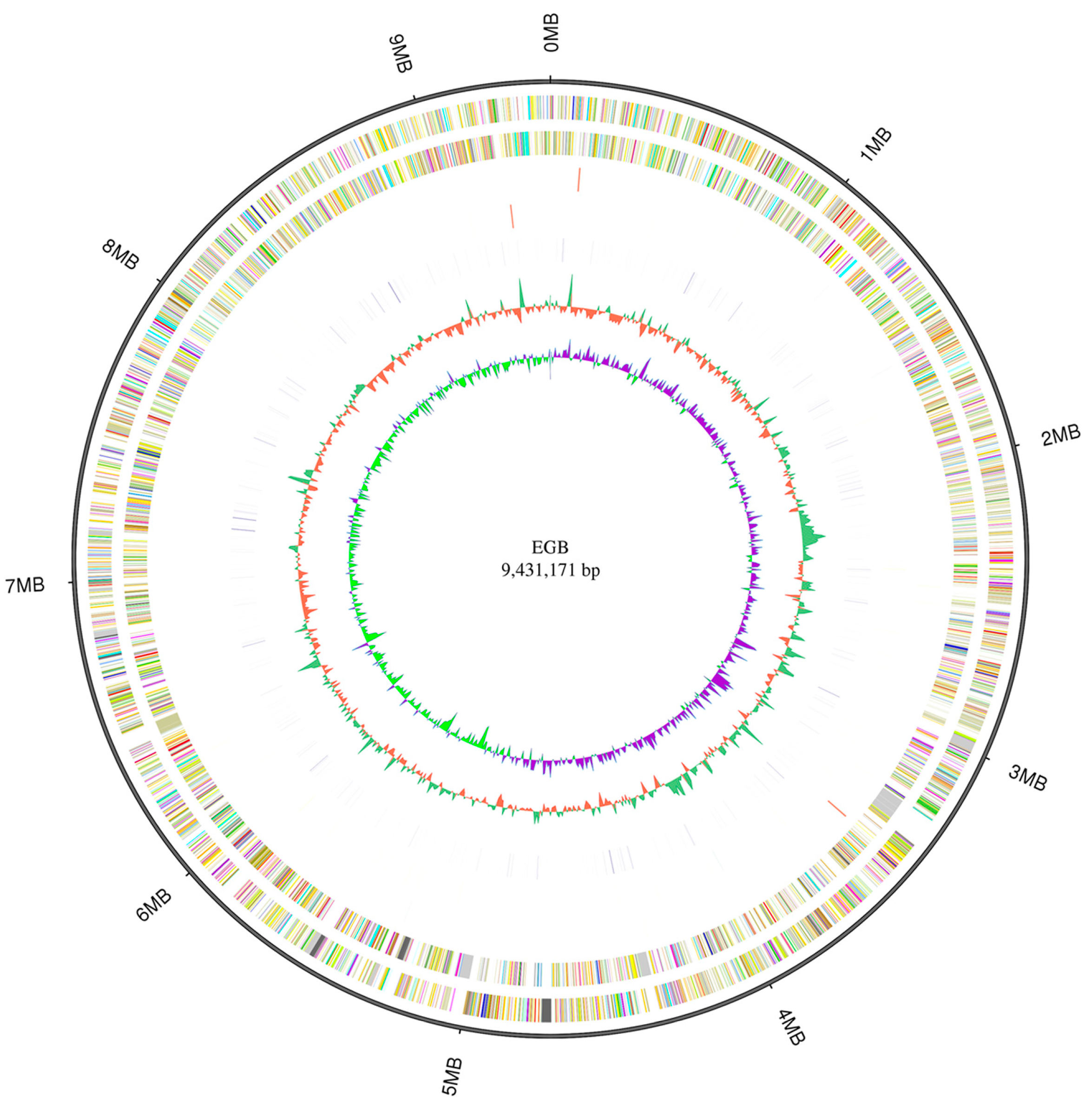
3.1. Genomic Characterization of Corallococcus sp. Strain EGB

The genome of Corallococcus sp. strain EGB was sequenced using a hybrid strategy that combined sequences from PacBio long reads and Illumina short reads. The genome was assembled as a single circular chromosome of 9.4 Mb with a high GC content of 70.4% (Figure 1; Table 1). In contrast to the genomes of 11 reported Corallococcus species, only C. coralloides strain DSM 2259 had a chromosome level genome that was similar to strain EGB, while the remaining 10 Corallococcus species only had draft genomes containing from 8 to 1491 contigs in each genome (Table 1), suggesting that strain EGB had a high-quality genome in genus Corallococcus. Subsequently, RAST-based annotation identified 7916 coding genes in the strain EGB genome, and 61.5% of the proteins were functionally annotated, while the remaining 38.5% of the proteins were hypothetical proteins. The nonrepetitive gene density in the strain EGB genome was 842 genes/Mb with an average gene length of 1082 bp. By contrast, the genome size and gene number of strain EGB were slightly smaller than the majority of other Corallococcus genomes (Table 1).

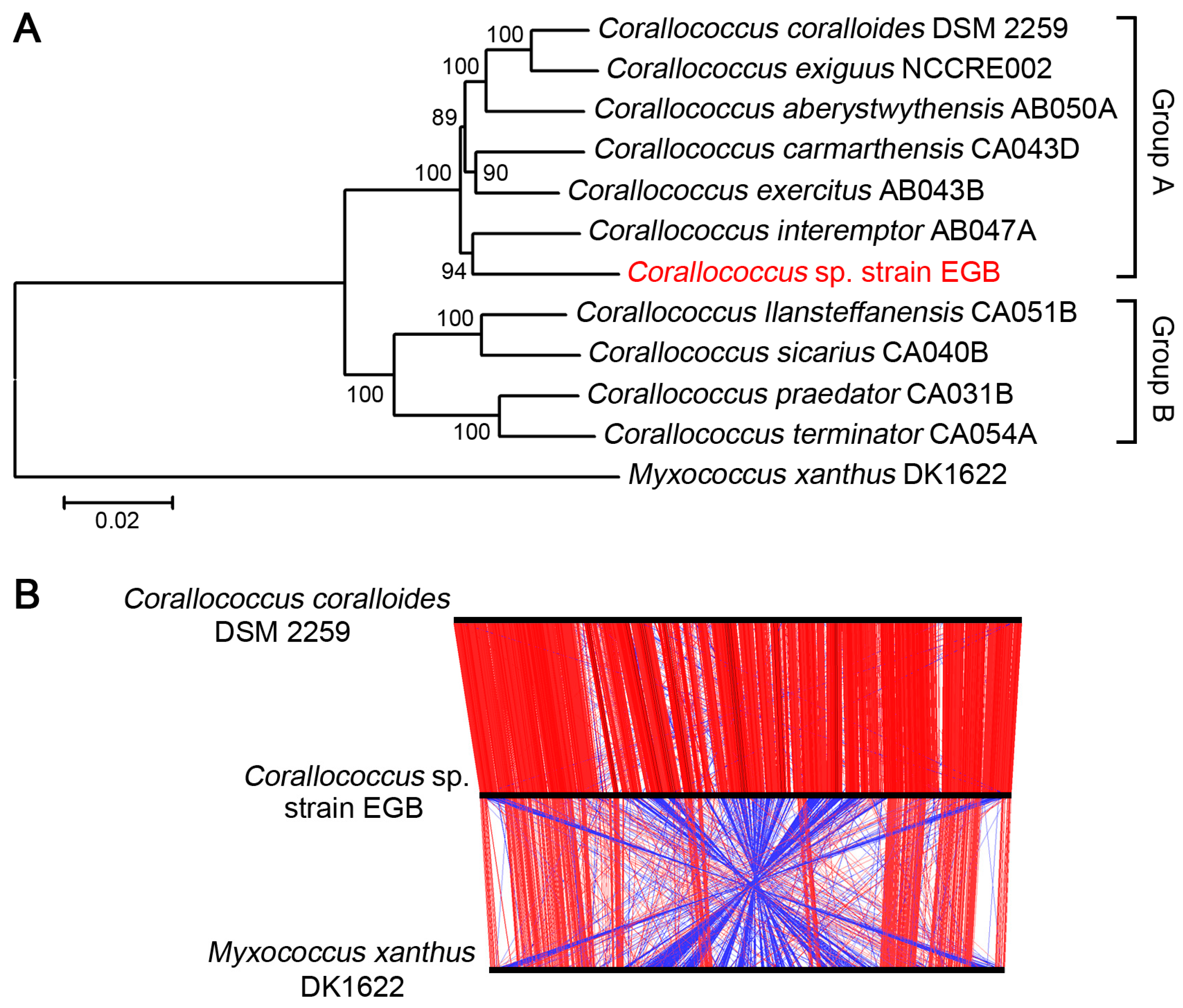
3.2. Strain EGB was Inferred as A Novel Species

In genus Corallococcus, more than ten species were reported. To establish the phylogenetic relationship of strain EGB with other related Corallococcus species, the phylogenetic tree was constructed using the concatenated sequences of 135 single-copy genes, which were conserved in all the analyzed genomes. The tree clearly showed that Corallococcus species grouped into two major clades, Group A and Group B (Figure 2A), which was consistent with previous taxonomy [15]. The strain EGB was located in the larger clade Group A, and clustered closely with C. interemptor AB047A (Figure 2A). Furthermore, the alignment of three chromosomal sequences of strain EGB, C. coralloides DSM 2259, and M. xanthus DK1622 was analyzed using ACT software. The results showed that strain EGB exhibited high synteny with C. coralloides DSM 2259; however, a relatively poor synteny with significant rearrangements was clearly observed between the genomes of strain EGB and M. xanthus DK1622 (Figure 2B).
The average nucleotide identity (ANI) and dDDH values between the above genomes were calculated. The results showed that ANI values ranged from 85.82% to 94.32% between the Corallococcus genomes. Notably, strain EGB had higher ANI values (90.79–91.51%) and dDDH values (41.2–43.7%) with C. interemptor AB047A, C. exercitus AB043B, C. carmarthensis CA043D, C. aberystwythensis AB050A, C. exiguus NCCRE002, and C. coralloides DSM 2259 (Table 2), which was consistent with the relationship obtained by the phylogenetic tree. Strains with dDDH values below 70% and ANI values below 95% were considered to be members of different species [29]. All comparisons of strain EGB and other Corallococcus genomes gave ANI values below 95% and dDDH values below 70% (Table 2), indicating that strain EGB was a novel species within Group A of Corallococcus genus.

3.3. Corallococcus Pan-Genome Was Large and Open

In order to explore the pan-genome of Corallococcus sp. strain EGB and ten other Corallococcus species, the predicted protein sequences of all these genomes were used to perform all-against-all Blastp analysis and subsequent ortholog clustering using MCL software. A total of 10,546 orthologous protein clusters were identified (Supplementary Table S1). Among these, 4086 clusters were found to be conserved in all the detected Corallococcus genomes, and were treated as the core proteome for the Corallococcus genus. The core proteome accounted for 68.9–74.1% of the total proteins in each genome, while 70.5% of strain EGB proteins belonged to the core proteome (Figure 3A, Supplementary Table S1). Based on the COG annotation, 10.6% of the core proteome participated in signal transduction, and 43.4% of the core proteome were responsible for essential biological functions such as transcription (category K), cell wall/membrane/envelope biogenesis (category M), amino acid transport and metabolism (category E), translation, ribosomal structure and biogenesis (category J), lipid transport and metabolism (category I), posttranslational modification (category O), energy production and conversion (category C), coenzyme transport and metabolism (category H), and carbohydrate transport and metabolism (category G) (Figure 3B). Notably, 15.9% of the core proteome could not be attributed to any known function. Despite the core proteome, around one quarter of the proteins in each Corallococcus genome were defined as the dispensable proteome, which shared an orthology with two or more genomes, but not in all genomes. These dispensable proteins were enriched in transcription (category K), mobilome: prophages and transposons (category X), and cell-wall/membrane/envelope biogenesis (category M) (Figure 3B). Moreover, from 157 to 495 unique proteins were found only in one genome, with no homology in other genomes (Supplementary Table S1). Of these genomes, EGB contained the largest number of unique proteins (Figure 3A), and only 18 of the 495 unique proteins could map to diverse COG categories, while the remaining unique proteins were annotated as hypothetical proteins with unknown functions, which might be related to adaptation to the specific ecological niche of strain EGB.

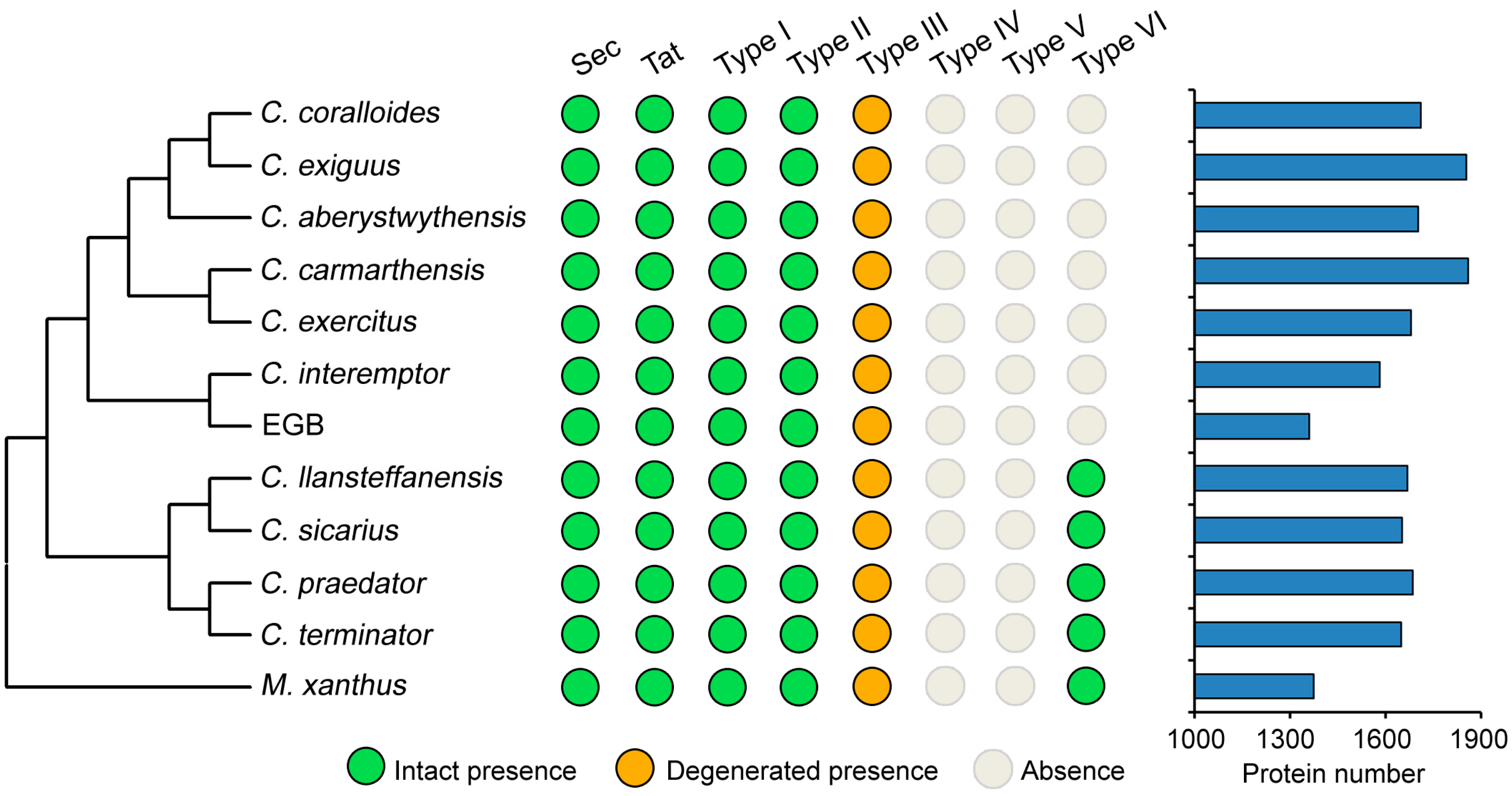
3.4. Genes Involved in Secretion

Protein secretion has not been well-studied in the Corallococcus genus. To explore the genomic potential in strain EGB for extracellular functions, the genome-wide identification of protein secretion systems was undertaken. The Sec and twin-arginine translocation (Tat) pathways are the most commonly used secretion systems for bacteria to transport unfolded and folded proteins across the cytoplasmic membrane [30]. In the Sec system, strain EGB contained all the key proteins except the chaperone SecB, which was dispensable (Supplementary Table S2). The Tat pathway consisted of three components: TatA, TatB, and TatC, and all of them were present in the strain EGB genome. Additionally, many other proteins are secreted through secretion systems numbered Type I through Type VI, with each system transporting specific proteins [31]. Genome mining showed that the primary proteins constituting the intact Type I and Type II secretion systems existed in the strain EGB genome (Figure 4, Supplementary Table S2). We also found two degenerated gene clusters encoding the Type III secretion system in the strain EGB (Supplementary Table S2), which was similar to that in M. xanthus. In contrast, the majority of proteins making up Type IV, Type V, and Type VI secretion systems were lost in strain EGB (Figure 4). An investigation of other Corallococcus genomes revealed that their distributions of the above secretion systems were consistent with strain EGB, except for the Type VI secretion system, which was present in C. llansteffanensis, C. sicarius, C. praedator, and C. terminator, whereas it was absent in the remaining Corallococcus species. A SignalP analysis showed that strain EGB possessed 1361 proteins for secretion via the Sec pathway, while 93 proteins were secreted by the Tat pathway (Figure 4, Supplementary Table S3). In contrast, other Corallococcus species contained more secreted proteins. These results suggested that strain EGB may utilize distinct secretion systems for protein transport compared to other Gram-negative bacteria.

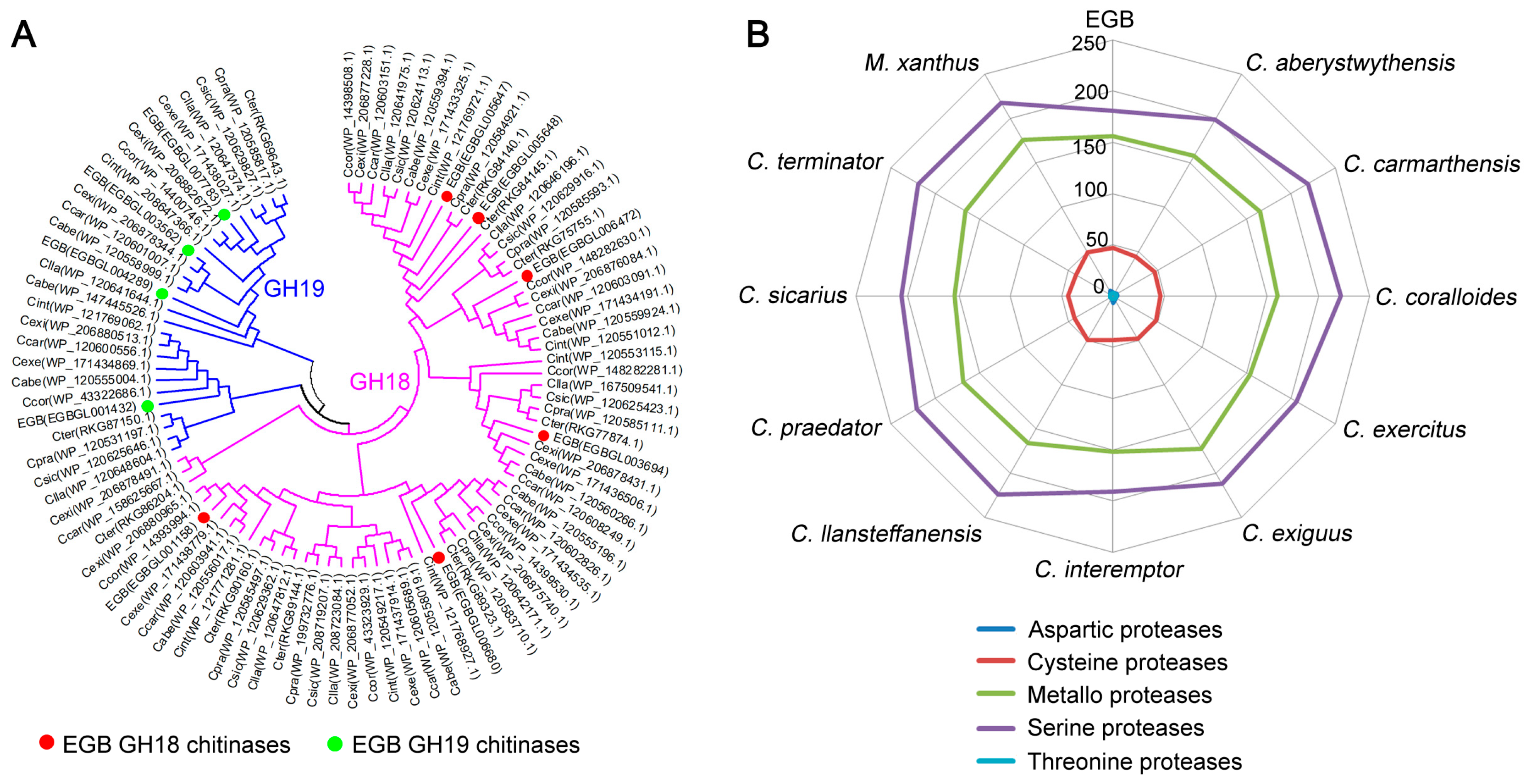
3.5. Expansion of Microorganism Surface Degrading Enzymes

Corallococcus sp. strain EGB is known to exhibit antifungal activity against numerous phytopathogenic fungi and, as expected, it would produce various biochemically distinct extracellular enzymes such as chitinases, glucanases, and proteases to lyse other fungi. Based on the CAZy annotation, the strain EGB genome contained 62 genes encoding glycoside hydrolases (Supplementary Table S4), while other Corallococcus species possessed 67–80 glycoside hydrolases. We further focused on specific glycoside hydrolases known to degrade chitin and glucan, which were the main components of the fungal cell wall. Strain EGB was reported to show chitinase activity and β-(1,3)-glucanase activity. A total of 10 chitinases were identified in strain EGB, and they were divided into the two glycoside hydrolase (GH) families 18 and 19 (Supplementary Table S4). GH18 is a well-known chitinase family and strain EGB contained six GH18 chitinases, which was similar to the number in other Corallococcus genomes. Interestingly, we found four GH19 chitinases in strain EGB, whereas three such chitinases at most were predicted in other Corallococcus genomes. Phylogenetic analysis showed that one GH18 chitinase and two GH19 chitinases derived from strain EGB were species specifically expanded (Figure 5A). In addition, the strain EGB genome contained four β-(1,3)-glucanase-encoding genes belonging to the GH16 family (Supplementary Table S4), which resemble other Corallococcus species. Nearly all the identified chitinases and β-(1,3)-glucanases were distributed in the core proteome of the Corallococcus genus, suggesting that Corallococcus spp. had universal antifungal activity.
Protease is a large family of extracellular enzymes that participate in degrading components on the surface of the microorganisms. A total of 4.9–5.5% of the total genes were predicted to encode proteases in the Corallococcus genomes. The strain EGB genome contained 391 proteases, a slightly fewer than other Corallococcus species. The majority of identified proteases belonged to serine proteases, followed by metallo proteases (Figure 5B). These proteases of strain EGB were further classified into 70 families according to the MEROPS database (Supplementary Table S5). The three largest families, S09, S33, and S01, comprised 30% of the total proteases. We also identified an M36 metalloprotease with high similarity to M. xanthus MepA, which enables the extracellular digestion of proteins during predatory behavior [32].

3.6. Secondary Metabolite Gene Clusters Are Enriched in the Genome

Corallococcus sp. strain EGB and other Corallococcus species exhibited different antibacterial and antifungal activities, indicating that they have different or multiple biosynthetic gene clusters (BGCs) coding for antimicrobial compounds. A survey of the 11 Corallococcus genomes produced 654 BGCs, and each genome possessed an average of 57 BGCs. C. llansteffanensis CA051B contained the largest number of 97 BGCs, while strain EGB had the smallest number of 35 BGCs (Supplementary Table S6). It was noteworthy that nonribosomal peptide synthetases (NRPS) were the most prevalent BGCs, and each Corallococcus genome possessed an average of 25 NRPS gene clusters. There were also, on average, 10 hybrid NRPS-T1PKS gene clusters per genome. Compared to other Corallococcus genomes, strain EGB had fewer NRPS (9 BGCs) and NRPS-T1PKS (8 BGCs) gene clusters (Supplementary Table S6).
Of the 654 predicted BGCs in the Corallococcus genomes, 119 possessed sequence similarities ≥ 50% with annotated BGCs in the antiSMASH database by KnownClusterBlast analysis, indicating that many predicted BGCs were similar to BGCs producing known metabolites. All of the 11 Corallococcus contained the BGCs with high similarity to the carotenoid biosynthetic gene cluster from M. xanthus [33], the BGCs associated with VEPE biosynthetic pathway from M. xanthus DK1622 [34], the BGCs homologous to the myxochelin A biosynthetic gene cluster from Stigmatella aurantiaca Sg a15 [35], the BGCs similar to the geosmin biosynthetic gene cluster from Nostoc punctiforme PCC 73102 [36], and BGCs associated with alkylpyrone-407 biosynthetic gene cluster from Cystobacterineae bacterium [7]. These known metabolites exhibited potent antibacterial or antifungal activities. As well as the BGCs encoding known metabolites, there was also a high number of unknown BGCs present in each Corallococcus genome. Of the 35 BGCs identified in the genome of strain EGB, 28 BGCs were present in part of 10 other Corallococcus genomes, suggesting that a vast number of BGCs were restricted to specific Corallococcus species. These gene clusters could be new resources for novel metabolites with antifungal or antibacterial activity, and an in-depth functional analysis was required to investigate this.

3.7. Genomic Evidence for Signal Transduction and Regulation

Signaling proteins regulate diverse and complex developmental and predatory behaviors of Myxobacteria. The main regulatory proteins include serine/threonine (Ser/Thr) kinases, one-component systems (OCSs), two-component systems (TCSs), and transcription factors (TFs). Based on genome mining, a large number of regulatory proteins were identified in strain EGB and other Corallococcus species (Table 3). The strain EGB contained 96 Ser/Thr kinases, which was smaller than other Corallococcus genomes. Interestingly, 22 of the 96 Ser/Thr kinases were predicted to be integral membrane proteins, indicating that they were involved in sensing external signals. TCS, consisting of a histidine kinase sensor (HK) and a response regulator (RR), is the dominant sense–response mechanism to regulate a wide array of physiological pathways. We identified 78 HKs, 139 RRs, and 36 hybrid HKs in the strain EGB. The strain EGB also possessed 60 OCS proteins, which linked environmental signals to cellular responses. In addition, strain EGB genome encoded numerous TFs, including DNA-binding transcriptional regulators and alternative sigma factors. A total of 51 response regulators, 53 sigma factors, and 156 transcriptional regulators were identified in the strain EGB, while more TFs were predicted in the majority of other Corallococcus genomes (Table 3).

4. Discussion

Corallococcus sp. strain EGB has a complex life-cycle, characterized by some social behaviors, particularly predatory activity. Strain EGB shows excellent biocontrol activity against various phytopathogenic fungi; however, the underlying mechanisms of these behaviors are still unclear. The popularization of genome sequencing allows us to uncover the potential mechanisms for a better understanding of adaptative evolution. In this study, we reported the complete genome sequence and functional annotation of strain EGB. Notably, the quality of the chromosome-level genome of strain EGB was significantly better than that of other Corallococcus genomes.
Due to the similar morphologies and behaviors of Myxobacteria, the taxonomic status of many Myxobacteria species is unclear. 16S rRNA gene is the most commonly used method for bacterial taxonomic classification [37]. C. macrosporus was recently reassigned to the Myxococcus genus. The type strains of C. coralloides and C. exiguus represented only one species, which was supported by 16S rDNA similarity [38]. Recently, the whole-genome sequence has been proved to be more reliable and have a greater discriminatory power than the 16S rRNA gene for taxonomic assignment. Nine species have been verified in Corallococcus using such a method [15]. Here, we applied three genome-based methods to confirm the taxonomic status of strain EGB. The phylogenetic tree based on 135 single-copy genes showed that strain EGB belonged to Group A of Corallococcus genus, and it was closer to C. interemptor AB047A. A further analysis of dDDH and ANI values revealed that strain EGB did not exhibit sufficient genomic similarity with existing Corallococcus species. Thus, strain EGB is supposed to be a novel species, and further phenotypic and biochemical analyses are required to support this. In general, phylogenomics has surpassed the 16S rRNA gene-based method in the field of clarifying the accurate taxonomic classification of bacteria [39].
As a predatory member, strain EGB is known to produce a vast number of extracellular enzymes to degrade the cell-wall components of phytopathogenic fungi. Accordingly, the strain EGB genome contained a repertoire of genes encoding glycoside hydrolases and proteases. Chitinase is the most commonly reported extracellular enzyme that is widely distributed in many biocontrol bacteria, such as Pseudomonas and Bacillus species, and is involved in the biocontrol activity against diver fungi. The strain EGB genome has 10 genes encoding for chitinases. Notably, one chitinase gene CcCti1 was cloned from strain EGB and exhibited obvious antifungal activity against rice blast fungus. In addition, five β-(1,3)-glucanase-encoding genes belonging to the GH6 and GH16 families were identified in the strain EGB genome. One β-(1,3)-glucanase gene lamC deriving from strain EGB showed degradation activity toward a broad range of β-linked polysaccharides, and was confirmed to inhibit the growth of M. oryzae [12]. The presence of both chitinases and β-(1,3)-glucanases in the strain EGB genome supports the antifungal activity of this myxobacter to attack and degrade the cell wall of plant pathogenic fungi.
In contrast with extracellular enzymes, secondary metabolites have received more attention. Myxobacter are known to produce abundant secondary metabolites, and some of these have shown toxic activity to phytopathogenic fungi. In the Corallococcus genus, each genome possessed an average of 57 BGCs, and strain EGB contained the smallest number of 35 BGCs. Even so, stain EGB had a larger number of BGCs than M. xanthus DK1622. Within the Corallococcus genus, only a small proportion of BGCs were found to be conserved in all the analyzed Coralloccus genomes, and most BGCs were present in part of the selected species. Notably, 28 of 35 strain EGB BGCs were relatively enriched in the dispensable pan-genome, implying the presence of selective pressures for the evolutionary retention of BGCs. In contrast to previously characterized BGCs producing named metabolites, the strain EGB had five BGCs with high similarity to BGCs producing carotenoid, VEPE, myxochelin A, geosmin, and alkylpyrone-407. Some of these known metabolites have exhibited potent antimicrobial properties. Most BGCs of strain EGB were not found to be similar to annotated BGCs, indicating that there is considerable BGC diversity that remains unexplored. Such BGCs could be new resources for novel metabolites with antifungal activity, and more experiments are required for further investigation.
In summary, this study describes the genome of Corallococcus sp. strain EGB. Phylogenomic analysis exhibits a phylogenetic relationship and the diversity of strain EGB compared to other Corallococcus species. Further comparative genomic analyses provided diverse evidence that supported strain EGB as a predatory member. These results should encourage the further investigation of detailed mechanisms related to complex social behaviors, especially for predatory activity.

Supplementary Materials

The following are available online at https://www.mdpi.com/article/10.3390/genes12091421/s1, Table S1: Ortholog clustering of proteins between strain EGB and ten other Corallococcus species. Table S2: Proteins of secretion systems encoded by the strain EGB genome. Table S3: Secreted proteins of strain EGB predicted by SignalP v5 server. Table S4: Glycoside hydrolases encoded by the strain EGB genome. Table S5: Classificaton of the strain EGB genes encoding proteases by MEROPS database. Table S6: List of secondary metabolite gene clusters in the strain EGB genome.

Author Contributions

Conceptualization, Z.C. and Y.Z.; methodology, Y.Z.; software, Y.W.; formal analysis, C.X., X.L., X.Y. and Q.F.; investigation, Y.H., Z.L. and C.Z.; writing—original draft preparation, Y.Z.; writing—review and editing, Z.C.; visualization, Y.Z.; supervision, Z.C.; project administration, Z.C.; funding acquisition, Z.C. All authors have read and agreed to the published version of the manuscript.

Funding

This research was funded by the Natural Science Foundation of China (32170123, 32000101 and 32070027), National Key Research and Development Project of China (2019YFA0905203), China Postdoctoral Science Foundation (2020M671513), and Key Research & Development Plan of Jiangsu Province (BE2020340).

Institutional Review Board Statement

Not applicable.

Informed Consent Statement

Not applicable.

Data Availability Statement

The genome presented in this study is openly available in NCBI GenBank with the accession number CP079946.

Conflicts of Interest

The authors declare no conflict of interest.

References

  1. Reichenbach, H. The ecology of the myxobacteria. Environ. Microbiol. 1999, 1, 15–21. [Google Scholar] [CrossRef]
  2. Konovalova, A.; Petters, T.; Sogaard-Andersen, L. Extracellular biology of Myxococcus xanthus. FEMS Microbiol. Rev. 2010, 34, 89–106. [Google Scholar] [CrossRef] [Green Version]
  3. Whitworth, D.E.; Zwarycz, A. A genomic survey of signalling in the Myxococcaceae. Microorganisms 2020, 8, 1739. [Google Scholar] [CrossRef]
  4. Morgan, A.D.; MacLean, R.C.; Hillesland, K.L.; Velicer, G.J. Comparative analysis of Myxococcus predation on Soil Bacteria. Appl. Environ. Microb. 2010, 76, 6920–6927. [Google Scholar] [CrossRef] [Green Version]
  5. Livingstone, P.G.; Morphew, R.M.; Whitworth, D.E. Myxobacteria are able to prey broadly upon clinically-relevant pathogens, exhibiting a prey range which cannot be explained by phylogeny. Front. Microbiol. 2017, 8, 1593. [Google Scholar] [CrossRef] [Green Version]
  6. Xiao, Y.; Wei, X.M.; Ebright, R.; Wall, D. Antibiotic production by myxobacteria plays a role in predation. J. Bacteriol. 2011, 193, 4626–4633. [Google Scholar] [CrossRef] [PubMed] [Green Version]
  7. Hug, J.J.; Panter, F.; Krug, D.; Muller, R. Genome mining reveals uncommon alkylpyrones as type III PKS products from myxobacteria. J. Ind. Microbiol. Biotechnol. 2019, 46, 319–334. [Google Scholar] [CrossRef]
  8. Chambers, J.; Sparks, N.; Sydney, N.; Livingstone, P.G.; Cookson, A.R.; Whitworth, D.E. Comparative genomics and pan-genomics of the Myxococcaceae, including a description of five novel species: Myxococcus eversor sp. nov., Myxococcus IlanfairpwlIg-wyngyllgogetychwymdrobwIlllantysiliogogogochensis sp. nov., Myxococcus vastator sp. nov., Pyxidicoccus caerfyrddinensis sp. nov., and Pyxidicoccus trucidator sp. nov. Genome Biol. Evol. 2020, 12, 2279–2302. [Google Scholar]
  9. Perez-Burgos, M.; Garcia-Romero, I.; Jung, J.; Schander, E.; Valvano, M.A.; Sogaard-Andersen, L. Characterization of the exopolysaccharide biosynthesis pathway in Myxococcus xanthus. J. Bacteriol. 2020, 202, e00335-20. [Google Scholar] [CrossRef]
  10. Li, Z.K.; Ye, X.F.; Chen, P.L.; Ji, K.; Zhou, J.; Wang, F.; Dong, W.L.; Huang, Y.; Zhang, Z.G.; Cui, Z.L. Antifungal potential of Corallococcus sp strain EGB against plant pathogenic fungi. Biol. Control 2017, 110, 10–17. [Google Scholar] [CrossRef]
  11. Zhou, J.; Li, Z.K.; Wu, J.L.; Li, L.F.; Li, D.; Ye, X.F.; Luo, X.; Huang, Y.; Cui, Z.L.; Cao, H. Functional analysis of a novel beta-(1,3)-glucanase from Corallococcus sp strain EGB containing a fascin-like module. Appl. Environ. Microb. 2017, 83, e01016-17. [Google Scholar] [CrossRef] [Green Version]
  12. Zhou, J.; Chen, J.H.; Li, Z.K.; Ye, X.F.; Dong, W.L.; Jiang, M.; Huang, Y.; Cui, Z.L. Enzymatic properties of a multi-specific beta-(1,3)-glucanase from Corallococcus sp. EGB and its potential antifungal applications. Protein Expres Purif 2019, 164, 105481. [Google Scholar] [CrossRef]
  13. Li, Z.K.; Xia, C.Y.; Wang, Y.X.; Li, X.; Qiao, Y.; Li, C.Y.; Zhou, J.; Zhang, L.; Ye, X.F.; Huang, Y.; et al. Identification of an endo-chitinase from Corallococcus sp. EGB and evaluation of its antifungal properties. Int. J. Biol. Macromol. 2019, 132, 1235–1243. [Google Scholar] [CrossRef] [PubMed]
  14. Ye, X.F.; Chen, Y.; Ma, S.Y.; Yuan, T.; Wu, Y.X.; Li, Y.X.; Zhao, Y.Q.; Chen, S.Y.; Zhang, Y.W.; Li, L.Y.; et al. Biocidal effects of volatile organic compounds produced by the myxobacterium Corrallococcus sp. EGB against fungal phytopathogens. Food Microbiol. 2020, 91, 103502. [Google Scholar] [CrossRef]
  15. Livingstone, P.G.; Morphew, R.M.; Whitworth, D.E. Genome sequencing and pan-genome analysis of 23 Corallococcus spp. strains reveal unexpected diversity, with particular plasticity of predatory gene sets. Front. Microbiol. 2018, 9, 3187. [Google Scholar] [CrossRef] [Green Version]
  16. Chen, X.J.; Han, K.; Feng, J.; Zhuo, L.; Li, Y.J.; Li, Y.Z. The complete genome sequence and analysis of a plasmid-bearing myxobacterial strain Myxococcus fulvus 124B02 (M 206081). Stand Genom. Sci. 2016, 11, 1. [Google Scholar] [CrossRef] [Green Version]
  17. Aziz, R.K.; Bartels, D.; Best, A.A.; DeJongh, M.; Disz, T.; Edwards, R.A.; Formsma, K.; Gerdes, S.; Glass, E.M.; Kubal, M.; et al. The RAST server: Rapid annotations using subsystems technology. BMC Genom. 2008, 9, 1–15. [Google Scholar] [CrossRef] [PubMed] [Green Version]
  18. Tatusov, R.L.; Galperin, M.Y.; Natale, D.A.; Koonin, E.V. The COG database: A tool for genome-scale analysis of protein functions and evolution. Nucleic Acids Res. 2000, 28, 33–36. [Google Scholar] [CrossRef] [PubMed] [Green Version]
  19. Enright, A.J.; Van Dongen, S.; Ouzounis, C.A. An efficient algorithm for large-scale detection of protein families. Nucleic Acids Res. 2002, 30, 1575–1584. [Google Scholar] [CrossRef]
  20. Edgar, R.C. MUSCLE: A multiple sequence alignment method with reduced time and space complexity. BMC Bioinform. 2004, 5, 1–19. [Google Scholar] [CrossRef] [Green Version]
  21. Kumar, S.; Stecher, G.; Tamura, K. MEGA7: Molecular evolutionary genetics analysis version 7.0 for bigger datasets. Mol. Biol. Evol. 2016, 33, 1870–1874. [Google Scholar] [CrossRef] [PubMed] [Green Version]
  22. Carver, T.J.; Rutherford, K.M.; Berriman, M.; Rajandream, M.A.; Barrell, B.G.; Parkhill, J. ACT: The Artemis comparison tool. Bioinformatics 2005, 21, 3422–3423. [Google Scholar] [CrossRef] [Green Version]
  23. Yoon, S.H.; Ha, S.M.; Lim, J.; Kwon, S.; Chun, J. A large-scale evaluation of algorithms to calculate average nucleotide identity. Anton Leeuw Int. J. G 2017, 110, 1281–1286. [Google Scholar] [CrossRef]
  24. Meier-Kolthoff, J.P.; Auch, A.F.; Klenk, H.P.; Goker, M. Genome sequence-based species delimitation with confidence intervals and improved distance functions. BMC Bioinform. 2013, 14, 60. [Google Scholar] [CrossRef] [PubMed] [Green Version]
  25. Almagro Armenteros, J.J.; Tsirigos, K.D.; Sonderby, C.K.; Petersen, T.N.; Winther, O.; Brunak, S.; von Heijne, G.; Nielsen, H. SignalP 5.0 improves signal peptide predictions using deep neural networks. Nat. Biotechnol. 2019, 37, 420–423. [Google Scholar] [CrossRef]
  26. Zhang, H.; Yohe, T.; Huang, L.; Entwistle, S.; Wu, P.; Yang, Z.; Busk, P.K.; Xu, Y.; Yin, Y. dbCAN2: A meta server for automated carbohydrate-active enzyme annotation. Nucleic Acids Res. 2018, 46, W95–W101. [Google Scholar] [CrossRef] [Green Version]
  27. Rawlings, N.D.; Waller, M.; Barrett, A.J.; Bateman, A. MEROPS: The database of proteolytic enzymes, their substrates and inhibitors. Nucleic Acids Res. 2014, 42, D503–D509. [Google Scholar] [CrossRef] [PubMed] [Green Version]
  28. Blin, K.; Shaw, S.; Kloosterman, A.M.; Charlop-Powers, Z.; van Wezel, G.P.; Medema, M.H.; Weber, T. antiSMASH 6.0: Improving cluster detection and comparison capabilities. Nucleic Acids Res 2021, 49, W29–W35. [Google Scholar] [CrossRef]
  29. Chun, J.; Oren, A.; Ventosa, A.; Christensen, H.; Arahal, D.R.; da Costa, M.S.; Rooney, A.P.; Yi, H.; Xu, X.W.; De Meyer, S.; et al. Proposed minimal standards for the use of genome data for the taxonomy of prokaryotes. Int. J. Syst. Evol. Micr. 2018, 68, 461–466. [Google Scholar] [CrossRef]
  30. Natale, P.; Bruser, T.; Driessen, A.J. Sec- and Tat-mediated protein secretion across the bacterial cytoplasmic membrane--distinct translocases and mechanisms. Biochim. Biophys. Acta 2008, 1778, 1735–1756. [Google Scholar] [CrossRef] [PubMed] [Green Version]
  31. Green, E.R.; Mecsas, J. Bacterial Secretion Systems: An Overview. Microbiol. Spectr. 2016, 4. [Google Scholar] [CrossRef] [Green Version]
  32. Berleman, J.E.; Allen, S.; Danielewicz, M.A.; Remis, J.P.; Gorur, A.; Cunha, J.; Hadi, M.Z.; Zusman, D.R.; Northen, T.R.; Witkowska, H.E.; et al. The lethal cargo of Myxococcus xanthus outer membrane vesicles. Front. Microbiol. 2014, 5, 474. [Google Scholar] [CrossRef]
  33. Botella, J.A.; Murillo, F.J.; Ruizvazquez, R. A cluster of structural and regulatory genes for light-induced carotenogenesis in Myxococcus xanthus. Eur. J. Biochem. 1995, 233, 238–248. [Google Scholar] [CrossRef]
  34. Lorenzen, W.; Ahrendt, T.; Bozhuyuk, K.A.J.; Bode, H.B. A multifunctional enzyme is involved in bacterial ether lipid biosynthesis. Nat. Chem. Biol. 2014, 10, 425–427. [Google Scholar] [CrossRef]
  35. Gaitatzis, N.; Kunze, B.; Muller, R. In vitro reconstitution of the myxochelin biosynthetic machinery of Stigmatella aurantiaca Sg a15: Biochemical characterization of a reductive release mechanism from nonribosomal peptide synthetases. Proc. Natl. Acad. Sci. USA 2001, 98, 11136–11141. [Google Scholar] [CrossRef] [Green Version]
  36. Giglio, S.; Jiang, J.Y.; Saint, C.P.; Cane, D.E.; Monis, P.T. Isolation and characterization of the gene associated with geosmin production in cyanobacteria. Environ. Sci. Technol. 2008, 42, 8027–8032. [Google Scholar] [PubMed] [Green Version]
  37. Lang, E.; Stackebrandt, E. Emended descriptions of the genera Myxococcus and Corallococcus, typification of the species Myxococcus stipitatus and Myxococcus macrosporus and a proposal that they be represented by neotype strains. Request for an Opinion. Int. J. Syst. Evol. Microbiol. 2009, 59, 2122–2128. [Google Scholar] [CrossRef]
  38. Garcia, R.; Gerth, K.; Stadler, M.; Dogma, I.J., Jr.; Muller, R. Expanded phylogeny of myxobacteria and evidence for cultivation of the ‘unculturables’. Mol. Phylogenet Evol. 2010, 57, 878–887. [Google Scholar] [CrossRef] [PubMed]
  39. Hinger, I.; Ansorge, R.; Mussmann, M.; Romano, S. Phylogenomic analyses of members of the widespread marine heterotrophic genus pseudovibrio suggest distinct evolutionary trajectories and a novel genus, Polycladidibacter gen. nov. Appl. Environ. Microbiol. 2020, 86, e02395-19. [Google Scholar] [CrossRef]
Figure 1. Circular representation of the complete genome of Corallococcus sp. strain EGB. The outermost circle is the coordinate of the genome position. From the outside to the inside, the first circle represents COG annotation gene distribution of the forwarding strand, colored according to cluster; the second circle illustrates COG annotation of the reverse strand; the third circle depicts the ncRNA distribution of the forwarding strand; the fourth circle represents the ncRNA distribution of the reverse strand; the fifth circle illustrates the repeat; the sixth circle depicts the GC content; the seventh circle denotes the GC skew.
Genes 12 01421 g001
Figure 2. Phylogenetic relationships of strain EGB and related bacteria. (A) Phylogenetic tree constructed using MEGA 7 with the neighbor-joining algorithm based on concatenated sequences of 135 single-copy genes. (B) Linear genomic comparison of strain EGB, C. coralloides DSM 2259, and M. xanthus DK1622. The red lines in between the genomes represent matching regions, and blue lines denote inverted matching regions.
Genes 12 01421 g002
Figure 3. Comparative analysis of proteins between strain EGB and other Corallococcus species. (A) Number of protein families shared by different species. (B) Proportion of proteins enriched in the clusters of orthologous groups (COG) categories in the core, dispensable, and unique proteome. A, RNA processing and modification; B, chromatin structure and dynamics; C, energy production and conversion; D, cell-cycle control, cell division, chromosome partitioning; E, amino acid transport and metabolism; F, nucleotide transport and metabolism; G, carbohydrate transport and metabolism; H, coenzyme transport and metabolism; I, lipid transport and metabolism; J, translation, ribosomal structure and biogenesis; K, transcription; L, replication, recombination and repair; M, cell-wall/membrane/envelope biogenesis; N, cell motility; O, posttranslational modification, protein turnover, chaperones; P, inorganic ion transport and metabolism; Q, secondary metabolites biosynthesis, transport and catabolism; R, general function prediction only; S, function unknown; T, signal transduction mechanisms; U, intracellular trafficking, secretion, and vesicular transport; V, defense mechanisms; W, extracellular structures; X, mobilome: prophages, transposons; Z, cytoskeleton.
Genes 12 01421 g003
Figure 4. Distribution of secretion systems. The left part lists the relationship among species. The middle part shows presence or absence of each type of secretion system in each species. The right part represents the comparison of numbers of secreted proteins.
Genes 12 01421 g004
Figure 5. Comparison of chitinases and proteases. (A) Phylogenetic tree of chitinases derived from strain EGB, C. coralloides, C. praedator, and M. xanthus. (B) Comparison of protease numbers among Corallococcus species.
Genes 12 01421 g005
Table 1. Genome properties of Corallococcus species.
SpeciesStrainGenome Size (Mb)%GCContigsCDS * NumberAccession
Corallococcus sp.EGB9.470.417916CP079946
C. coralloidesDSM 225910.0869.917952CP003389.1
C. exiguusNCCRE00210.5469.788283GCA_017302975.1
C. interemptorAB047A9.47704597569GCA_003668875.1
C. praedatorCA031B10.5169.714918176GCA_003612125.1
C. aberystwythensisAB050A9.98706258319GCA_003612165.1
C. llansteffanensisCA051B10.5370.312448140GCA_003612055.1
C. sicariusCA040B10.3970.28028410GCA_003611735.1
C. carmarthensisCA043D10.7969.95308523GCA_003611695.1
C. exercitusAB043B10.2670.26908102GCA_013116705.1
C. terminatorCA054A10.3569.58638454GCA_003611635.1
* CDS: Coding sequence.
Table 2. ANI and dDDH values for pairwise comparisons between Corallococcus species and M. xanthus DK1622.
dDDH\ANIEGBCintCexeCcarCabeCexiCcorCllaCsicCpraCterMxan
EGB10091.1991.1190.8890.7991.3491.5186.6386.2186.1785.8378.37
Cint42.410091.2991.1291.3191.7292.2786.5386.386.1885.978.35
Cexe41.942.710092.7392.691.5691.8387.3787.0386.8786.5278.43
Ccar41.542.147.510092.5791.6291.7887.1886.8786.8186.3878.41
Cabe41.242.747.147.610091.5591.8487.0486.7386.786.278.41
Cexi42.443.143.143.643.210094.3286.6186.1386.1285.8278.15
Ccor43.746.143.944.444.354.110086.8286.4686.3885.9578.25
Clla30.930.832.432.131.730.731.110093.4289.2788.8278.78
Csic29.93031.631.33129.830.250.410088.8788.4578.65
Cpra29.930.131.531.4313030.536.635.910093.2378.68
Cter29.329.730.830.630.229.429.835.23549.610078.27
Mxan21.221.321.621.621.521.221.221.721.521.621.2100
Average nucleotide identity (ANI) values were shown above the diagonal and the digital DNA-DNA hybridization (dDDH) values were present below the diagonal. Cint, C. interemptor AB047A; Cexe, C. exercitus AB043B; Ccar, C. carmarthensis CA043D; Cabe, C. aberystwythensis AB050A; Cexi, C. exiguus NCCRE002; Ccor, C. coralloides DSM2259; Clla, C. llansteffanensis CA051B; Csic, C. sicarius CA040B; Cpra, C. praedator CA031B; Cter, C. terminator CA054A; Mxan, M. xanthus DK1622.
Table 3. Variability in the numbers of regulatory genes per genome.
EGBCabeCcarCcorCexeCexiCintCllaCpraCsicCter
Ser/Thr kinases96106123103112105108113101116106
Two-component system proteins289299307303318297284322323312311
Histidine kinases146156157160167153151170162167163
Phosphotransfer proteins55324225744
Response regulators138138147141147142131147154141144
Transcription factors320322380351353378306344352303334
One-component system proteins6068867978846175696168
Response regulators5142544847524246514450
sigma factors5358585657565158565350
Transcriptional regulators156154182168171186152165176145166
Publisher’s Note: MDPI stays neutral with regard to jurisdictional claims in published maps and institutional affiliations.

