Published online Aug 16, 2021. doi: 10.12998/wjcc.v9.i23.6907
Peer-review started: April 25, 2021
First decision: May 24, 2021
Revised: June 2, 2021
Accepted: June 17, 2021
Article in press: June 17, 2021
Published online: August 16, 2021
Endometrial stromal tumors originate from the endometrial stroma and account for < 2% of all uterine tumors. Uterine tumor resembling an ovarian sex cord tumor (UTROSCT) is a rare histological class of endometrial stromal and related tumors according to the latest World Health Organization classification of female genital tumors. Here, we report a case of UTROSCT in a 51-year-old woman.
A 51-year-old woman had irregular menses for 6 mo. The patient visited a local hospital for vaginal bleeding. Pelvic computed tomography (CT) showed a mass in the pelvic cavity. Five days later, she came to our hospital for further diagnosis. The results of contrast-enhanced CT and pelvic ultrasound at our hospital suggested a malignant pelvic tumor. She then underwent total removal of the uterus with bilateral salpingectomy. Postoperative histological examination showed that the tumor cells had abundant cytoplasm, ovoid and spindle-shaped nuclei, fine chromatin, a high nucleoplasm ratio, and a lamellar distribution. The findings were consistent with UTROSCT, and the results of immunohistochemical analysis supported that diagnosis. The tumor was International Federation of Gynecology and Obstetrics stage IB. No adjuvant therapy was administered after radical surgery. The patient was followed up for 58 mo, and no recurrence was found.
We report a case of UTROSCT with abnormal menstruation as a symptom, which is one of the most common symptoms. In patients with vaginal bleeding, ultrasonography can be used as a screening test because of its convenience, speed, and lack of radiation exposure. For patients with long-term tamoxifen use, routine monitoring of the endometrium is recommended. As UTROSCT may have low malignant potential, surgery remains the primary management strategy. Additionally, fertility preservation in patients of childbearing age is a vital consideration.
Core Tip: Uterine tumor resembling an ovarian sex cord tumor (UTROSCT) is a rare histological form of endometrial stromal and related tumors. Limited knowledge of the disease can make diagnosis difficult. Here, we present the case of a 51-year-old woman with UTROSCT.
- Citation: Zhou FF, He YT, Li Y, Zhang M, Chen FH. Uterine tumor resembling an ovarian sex cord tumor: A case report and review of literature. World J Clin Cases 2021; 9(23): 6907-6915
- URL: https://www.wjgnet.com/2307-8960/full/v9/i23/6907.htm
- DOI: https://dx.doi.org/10.12998/wjcc.v9.i23.6907
Endometrial stromal tumors originate from the endometrial stroma and account for < 2% of all uterine tumors[1]. The latest World Health Organization classification of female genital tumors recognizes uterine tumors resembling an ovarian sex cord tumor (UTROSCT) as a rare histological form of endometrial stromal and related tumors[2]. It was first described by Clement and Scully in 1976[3]. The currently available literature focuses on the diagnosis of UTROSCT and lacks information on the characteristic symptoms and radiological findings. Here, we report a case of UTROSCT in a 51-year-old woman. An attempt was made to review the literature to elucidate the clinical features, treatment options and outcomes of this rare disease to avoid missing or misdiagnosing the disease in clinical practice.
A 51-year-old woman had irregular menses for 6 mo.
The patient had irregular menses for 6 mo. She had pelvic pain and prolonged menstruation but no nausea, vomiting, diarrhea, abdominal bloating, or fever. Pelvic computed tomography (CT) at a local hospital revealed a mass in her pelvis. Five days later, she came to our hospital for further treatment. We investigated whether the patient experienced mild limitations in performing life activities and societal participation, congruent with the domains on the International Classification of Functioning, Disability, and Health.
She had been suffering from hypertension for 2 years, and had been treated with nifedipine extended-release tablets. She underwent surgery to remove a fibroadenoma from her right breast 4 years prior to presentation. The patient had no history of diabetes, heart disease, alcohol consumption, or smoking.
The patient denied a relevant family history.
Gynecological examination revealed a cervical nodule, approximately 7 cm × 9 cm in size, with a soft texture, no tenderness, a clear boundary, and blood vessel pulsation on the surface without adnexal masses; the vulva, urethra and vagina were normal.
Her blood potassium level was 3.20 mmol/L. All tumor marker results were within the normal range. The reactivity tests for hepatitis B virus, human immunodeficiency virus, syphilis, and hepatitis C virus were all negative.
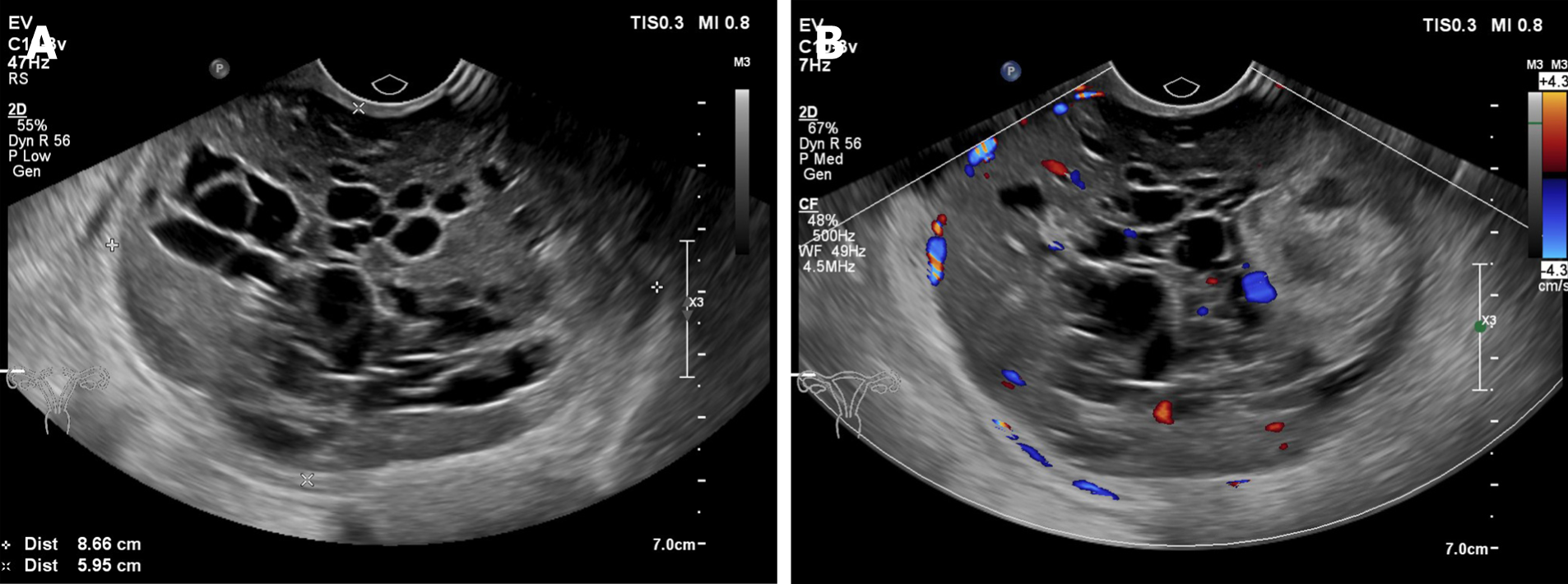
A pelvic ultrasound (US) revealed an 87 mm × 60 mm mass with heterogeneous echo on the right side of the pelvic cavity, consisting of hypoechoic intracystic effusion and a hypoechoic intracystic tumor with local honeycomb changes. Color Doppler imaging showed high degrees of vascularity. The mass had no clear boundary with the cervix and seemed to adhere to it (Figure 1). Pelvic US also suggested multiple uterine fibroids. Pelvic CT revealed a round, low-density mass in the pelvic cavity with uneven internal density. Contrast-enhanced CT showed uneven enhancement in the arterial and venous phases of the scan and no enhancement in low-density areas (Figure 2). The results suggested a malignant pelvic tumor. No enlarged lymph nodes were found in the pelvic cavity.
The diagnosis was UTROSCT.
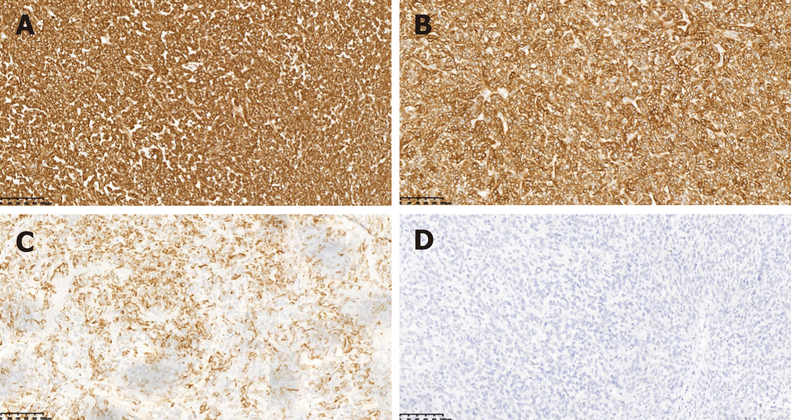
The patient underwent total removal of the uterus with bilateral salpingectomy. During the surgery, a 100 mm × 80 mm solid mass was detected in the cervix. It had a smooth surface with an intact capsule. The cut surface of the lesion had a solid grayish-yellow appearance, soft cystic areas with hemorrhage and necrosis, and a fish-flesh texture. The intraoperative rapid pathology report suggested a small round cell tumor, and the definite diagnosis was pending routine examination and immunohistochemistry. Postoperative histological examination showed that the tumor cells had abundant cytoplasm, ovoid and spindle-shaped nuclei, fine chromatin, a high nucleoplasm ratio, and a lamellar distribution. These findings were consistent with UTROSCT (Figure 3). Immunohistochemical staining showed that the tumor cells were positive for Ki67 (8% positive), Vim, CD99, CK, and Syn but negative for SMA, ER, PR, myogenin, EMA, HMB45, α-inhibin, CD31, PLAP, CK7, CD56, CD10, WT-1, and caldesmon (Figure 4). No metastasis to the omentum or lymph nodes was observed. To ensure the accuracy of the diagnosis, senior pathologists from other organizations were invited for consultation, and they confirmed the diagnosis of UTROSCT. The International Federation of Gynecology and Obstetrics tumor stage was IB. No adjuvant therapy was administered after radical surgery.
The patient was followed up every 3 mo, and each follow-up examination included a medical history, a physical examination, comprehensive biochemical tests, a chest CT, a vaginal US, and a routine blood examination. An abdominal CT was performed every 6 mo for 58 mo after surgery, and no signs of recurrence or metastasis were detected. The patient’s performance of life activities and societal participation had improved.
UTROSCT is a rare class of uterine tumors first reported by Clement and Scully in 1976[3]. They reported 14 cases of UTROSCT, which were divided into two groups based on the proportion and the appearance of the stromal cell component. Type 1 tumors were endometrial stromal tumors that showed focal epithelial-like differentiation of the type seen in ovarian sex cord tumors, and type 2 tumors—designated UTROSCT—were uterine mural masses with a predominant or exclusive histological appearance of sex cord elements[2]. The origin of UTROSCT is not clear, and the lack of a JAZF1-SUZ12 gene fusion suggests that the origin is the endometrial stroma[4].
To date, including this patient, approximately 90[3-48] cases have been reported in the literature. According to previous reports, the age of onset is 20 to 86 years; the average age is 50.6 years, the median age is 51 years, and the tumor size is 4 to 135 (average 47.6) mm. The patient in this case was a 51-year-old woman, which is consis
| Characteristic | n (%) | |
| Age in yr | 90 | |
| ≤ 30 | 12 (13.3) | |
| 30-60 | 47 (52.2) | |
| ≥ 60 | 31 (34.4) | |
| Location | 53 | |
| Uterine wall | 35 (66.1) | |
| Uterine cavity | 12 (22.6) | |
| Uterine wall and cavity | 6 (11.3) | |
| Tumor size in mm | 72 | |
| ≤ 40 | 36 (50.0) | |
| 40-80 | 20 (27.8) | |
| ≥ 80 | 16 (22.2) | |
| Presenting symptom | 59 | |
| Postmenopausal bleeding | 20 (33.9) | |
| Abnormal menstruation | 20 (33.9) | |
| Pelvic pain | 11 (18.6) | |
| Elevated prolactin | 2 (3.4) | |
| Incidental | 11 (18.6) | |
| Diagnostic modality | 30 | |
| US | 22 (73.3) | |
| CT | 3 (10.0) | |
| MRI | 5 (16.7) | |
| Accompanied diseases | 20 | |
| Leiomyoma | 12 (60.0) | |
| Adenomyosis | 4 (20.0) | |
| Endometrial hyperplasia | 2 (10.0) | |
| Uterine prolapse | 2 (10.0) | |
| Surgical approach | 75 | |
| TAH+BSO | 57 (76.0) | |
| TAH alone | 8 (10.7) | |
| Mass resection alone | 3 (4.0) | |
| Hysteroscopic mass resection | 7 (9.3) | |
| Recurrence/metastasis | 52 | |
| Yes | 10 (19.2) | |
| No | 42 (80.8) | |
| Status | 50 | |
| ANED | 44 (88.0) | |
| AWD | 5 (10.0) | |
| DOD | 1 (2.0) | |
In our literature search, we found six patients with UTROSCT with previous or current use of tamoxifen. We therefore suspect that tamoxifen was a causative factor. The effect of tamoxifen on the endometrium has been reported in the literature. Tamoxifen intake can lead to extensive senile cystic atrophy of the human endome
The diagnosis of UTROSCT is incredibly difficult. The symptoms vary among patients and are not typical in some cases. Therefore, it is easy to miss or misdiagnose the disease. Common symptoms of UTROSCT include postmenopausal bleeding (33.9%)[5], abnormal menstruation, menorrhagia and extended menstruation (33.9%)[14,17], and pelvic pain (18.6%)[19]. We found reports of two patients with UTROSCT who presented with symptoms of hyperprolactinemia, and we found that postmenopausal bleeding and menstrual abnormalities are the most commonly reported symptoms of UTROSCT. The possible mechanism is overgrowth of sex cord elements in endometrial stromal neoplasms or of foci of adenomyosis and endometriosis[50]. The mechanism of hyperprolactinemia remains unclear. However, other reported cases of ectopic hyperprolactinemia with uterine tumors have features in common with this case, and it is possible that they belong to the same tumor category[51].
US, CT, and magnetic resonance imaging (MRI) are useful for detecting UTROSCT[5-11]. Among these techniques, vaginal US is more convenient and less costly for primary diagnosis, while avoiding radiation. According to the literature, 22 (73.3%) cases of UTROSCT were discovered through US. Of the affected patients, ten presented with uterine cavity lesions and five presented with an enlarged uterus. Nine cases included intratumoral cystic components. Of the five reports including MRI, three reported masses described as slightly hyperintense on T2-weighted images and intermediate signal intensity on T1-weighted images.
The diagnosis of UTROSCT is mainly based on the morphological features following hematoxylin-eosin (HE) staining and confirmed by immunohistochemical staining. UTROSCT predominantly has the morphological features of sex cord-like elements wherein tumor cells form cords, trabeculae, tubules, clusters, sheets, and retiform structures. Immunohistochemically, UTROSCT is multiphenotypic, coexpressing smooth muscle markers, cytokeratins, and hormone receptor markers that are usually positive in ovarian sex cord-mesenchymal tumors, including calretinin, inhibin, CD99, CD56, Melan-A, FOXL2, and steroidogenic factor-1 (SF-1)[52,53]. In addition to being positive for calreticulin, the tumors are positive for at least one sex cord marker[54-56]. In the case reported here, the immunohistochemical results showed that the tumor cells were positive for Ki67 (8% positive), Vim, CD99, CK, and Syn; but negative for SMA, ER, PR, myogenin, EMA, HMB45, α-inhibin, CD31, PLAP, CK7, CD56, CD10, WT-1 and caldesmon, consistent with previous published reports.
Standardized treatments for UTROSCT are lacking, perhaps due to its rarity. At present, the preferred treatment method is surgery. The surgical treatment options are either total abdominal hysterectomy with bilateral salpingo-oophorectomy (TAH+BCS), TAH alone and mass resection alone. Of the 75 patients in Table 1 who underwent surgery; 57 were treated with TAH+BCS and eight with TAH alone. Hysterectomy is a radical cure, but it is not suitable for patients with fertility requirements. Seven patients with reproductive requirements were treated by hysteroscopic mass resection. Following treatment, three had spontaneous conception and uncomplicated pregnancies 17 mo, 24 mo, and 13 mo after surgery and all three delivered healthy babies.
Of the 90 patients reported thus far, 52 were followed up for periods of 1 to 384 (mean 56.3) mo; 42 (80.8%) had no recurrence, and 10 (19.2%) had recurrences or distant metastases (lung, abdominal, bladder, mesentery, or pelvic). One patient developed lung and abdominal metastases and died of the disease 9 mo after diagnosis from complications of intra-abdominal tumor spread. In our UTROSCT case, the patient had no fertility requirements, so she underwent total abdominal hysterectomy with bilateral salpingo-oophorectomy, and the treatment was effective.
This UTROSCT patient presented with abnormal menstruation, which is one of the most common symptoms. In patients with vaginal bleeding, ultrasonography can be used as a screening test because it is convenient, rapid, and lacks radiation invol
Manuscript source: Unsolicited manuscript
Specialty type: Obstetrics and gynecology
Country/Territory of origin: China
Peer-review report’s scientific quality classification
Grade A (Excellent): 0
Grade B (Very good): B
Grade C (Good): 0
Grade D (Fair): 0
Grade E (Poor): 0
P-Reviewer: Jurys T S-Editor: Ma YJ L-Editor: Filipodia P-Editor: Zhang YL
| 1. | Koss LG, Spiro RH, Brunschwig A. Endometrial stromal sarcoma. Surg Gynecol Obstet. 1965;121:531-537. [PubMed] [Cited in This Article: ] |
| 2. | Böcker W. [WHO classification of breast tumors and tumors of the female genital organs: pathology and genetics]. Verh Dtsch Ges Pathol. 2002;86:116-119. [PubMed] [Cited in This Article: ] |
| 3. | Clement PB, Scully RE. Uterine tumors resembling ovarian sex-cord tumors. A clinicopathologic analysis of fourteen cases. Am J Clin Pathol. 1976;66:512-525. [PubMed] [DOI] [Cited in This Article: ] [Cited by in Crossref: 224] [Cited by in F6Publishing: 232] [Article Influence: 4.8] [Reference Citation Analysis (0)] |
| 4. | Staats PN, Garcia JJ, Dias-Santagata DC, Kuhlmann G, Stubbs H, McCluggage WG, De Nictolis M, Kommoss F, Soslow RA, Iafrate AJ, Oliva E. Uterine tumors resembling ovarian sex cord tumors (UTROSCT) lack the JAZF1-JJAZ1 translocation frequently seen in endometrial stromal tumors. Am J Surg Pathol. 2009;33:1206-1212. [PubMed] [DOI] [Cited in This Article: ] [Cited by in Crossref: 59] [Cited by in F6Publishing: 61] [Article Influence: 4.1] [Reference Citation Analysis (0)] |
| 5. | Czernobilsky B, Mamet Y, David MB, Atlas I, Gitstein G, Lifschitz-Mercer B. Uterine retiform sertoli-leydig cell tumor: report of a case providing additional evidence that uterine tumors resembling ovarian sex cord tumors have a histologic and immunohistochemical phenotype of genuine sex cord tumors. Int J Gynecol Pathol. 2005;24:335-340. [PubMed] [DOI] [Cited in This Article: ] [Cited by in Crossref: 28] [Cited by in F6Publishing: 30] [Article Influence: 1.6] [Reference Citation Analysis (0)] |
| 6. | Abdullazade S, Kosemehmetoglu K, Adanir I, Kutluay L, Usubutun A. Uterine tumors resembling ovarian sex cord-stromal tumors: synchronous uterine tumors resembling ovarian sex cord-stromal tumors and ovarian sex cord tumor. Ann Diagn Pathol. 2010;14:432-437. [PubMed] [DOI] [Cited in This Article: ] [Cited by in Crossref: 10] [Cited by in F6Publishing: 9] [Article Influence: 0.7] [Reference Citation Analysis (0)] |
| 7. | Anastasakis E, Magos AL, Mould T, Economides DL. Uterine tumor resembling ovarian sex cord tumors treated by hysteroscopy. Int J Gynaecol Obstet. 2008;101:194-195. [PubMed] [DOI] [Cited in This Article: ] [Cited by in Crossref: 13] [Cited by in F6Publishing: 13] [Article Influence: 0.8] [Reference Citation Analysis (0)] |
| 8. | Aziz O, Giles J, Knowles S. Uterine tumours resembling ovarian sex cord tumours: a case report. Cases J. 2009;2:55. [PubMed] [DOI] [Cited in This Article: ] [Cited by in Crossref: 7] [Cited by in F6Publishing: 7] [Article Influence: 0.5] [Reference Citation Analysis (0)] |
| 9. | Berretta R, Patrelli TS, Fadda GM, Merisio C, Gramellini D, Nardelli GB. Uterine tumors resembling ovarian sex cord tumors: a case report of conservative management in young women. Int J Gynecol Cancer. 2009;19:808-810. [PubMed] [DOI] [Cited in This Article: ] [Cited by in Crossref: 15] [Cited by in F6Publishing: 15] [Article Influence: 1.0] [Reference Citation Analysis (0)] |
| 10. | Biermann K, Heukamp LC, Büttner R, Zhou H. Uterine tumor resembling an ovarian sex cord tumor associated with metastasis. Int J Gynecol Pathol. 2008;27:58-60. [PubMed] [DOI] [Cited in This Article: ] [Cited by in Crossref: 35] [Cited by in F6Publishing: 36] [Article Influence: 2.3] [Reference Citation Analysis (0)] |
| 11. | Calisir C, Inan U, Yavas US, Isiksoy S, Kaya T. Mazabraud's syndrome coexisting with a uterine tumor resembling an ovarian sex cord tumor (UTROSCT): a case report. Korean J Radiol. 2007;8:438-442. [PubMed] [DOI] [Cited in This Article: ] [Cited by in Crossref: 8] [Cited by in F6Publishing: 7] [Article Influence: 0.4] [Reference Citation Analysis (0)] |
| 12. | Erhan Y, Baygün M, Ozdemir N. The coexistence of stromomyoma and uterine tumor resembling ovarian sex-cord tumors. Report of a case and immunohistochemical study. Acta Obstet Gynecol Scand. 1992;71:390-393. [PubMed] [DOI] [Cited in This Article: ] [Cited by in Crossref: 7] [Cited by in F6Publishing: 8] [Article Influence: 0.3] [Reference Citation Analysis (0)] |
| 13. | Garuti G, Gonfiantini C, Mirra M, Galli C, Luerti M. Uterine tumor resembling ovarian sex cord tumors treated by resectoscopic surgery. J Minim Invasive Gynecol. 2009;16:236-240. [PubMed] [DOI] [Cited in This Article: ] [Cited by in Crossref: 18] [Cited by in F6Publishing: 19] [Article Influence: 1.3] [Reference Citation Analysis (0)] |
| 14. | Giordano G, Lombardi M, Brigati F, Mancini C, Silini EM. Clinicopathologic features of 2 new cases of uterine tumors resembling ovarian sex cord tumors. Int J Gynecol Pathol. 2010;29:459-467. [PubMed] [DOI] [Cited in This Article: ] [Cited by in Crossref: 15] [Cited by in F6Publishing: 16] [Article Influence: 1.1] [Reference Citation Analysis (0)] |
| 15. | Hillard JB, Malpica A, Ramirez PT. Conservative management of a uterine tumor resembling an ovarian sex cord-stromal tumor. Gynecol Oncol. 2004;92:347-352. [PubMed] [DOI] [Cited in This Article: ] [Cited by in Crossref: 29] [Cited by in F6Publishing: 25] [Article Influence: 1.3] [Reference Citation Analysis (0)] |
| 16. | Kabbani W, Deavers MT, Malpica A, Burke TW, Liu J, Ordoñez NG, Jhingran A, Silva EG. Uterine tumor resembling ovarian sex-cord tumor: report of a case mimicking cervical adenocarcinoma. Int J Gynecol Pathol. 2003;22:297-302. [PubMed] [DOI] [Cited in This Article: ] [Cited by in Crossref: 31] [Cited by in F6Publishing: 34] [Article Influence: 1.6] [Reference Citation Analysis (0)] |
| 17. | Nogales FF, Stolnicu S, Harilal KR, Mooney E, García-Galvis OF. Retiform uterine tumours resembling ovarian sex cord tumours. A comparative immunohistochemical study with retiform structures of the female genital tract. Histopathology. 2009;54:471-477. [PubMed] [DOI] [Cited in This Article: ] [Cited by in Crossref: 21] [Cited by in F6Publishing: 23] [Article Influence: 1.5] [Reference Citation Analysis (0)] |
| 18. | O'Meara AC, Giger OT, Kurrer M, Schaer G. Case report: Recurrence of a uterine tumor resembling ovarian sex-cord tumor. Gynecol Oncol. 2009;114:140-142. [PubMed] [DOI] [Cited in This Article: ] [Cited by in Crossref: 24] [Cited by in F6Publishing: 26] [Article Influence: 1.7] [Reference Citation Analysis (0)] |
| 19. | Oztekin O, Soylu F, Yigit S, Sarica E. Uterine tumor resembling ovarian sex cord tumors in a patient using tamoxifen: report of a case and review of literature. Int J Gynecol Cancer. 2006;16:1694-1697. [PubMed] [DOI] [Cited in This Article: ] [Cited by in Crossref: 9] [Cited by in F6Publishing: 10] [Article Influence: 0.6] [Reference Citation Analysis (0)] |
| 20. | Sitic S, Korac P, Peharec P, Zovko G, Perisa MM, Gasparov S. Bcl-2 and MALT1 Genes are not involved in the oncogenesis of uterine tumors resembling ovarian sex cord tumors. Pathol Oncol Res. 2007;13:153-156. [PubMed] [DOI] [Cited in This Article: ] [Cited by in Crossref: 10] [Cited by in F6Publishing: 10] [Article Influence: 0.6] [Reference Citation Analysis (0)] |
| 21. | Stolnicu S, Balachandran K, Aleykutty MA, Loghin A, Preda O, Goez E, Nogales FF. Uterine adenosarcomas overgrown by sex-cord-like tumour: report of two cases. J Clin Pathol. 2009;62:942-944. [PubMed] [DOI] [Cited in This Article: ] [Cited by in Crossref: 14] [Cited by in F6Publishing: 15] [Article Influence: 1.1] [Reference Citation Analysis (0)] |
| 22. | Sutak J, Lazic D, Cullimore JE. Uterine tumour resembling an ovarian sex cord tumour. J Clin Pathol. 2005;58:888-890. [PubMed] [DOI] [Cited in This Article: ] [Cited by in Crossref: 26] [Cited by in F6Publishing: 28] [Article Influence: 1.5] [Reference Citation Analysis (0)] |
| 23. | Wang J, Blakey GL, Zhang L, Bane B, Torbenson M, Li S. Uterine tumor resembling ovarian sex cord tumor: report of a case with t(X;6)(p22.3;q23.1) and t(4;18)(q21.1;q21.3). Diagn Mol Pathol. 2003;12:174-180. [PubMed] [DOI] [Cited in This Article: ] [Cited by in Crossref: 26] [Cited by in F6Publishing: 28] [Article Influence: 1.3] [Reference Citation Analysis (0)] |
| 24. | Hauptmann S, Nadjari B, Kraus J, Turnwald W, Dietel M. Uterine tumor resembling ovarian sex-cord tumor--a case report and review of the literature. Virchows Arch. 2001;439:97-101. [PubMed] [DOI] [Cited in This Article: ] [Cited by in Crossref: 32] [Cited by in F6Publishing: 36] [Article Influence: 1.6] [Reference Citation Analysis (0)] |
| 25. | Kantelip B, Cloup N, Dechelotte P. Uterine tumor resembling ovarian sex cord tumors: report of a case with ultrastructural study. Hum Pathol. 1986;17:91-94. [PubMed] [DOI] [Cited in This Article: ] [Cited by in Crossref: 44] [Cited by in F6Publishing: 45] [Article Influence: 1.2] [Reference Citation Analysis (0)] |
| 26. | Iwasaki I, Yu TJ, Takahashi A, Asanuma K, Izawa Y. Uterine tumor resembling ovarian sex-cord tumor with osteoid metaplasia. Acta Pathol Jpn. 1986;36:1391-1395. [PubMed] [DOI] [Cited in This Article: ] [Cited by in Crossref: 1] [Cited by in F6Publishing: 1] [Article Influence: 0.0] [Reference Citation Analysis (0)] |
| 27. | Moll M, Horn T. An unusual uterine tumor resembling sex-cord-tumor of the ovary with papillomatous features. Immunohistochemical and electron microscopic observations. Acta Obstet Gynecol Scand. 1992;71:550-554. [PubMed] [DOI] [Cited in This Article: ] [Cited by in Crossref: 4] [Cited by in F6Publishing: 4] [Article Influence: 0.1] [Reference Citation Analysis (0)] |
| 28. | Fukunaga M. Adenomyosis with a sex cord-like stromal element. Pathol Int. 2000;50:336-339. [PubMed] [DOI] [Cited in This Article: ] [Cited by in Crossref: 6] [Cited by in F6Publishing: 6] [Article Influence: 0.3] [Reference Citation Analysis (0)] |
| 29. | De Quintal MM, De Angelo Andrade LA. Endometrial polyp with sex cord-like pattern. Int J Gynecol Pathol. 2006;25:170-172. [PubMed] [DOI] [Cited in This Article: ] [Cited by in Crossref: 12] [Cited by in F6Publishing: 12] [Article Influence: 0.7] [Reference Citation Analysis (0)] |
| 30. | Lee FY, Wen MC, Wang J. Epithelioid leiomyosarcoma of the uterus containing sex cord-like elements. Int J Gynecol Pathol. 2010;29:67-68. [PubMed] [DOI] [Cited in This Article: ] [Cited by in Crossref: 7] [Cited by in F6Publishing: 8] [Article Influence: 0.6] [Reference Citation Analysis (0)] |
| 31. | Ulker V, Yavuz E, Gedikbasi A, Numanoglu C, Sudolmus S, Gulkilik A. Uterine adenosarcoma with ovarian sex cord-like differentiation: a case report and review of the literature. Taiwan J Obstet Gynecol. 2011;50:518-521. [PubMed] [DOI] [Cited in This Article: ] [Cited by in Crossref: 4] [Cited by in F6Publishing: 3] [Article Influence: 0.3] [Reference Citation Analysis (0)] |
| 32. | Dickson BC, Childs TJ, Colgan TJ, Sung YS, Swanson D, Zhang L, Antonescu CR. Uterine Tumor Resembling Ovarian Sex Cord Tumor: A Distinct Entity Characterized by Recurrent NCOA2/3 Gene Fusions. Am J Surg Pathol. 2019;43:178-186. [PubMed] [DOI] [Cited in This Article: ] [Cited by in Crossref: 49] [Cited by in F6Publishing: 52] [Article Influence: 10.4] [Reference Citation Analysis (0)] |
| 33. | Dubruc E, Alvarez Flores MT, Bernier Y, Gherasimiuc L, Ponti A, Mathevet P, Bongiovanni M. Cytological features of uterine tumors resembling ovarian sex-cord tumors in liquid-based cervical cytology: a potential pitfall. Report of a unique and rare case. Diagn Cytopathol. 2019;47:603-607. [PubMed] [DOI] [Cited in This Article: ] [Cited by in Crossref: 3] [Cited by in F6Publishing: 3] [Article Influence: 0.6] [Reference Citation Analysis (0)] |
| 34. | Marrucci O, Nicoletti P, Mauriello A, Facchetti S, Patrizi L, Ticconi C, Sesti F, Piccione E. Uterine Tumor Resembling Ovarian Sex Cord Tumors Type II with Vaginal Vault Recurrence. Case Rep Obstet Gynecol. 2019;2019:5231219. [PubMed] [DOI] [Cited in This Article: ] [Cited by in Crossref: 3] [Cited by in F6Publishing: 3] [Article Influence: 0.6] [Reference Citation Analysis (0)] |
| 35. | Segala D, Gobbo S, Pesci A, Martignoni G, Santoro A, Angelico G, Arciuolo D, Spadola S, Valente M, Scambia G, Zannoni GF. Tamoxifen related Uterine Tumor Resembling Ovarian Sex Cord Tumor (UTROSCT): A case report and literature review of this possible association. Pathol Res Pract. 2019;215:1089-1092. [PubMed] [DOI] [Cited in This Article: ] [Cited by in Crossref: 7] [Cited by in F6Publishing: 6] [Article Influence: 1.2] [Reference Citation Analysis (0)] |
| 36. | Takeuchi M, Matsuzaki K, Bando Y, Nishimura M, Hayashi A, Harada M. A Case of Uterine Tumor Resembling Ovarian Sex-cord Tumor (UTROSCT) Exhibiting Similar Imaging Characteristics to Those of Ovarian Sex-cord Tumor. Magn Reson Med Sci. 2019;18:113-114. [PubMed] [DOI] [Cited in This Article: ] [Cited by in Crossref: 5] [Cited by in F6Publishing: 5] [Article Influence: 0.8] [Reference Citation Analysis (0)] |
| 37. | Vilos AG, Zhu C, Abu-Rafea B, Ettler HC, Weir MM, Vilos GA. Uterine Tumors Resembling Ovarian Sex Cord Tumors Identified at Resectoscopic Endometrial Ablation: Report of 2 Cases. J Minim Invasive Gynecol. 2019;26:105-109. [PubMed] [DOI] [Cited in This Article: ] [Cited by in Crossref: 2] [Cited by in F6Publishing: 2] [Article Influence: 0.4] [Reference Citation Analysis (0)] |
| 38. | Zhang X, Zou S, Gao B, Qu W. Uterine tumor resembling ovarian sex cord tumor: a clinicopathological and immunohistochemical analysis of two cases and a literature review. J Int Med Res. 2019;47:1339-1347. [PubMed] [DOI] [Cited in This Article: ] [Cited by in Crossref: 5] [Cited by in F6Publishing: 6] [Article Influence: 1.2] [Reference Citation Analysis (0)] |
| 39. | Bennett JA, Lastra RR, Barroeta JE, Parilla M, Galbo F, Wanjari P, Young RH, Krausz T, Oliva E. Uterine Tumor Resembling Ovarian Sex Cord Stromal Tumor (UTROSCT): A Series of 3 Cases With Extensive Rhabdoid Differentiation, Malignant Behavior, and ESR1-NCOA2 Fusions. Am J Surg Pathol. 2020;44:1563-1572. [PubMed] [DOI] [Cited in This Article: ] [Cited by in Crossref: 14] [Cited by in F6Publishing: 16] [Article Influence: 4.0] [Reference Citation Analysis (0)] |
| 40. | Chang B, Bai Q, Liang L, Ge H, Yao Q. Recurrent uterine tumors resembling ovarian sex-cord tumors with the growth regulation by estrogen in breast cancer 1-nuclear receptor coactivator 2 fusion gene: a case report and literature review. Diagn Pathol. 2020;15:110. [PubMed] [DOI] [Cited in This Article: ] [Cited by in Crossref: 5] [Cited by in F6Publishing: 6] [Article Influence: 1.5] [Reference Citation Analysis (0)] |
| 41. | Dimitriadis GK, Wajman DS, Bidmead J, Diaz-Cano SJ, Arshad S, Bakhit M, Lewis D, Aylwin SJB. Ectopic hyperprolactinaemia due to a malignant uterine tumor resembling ovarian sex cord tumors (UTROCST). Pituitary. 2020;23:641-647. [PubMed] [DOI] [Cited in This Article: ] [Cited by in Crossref: 7] [Cited by in F6Publishing: 3] [Article Influence: 0.8] [Reference Citation Analysis (0)] |
| 42. | Goebel EA, Hernandez Bonilla S, Dong F, Dickson BC, Hoang LN, Hardisson D, Lacambra MD, Lu FI, Fletcher CDM, Crum CP, Antonescu CR, Nucci MR, Kolin DL. Uterine Tumor Resembling Ovarian Sex Cord Tumor (UTROSCT): A Morphologic and Molecular Study of 26 Cases Confirms Recurrent NCOA1-3 Rearrangement. Am J Surg Pathol. 2020;44:30-42. [PubMed] [DOI] [Cited in This Article: ] [Cited by in Crossref: 30] [Cited by in F6Publishing: 32] [Article Influence: 8.0] [Reference Citation Analysis (0)] |
| 43. | Grither WR, Dickson BC, Fuh KC, Hagemann IS. Detection of a somatic GREB1-NCOA1 gene fusion in a uterine tumor resembling ovarian sex cord tumor (UTROSCT). Gynecol Oncol Rep. 2020;34:100636. [PubMed] [DOI] [Cited in This Article: ] [Cited by in Crossref: 4] [Cited by in F6Publishing: 4] [Article Influence: 1.0] [Reference Citation Analysis (0)] |
| 44. | Nguyen CV, Phung HT, Dao LT, Ta DHH, Tran MN. Uterine Tumor Resembling Ovarian Sex Cord Tumor: Clinicopathological Characteristics of a Rare Case. Case Rep Oncol. 2020;13:807-812. [PubMed] [DOI] [Cited in This Article: ] [Cited by in Crossref: 2] [Cited by in F6Publishing: 2] [Article Influence: 0.5] [Reference Citation Analysis (0)] |
| 45. | Sato M, Yano M, Sato S, Aoyagi Y, Aso S, Matsumoto H, Yamamoto I, Nasu K. Uterine tumor resembling ovarian sex-cord tumor (UTROSCT) with sarcomatous features without recurrence after extended radical surgery: A case report. Medicine (Baltimore). 2020;99:e19166. [PubMed] [DOI] [Cited in This Article: ] [Cited by in Crossref: 5] [Cited by in F6Publishing: 4] [Article Influence: 1.0] [Reference Citation Analysis (0)] |
| 46. | Carbone MV, Cavaliere AF, Fedele C, Vidiri A, Aciuolo D, Zannoni G, Scambia G. Uterine tumor resembling ovarian sex-cord tumor: Conservative surgery with successful delivery and case series. Eur J Obstet Gynecol Reprod Biol. 2021;256:326-332. [PubMed] [DOI] [Cited in This Article: ] [Cited by in Crossref: 5] [Cited by in F6Publishing: 5] [Article Influence: 1.3] [Reference Citation Analysis (0)] |
| 47. | Chen Z, Lan J, Chen Q, Lin D, Hong Y. A novel case of uterine tumor resembling ovarian sex-cord tumor (UTROSCT) recurrent with GREB1-NCOA2 fusion. Int J Gynaecol Obstet. 2021;152:266-268. [PubMed] [DOI] [Cited in This Article: ] [Cited by in Crossref: 5] [Cited by in F6Publishing: 6] [Article Influence: 1.5] [Reference Citation Analysis (0)] |
| 48. | Kao YC, Lee JC. An update of molecular findings in uterine tumor resembling ovarian sex cord tumor and GREB1-rearranged uterine sarcoma with variable sex-cord differentiation. Genes Chromosomes Cancer. 2021;60:180-189. [PubMed] [DOI] [Cited in This Article: ] [Cited by in Crossref: 7] [Cited by in F6Publishing: 8] [Article Influence: 2.0] [Reference Citation Analysis (0)] |
| 49. | Neven P, Vernaeve H. Guidelines for monitoring patients taking tamoxifen treatment. Drug Saf. 2000;22:1-11. [PubMed] [DOI] [Cited in This Article: ] [Cited by in Crossref: 48] [Cited by in F6Publishing: 50] [Article Influence: 2.1] [Reference Citation Analysis (0)] |
| 50. | Sholl LM, Do K, Shivdasani P, Cerami E, Dubuc AM, Kuo FC, Garcia EP, Jia Y, Davineni P, Abo RP, Pugh TJ, van Hummelen P, Thorner AR, Ducar M, Berger AH, Nishino M, Janeway KA, Church A, Harris M, Ritterhouse LL, Campbell JD, Rojas-Rudilla V, Ligon AH, Ramkissoon S, Cleary JM, Matulonis U, Oxnard GR, Chao R, Tassell V, Christensen J, Hahn WC, Kantoff PW, Kwiatkowski DJ, Johnson BE, Meyerson M, Garraway LA, Shapiro GI, Rollins BJ, Lindeman NI, MacConaill LE. Institutional implementation of clinical tumor profiling on an unselected cancer population. JCI Insight. 2016;1:e87062. [PubMed] [DOI] [Cited in This Article: ] [Cited by in Crossref: 258] [Cited by in F6Publishing: 326] [Article Influence: 40.8] [Reference Citation Analysis (0)] |
| 51. | Hsu CT, Yu MH, Lee CY, Jong HL, Yeh MY. Ectopic production of prolactin in uterine cervical carcinoma. Gynecol Oncol. 1992;44:166-171. [PubMed] [DOI] [Cited in This Article: ] [Cited by in Crossref: 26] [Cited by in F6Publishing: 25] [Article Influence: 0.8] [Reference Citation Analysis (0)] |
| 52. | Stewart CJ, Crook M, Tan A. SF1 immunohistochemistry is useful in differentiating uterine tumours resembling sex cord-stromal tumours from potential histological mimics. Pathology. 2016;48:434-440. [PubMed] [DOI] [Cited in This Article: ] [Cited by in Crossref: 12] [Cited by in F6Publishing: 12] [Article Influence: 1.5] [Reference Citation Analysis (0)] |
| 53. | de Leval L, Lim GS, Waltregny D, Oliva E. Diverse phenotypic profile of uterine tumors resembling ovarian sex cord tumors: an immunohistochemical study of 12 cases. Am J Surg Pathol. 2010;34:1749-1761. [PubMed] [DOI] [Cited in This Article: ] [Cited by in Crossref: 45] [Cited by in F6Publishing: 47] [Article Influence: 3.4] [Reference Citation Analysis (0)] |
| 54. | Carta G, Crisman G, Margiotta G, Mastrocola N, Di Fonso A, Coletti G. Uterine tumors resembling ovarian sex cord tumors. A case report. Eur J Gynaecol Oncol. 2010;31:456-458. [PubMed] [Cited in This Article: ] |
| 55. | Pradhan D, Mohanty SK. Uterine tumors resembling ovarian sex cord tumors. Arch Pathol Lab Med. 2013;137:1832-1836. [PubMed] [DOI] [Cited in This Article: ] [Cited by in Crossref: 36] [Cited by in F6Publishing: 38] [Article Influence: 3.8] [Reference Citation Analysis (0)] |
| 56. | Irving JA, Carinelli S, Prat J. Uterine tumors resembling ovarian sex cord tumors are polyphenotypic neoplasms with true sex cord differentiation. Mod Pathol. 2006;19:17-24. [PubMed] [DOI] [Cited in This Article: ] [Cited by in Crossref: 127] [Cited by in F6Publishing: 131] [Article Influence: 7.3] [Reference Citation Analysis (0)] |


