Hostname: page-component-8448b6f56d-42gr6 Total loading time: 0 Render date: 2024-04-16T19:38:40.028Z Has data issue: false hasContentIssue false

Structural and white matter changes associated with duration of Braille education in early and late blind children

Published online by Cambridge University Press:  24 August 2021

A. Ankeeta
Affiliation:
Department of NMR & MRI Facility, All India Institute of Medical Sciences, New Delhi, India
S. Senthil Kumaran*
Affiliation:
Department of NMR & MRI Facility, All India Institute of Medical Sciences, New Delhi, India
Rohit Saxena
Affiliation:
Dr. R. P. Centre for Ophthalmic Sciences, All India Institute of Medical Sciences, New Delhi, India
N.R. Jagannathan
Affiliation:
Department of NMR & MRI Facility, All India Institute of Medical Sciences, New Delhi, India Department of Radiology, Chettinad Academy of Research & Education, Kelambakkam, India
*
Corresponding author:*S. Senthil Kumaran, email: senthil@aiims.edu; senthilssk@yahoo.com

Abstract

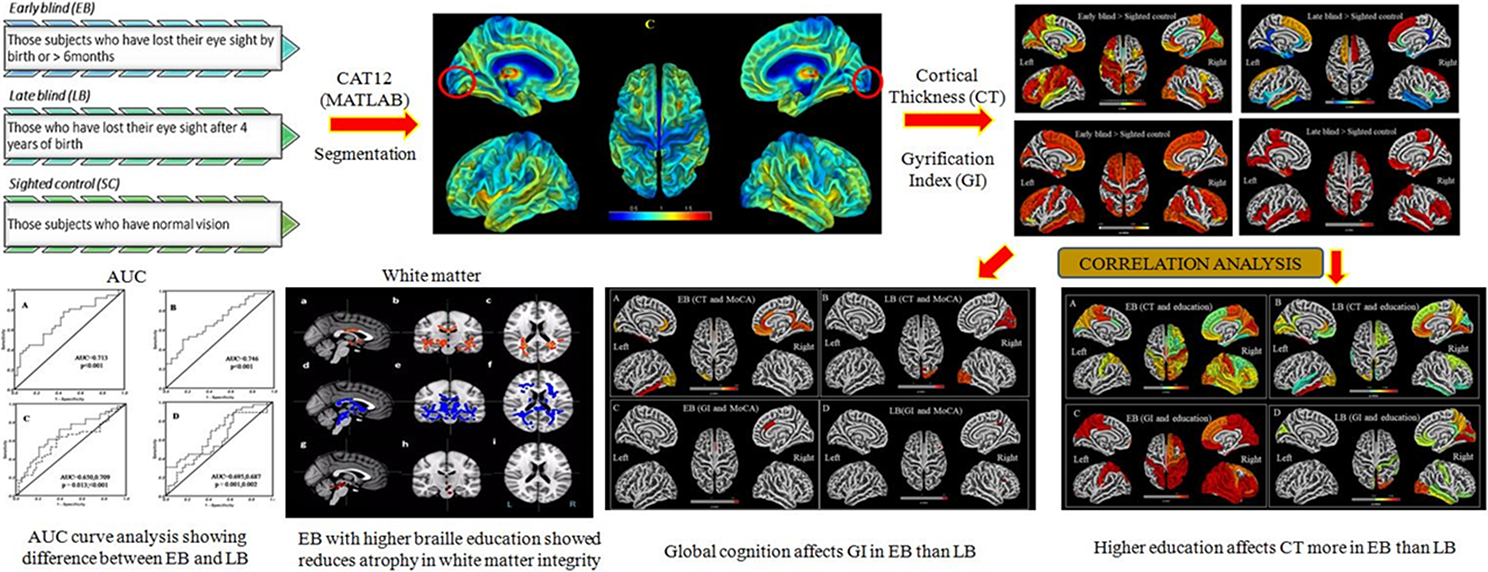
In early (EB) and late blind (LB) children, vision deprivation produces cross-modal plasticity in the visual cortex. The progression of structural- and tract-based spatial statistics changes in the visual cortex in EB and LB, as well as their impact on global cognition, have yet to be investigated. The purpose of this study was to determine the cortical thickness (CT), gyrification index (GI), and white matter (WM) integrity in EB and LB children, as well as their association to the duration of blindness and education. Structural and diffusion tensor imaging data were acquired in a 3T magnetic resonance imaging in EB and LB children (n = 40 each) and 30 sighted controls (SCs) and processed using CAT12 toolbox and FSL software. Two sample t-test was used for group analyses with P < 0.05 (false discovery rate-corrected). Increased CT in visual, sensory-motor, and auditory areas, and GI in bilateral visual cortex was observed in EB children. In LB children, the right visual cortex, anterior-cingulate, sensorimotor, and auditory areas showed increased GI. Structural- and tract-based spatial statistics changes were observed in anterior visual pathway, thalamo-cortical, and corticospinal tracts, and were correlated with education onset and global cognition in EB children. Reduced impairment in WM, increased CT and GI and its correlation with global cognitive functions in visually impaired children suggests cross-modal plasticity due to adaptive compensatory mechanism (as compared to SCs). Reduced CT and increased FA in thalamo-cortical areas in EB suggest synaptic pruning and alteration in WM integrity. In the visual cortical pathway, higher education and the development of blindness modify the morphology of brain areas and influence the probabilistic tractography in EB rather than LB.

Type
Research Article
Copyright
© The Author(s), 2021. Published by Cambridge University Press

Access options

Get access to the full version of this content by using one of the access options below. (Log in options will check for institutional or personal access. Content may require purchase if you do not have access.)

References

Amedi, A., Raz, N., Pianka, P., Malach, R., & Zohary, E. (2003). Early “visual” cortex activation correlates with superior verbal memory performance in the blind. Nature Neuroscience 6, 758766.CrossRefGoogle ScholarPubMed
Anurova, I., Renier, L.A., De Volder, A.G., Carlson, S., & Rauschecker, J.P. (2015). Relationship between cortical thickness and functional activation in the early blind. Cerebral Cortex 25, 20352048.CrossRefGoogle ScholarPubMed
Cohen, L.G., Celnik, P., Pascual-Leone, A., Corwell, B., Faiz, L., Dambrosia, J., Honda, M., Sadato, N., Gerloff, C., Catala, M.D., & Hallett, M. (1997). Functional relevance of cross-modal plasticity in blind humans. Nature 389, 180183.CrossRefGoogle ScholarPubMed
Dahnke, R., Ziegler, G. & Gaser, C. (2012). Local Adaptive Segmentation. Human Brain Mapping Conference, Peking, China. Poster #521.Google Scholar
De Volder, A.G., Bol, A., Blin, J., Robert, A., Arno, P., Grandin, C., Michel, C., & Veraart, C. (1997). Brain energy metabolism in early blind subjects: Neural activity in the visual cortex. Brain Research 750, 235244.CrossRefGoogle ScholarPubMed
Desikan, R.S., Ségonne, F., Fischl, B., Quinn, B.T., Dickerson, B.C., Blacker, D., Buckner, R.L., Dale, A.M., Maguire, R.P., Hyman, B.T., & Albert, M.S. (2006). An automated labeling system for subdividing the human cerebral cortex on, M.R.I scans into gyral based regions of interest. Neuroimage 31, 968980.CrossRefGoogle Scholar
Gougoux, F., Zatorre, R.J., Lassonde, M., Voss, P., & Lepore, F. (2005). A functional neuroimaging study of sound localization: Visual cortex activity predicts performance in early-blind individuals. PLoS Biology 3, 324333.CrossRefGoogle ScholarPubMed
Hofstetter, S., Sabbah, N., Mohand-Saïd, S., Sahel, J.A., Habas, C., Safran, A.B., & Amedi, A. (2019). The development of white matter structural changes during the process of deterioration of the visual field. Scientific Reports 9, 2085CrossRefGoogle ScholarPubMed
Hogstrom, L.J., Westlye, L.T., Walhovd, K.B., & Fjell, A.M. (2013). The structure of the cerebral cortex across adult life: Age-related patterns of surface area, thickness, and gyrification. Cerebral Cortex 23, 25212530.CrossRefGoogle ScholarPubMed
Jenkinson, M., Beckmann, C.F., Behrens, T.E.J., Woolrich, M.W., & Smith, S.M. (2012). FSL. Neuroimage 62, 782790.CrossRefGoogle ScholarPubMed
Jiang, J., Zhu, W., Shi, F., Liu, Y., Li, J., Qin, W., Li, K., Yu, C., & Jiang, T. (2009). Thick visual cortex in the early blind. Journal of Neuroscience 29, 22052211.CrossRefGoogle ScholarPubMed
Karlen, S.J., Kahn, D.M., & Krubitzer, L. (2006). Early blindness results in abnormal corticocortical and thalamocortical connections. Neuroscience 142, 843858.CrossRefGoogle ScholarPubMed
Kingsbury, M.A., Lettman, N.A., & Finlay, B.L. (2002). Reduction of early thalamic input alters adult corticocortical connectivity. Developmental Brain Research 138, 3543.CrossRefGoogle ScholarPubMed
Lamballais, S., Vinke, E.J., Vernooij, M.W., Ikram, M.A., & Muetzel, R.L. (2020). Cortical gyrification in relation to age and cognition in older adults. Neuroimage 212, 116637CrossRefGoogle ScholarPubMed
Leporé, N., Voss, P., Lepore, F., Chou, Y.Y., Fortin, M., Gougoux, F., Lee, A.D., Brun, C., Lassonde, M., Madsen, S.K., & Toga, A.W. (2010). Brain structure changes visualized in early- and late-onset blind subjects. Neuroimage 49, 134140.CrossRefGoogle ScholarPubMed
Li, J., Liu, Y., Qin, W., Jiang, J., Qiu, Z., Xu, J., Yu, C, & Jiang, T. (2013a). Age of onset of blindness affects brain anatomical networks constructed using diffusion tensor tractography. Cerebral Cortex 23, 542551.CrossRefGoogle Scholar
Li, Q., Jiang, Q., Guo, M., Li, Q., Cai, C., & Yin, X. (2013b). Grey and white matter changes in children with monocular amblyopia: Voxel-based morphometry and diffusion tensor imaging study. British Journal of Ophthalmology 97, 524529.CrossRefGoogle Scholar
Li, Q., Song, M., Xu, J., Qin, W., Yu, C., & Jiang, T. (2017). Cortical thickness development of human primary visual cortex related to the age of blindness onset. Brain Imaging and Behavior 11, 10291036.CrossRefGoogle ScholarPubMed
Luders, E., Thompson, P.M., Narr, K.L., Toga, A.W., Jancke, L., & Gaser, C. (2006). A curvature-based approach to estimate local gyrification on the cortical surface. Neuroimage 29, 12241230.CrossRefGoogle ScholarPubMed
Malone, I.B., Leung, K.K., Clegg, S., Barnes, J., Whitwell, J.L., Ashburner, J., Fox, N.C., & Ridgway, G.R. (2015). Accurate automatic estimation of total intracranial volume: A nuisance variable with less nuisance. Neuroimage 104, 366372.CrossRefGoogle ScholarPubMed
Merabet, L.B. & Pascual-Leone, A. (2010). Neural reorganization following sensory loss: The opportunity of change. Nature Reviews Neuroscience 11, 4452.CrossRefGoogle ScholarPubMed
Oldfield, R.C. (1971). The assessment and analysis of handedness: The Edinburgh inventory. Neuropsychologia 9, 97113.CrossRefGoogle ScholarPubMed
Pan, W.J., Wu, G., Li, C.X., Lin, F., Sun, J., & Lei, H. (2007). Progressive atrophy in the optic pathway and visual cortex of early blind Chinese adults: A voxel-based morphometry magnetic resonance imaging study. Neuroimage 37, 212220.CrossRefGoogle ScholarPubMed
Park, H.J., Lee, J.D., Kim, E.Y., Park, B., Oh, M.K., Lee, S.C., & Kim, J. J. (2009). Morphological alterations in the congenital blind based on the analysis of cortical thickness and surface area. Neuroimage 47, 98106.CrossRefGoogle ScholarPubMed
Petanjek, Z., Judaš, M., Šimić, G., Rašin, M.R., Uylings, H.B.M., Rakic, P., & Kostović, I. (2011). Extraordinary neoteny of synaptic spines in the human prefrontal cortex. Proceedings of the National Academy of Sciences of the United States of America 108, 1328113286.CrossRefGoogle ScholarPubMed
Rauschecker, J.P. (1995). Compensatory plasticity and sensory substitution in the cerebral cortex. Trends in Neurosciences 18, 3643.CrossRefGoogle ScholarPubMed
Röder, B., Teder-Sälejärvi, W., Sterr, A., Rösler, F., Hillyard, S.A., & Neville, H.J. (1999). Improved auditory spatial tuning in blind humans. Nature 400, 162166.CrossRefGoogle ScholarPubMed
Sadato, N., Okada, T., Honda, M., & Yonekura, Y. (2002). Critical period for cross-modal plasticity in blind humans: A functional MRI study. Neuroimage 16, 389400.CrossRefGoogle ScholarPubMed
Sadato, N., Pascual-Leone, A., Grafman, J., Ibañez, V., Deiber, M.P., Dold, G., & Hallett, M. (1996). Activation of the primary visual cortex by Braille reading in blind subjects. Nature 380, 526528.CrossRefGoogle ScholarPubMed
Striem-Amit, E., Cohen, L., Dehaene, S., & Amedi, A. (2012). Reading with sounds: Sensory substitution selectively activates the visual word form area in the blind. Neuron 76, 640652.CrossRefGoogle ScholarPubMed
Veraart, C., De Volder, A.G., Wanet-Defalque, M.C., Bol, A., Michel, C., & Goffinet, A.M. (1990). Glucose utilization in human visual cortex is abnormally elevated in blindness of early onset but decreased in blindness of late onset. Brain Research 510, 115121.CrossRefGoogle ScholarPubMed
Volcic, R. & Kappers, A.M.L. (2008). Allocentric and egocentric reference frames in the processing of three-dimensional haptic space. Experimental Brain Research 188, 199213.CrossRefGoogle ScholarPubMed
Voss, P., Gougoux, F., Zatorre, R.J., Lassonde, M., & Lepore, F. (2008). Differential occipital responses in early- and late-blind individuals during a sound-source discrimination task. Neuroimage 40, 746758.CrossRefGoogle ScholarPubMed
Voss, P., Lassonde, M., Gougoux, F., Fortin, M., Guillemot, J.P., & Lepore, F. (2004). Early- and late-onset blind individuals show supra-normal auditory abilities in far-space. Current Biology 14, 17341738.CrossRefGoogle ScholarPubMed
Voss, P. & Zatorre, R.J. (2012). Occipital cortical thickness predicts performance on pitch and musical tasks in blind individuals. Cereb Cortex 22, 24552465.CrossRefGoogle ScholarPubMed
Wang, D., Qin, W., Liu, Y., Zhang, Y., Jiang, T., & Yu, C. (2013). Altered white matter integrity in the congenital and late blind people. Neural Plasticity 2013, Article ID 128236.CrossRefGoogle ScholarPubMed
White, T., Su, S., Schmidt, M., Kao, C.Y., & Sapiro, G. (2010). The development of gyrification in childhood and adolescence. Brain and Cognition 72, 3645.CrossRefGoogle ScholarPubMed
Wittich, W., Phillips, N., Nasreddine, Z.S., & Chertkow, H. (2010). Sensitivity and specificity of the Montreal cognitive assessment modified for individuals who are visually impaired. Journal of Visual Impairment and Blindness 104, 360368.CrossRefGoogle Scholar
Woolrich, M.W., Jbabdi, S., Patenaude, B., Chappell, M., Makni, S., Behrens, T., Beckmann, C., Jenkinson, M., & Smith, S.M. (2009). Bayesian analysis of neuroimaging data in FSL. Neuroimage 45, S173S186.CrossRefGoogle ScholarPubMed
Yotter, R.A., Dahnke, R., Thompson, P.M., & Gaser, C. (2011a). Topological correction of brain surface meshes using spherical harmonics. Human Brain Mapping 32, 11091124.CrossRefGoogle Scholar
Yotter, R.A., Nenadic, I., Ziegler, G., Thompson, P.M., & Gaser, C. (2011b). Local cortical surface complexity maps from spherical harmonic reconstructions. Neuroimage 56, 961973.CrossRefGoogle Scholar
Yotter, R.A., Thompson, P.M., & Gaser, C. (2011c). Algorithms to improve the reparameterization of spherical mappings of brain surface meshes. Journal of Neuroimaging 21, e134e147.CrossRefGoogle Scholar
Yu, C., Shu, N., Li, J., Qin, W., Jiang, T., & Li, K. (2007). Plasticity of the corticospinal tract in early blindness revealed by quantitative analysis of fractional anisotropy based on diffusion tensor tractography. Neuroimage 36, 411417.CrossRefGoogle ScholarPubMed
Supplementary material: File

Ankeeta et al. supplementary material

Tables S1-S7 and Figures S1-S3

Download Ankeeta et al. supplementary material(File)
File 5.7 MB