
“Horsetail” Inclusions in the Ural Demantoids: Growth Formations


A.N. Zavaritsky Institute of Geology and Geochemistry, Ural Branch of Russian Academy of Scences, Vonsovskogo str. 15, 620016 Ekaterinburg, Russia
*
Author to whom correspondence should be addressed.
Minerals 2021, 11(8), 825; https://doi.org/10.3390/min11080825
Submission received: 9 July 2021
/
Revised: 23 July 2021
/
Accepted: 27 July 2021
/
Published: 29 July 2021
(This article belongs to the Special Issue Gems and Gem Minerals)
Abstract
:The term “demantoid”, first proposed in 1856 by the famous Finnish mineralogist Nils von Nordensheld, refers to a highly dispersed yellow-green mineral from the Central Urals placers. In 1874, it was found to be a gem variety of andradite garnet. “Horsetail” inclusions are considered a sign of the Ural type demantoid. Although these inclusions are large (visible to the naked eye), their diagnostics remains debatable: some researchers attribute them to byssolite (amphibole-asbestos), others consider them chrysotile. We investigated the horsetail inclusions in the Ural demantoids through various methods: optical microscopy, scanning electron microscopy (SEM), Raman spectrometry, X-ray powder diffraction, and thermal analysis. In most cases, “horsetail” inclusions in the Ural demantoid were represented by hollow channels and only the outcrops, on the demantoid surface, were occasionally filled with serpentine (established by SEM); in one case, magnetite was observed. Hollow canals were usually collected not in bundles, such as a “horsetail”, but in fans, sometimes curved into cones. The structure of the grains was spheroidal, sectorial, and sometimes had induction surfaces, which, to the periphery of the grain, were replaced by tubular channels assembled in a fan. The specifics of the growth of the “horsetail” inclusions of the demantoid grains can be explained by the decompression conditions that arose when the ultrabasites (a crust-mantle mixture) were squeezed upwards during collision.
1. Introduction
Demantoid is a jewelry variety of andradite. Its chemical formula is Ca3Fe2Si3O12, and it often contains admixtures of Cr, Ti, V, Al, and Mg. The refractive index of demantoid is 1.887, the dispersion is 0.057, the hardness is 6.5, and it is inert to UV-rays [1].
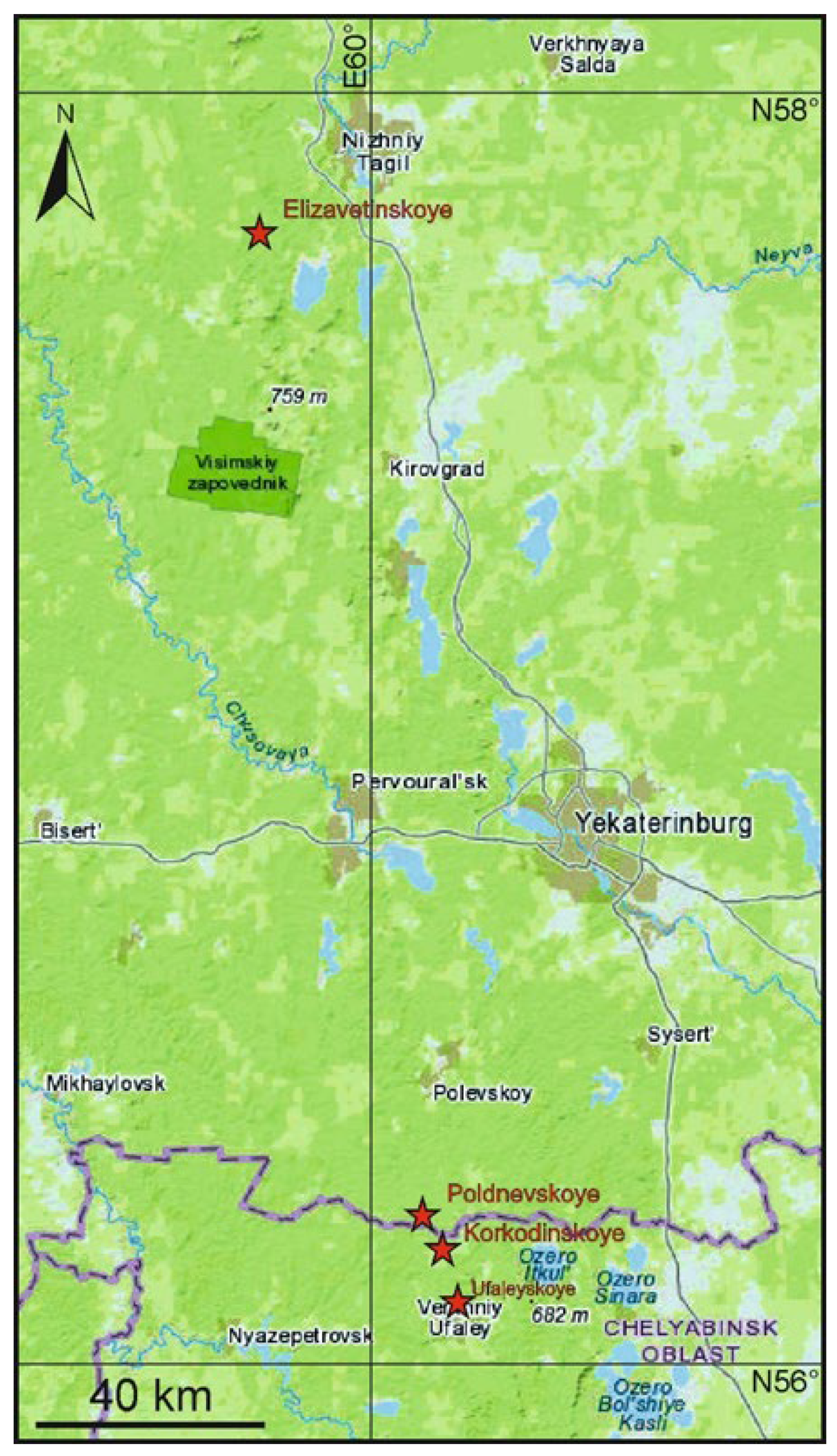
The demantoid was first discovered in gold-bearing alluvial placers of the Bobrovka river, 100 km NNW from Yekaterinburg (Figure 1). It was named in 1856 by the Finnish mineralogist Nils von Nordensheld for its strong dispersion (0.057), comparable with that of diamonds (0.044). The mineral, however, was not identified. In 1874, demantoids were discovered in gold-bearing placers near the Bobrovka river (a widespread name for small rivers in Russia) near the Poldnevaya village, 80 km south of Ekaterinburg (see Figure 1), by V.G. and A.V. Kalugin. In 1879, after the study of the chemical composition of the demantoid by mining engineer A.A. Loesch, the mineral was identified as the andradite garnet. The primary sources of the demantoid were identified in the bedrock of placers in the upper reaches of these rivers at the end of the 19th century. Political events in Russia at the beginning of the 20th century stopped the development of the demantoid deposits in the Urals for many years.
In 1978–1979, one of the authors of this study, A.K., conducted a study of the Poldnevskoye demantoid deposit. This served as the beginning of the systematic prospecting for the demantoid in the Middle Urals, which resulted in the discovery of the Korkodinskoye deposit. Later, the mineralization of the demantoid was established in the Verkhneivinskiy ultrabasic massif (the Central Urals, 50 km north-west of Ekaterinburg) [2], in the Polar Urals [3], on the Koryak highlands, and on the Kamchatka [4]. Demantoids are also known to be present in Italy [5,6], Azerbaijan [7], Iran [8], Pakistan [9], the United States of America, China [10], Slovakia [11], Namibia [12], and Madagascar [13,14].
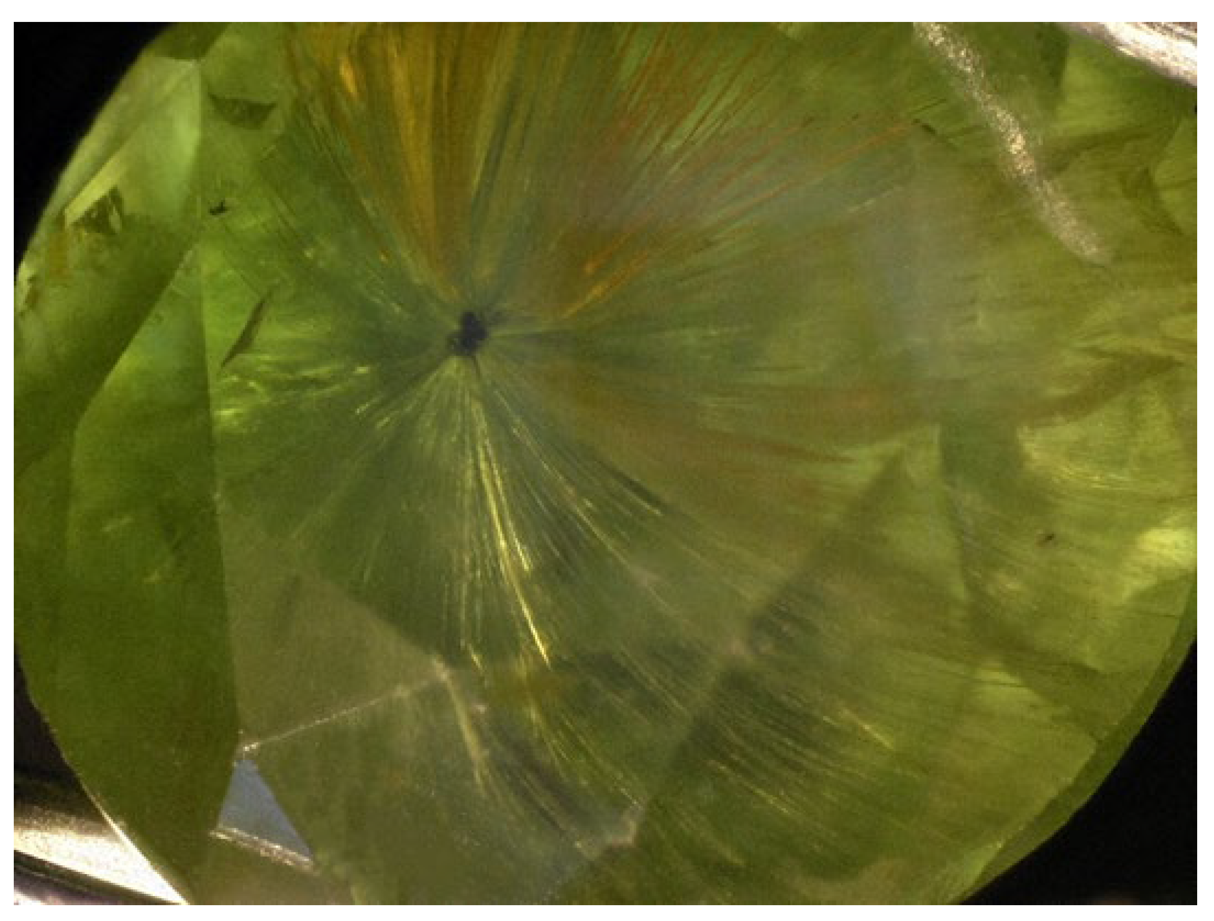
At present, the gem demantoids related to (1) the massif of ultrabasic rocks (the Urals type) and (2) the skarns (the Namibian type) are recognized. The Ural demantoids are characterized by horsetail inclusions, which are hair-like inclusions that diverge in bunches from the central part of the grain to its periphery (Figure 2). These inclusions became the key markers for identifying the demantoid and determining its geographical origin [15,16,17]. Horsetail inclusions were first discovered in the Ural demantoids, as these inclusions are: (1) always present, (2) numerous (the gem can completely lose its transparency), (3) often large and easily detected by the naked eye, and (4) sometimes colored in ochre and clearly visible. In spite of this, there is no consensus among researchers as to the origin or composition of these inclusions. Initially it was assumed that they were byssolite (actinolite-asbestos) and, under this name, they were mentioned in geological reports and articles [3,15,18]. In 1992, at the International Gemological Conference (Paris), Gübelin (Switzerland) reported that he managed to analyze these inclusions in a demantoid from Val Malenco (Italy) by the X-ray diffraction method, and determined their chemical composition to be chrysotile (a serpentine group mineral) [19]. These results were confirmed by microprobe analyses [20]. Chrysotile was also detected in the demantoid from Balochistan (Pakistan) [21].
At the beginning of the 1990s, two of the authors (A.K. and V.M.) studied the “horsetail” inclusions of Ural demantoids and came to the conclusion that they are tubular inclusions [22].
2. Geological Setting
All finds of the demantoid in the Central Ural were confined to the ultrabasite massifs of the Main Urals and the Serovsko-Maukskogo faults: the Nizhnetagilskiy massif (Elizavetinskoye deposit), the Verkhneivinskiy and Korkodinskiy massifs (the Poldnevskoye and Korkodinskoye deposits), and the Ufaleiskiy massif. These massif structures are composed of dunites, clinopyroxenites, and, more rarely, harzburgites and, very rarely, verlites and websterites. The rocks are largely serpentinized and deformed.
Veins with demantoid mineralization are rare: along the strike, they are traced for 2–3 m and are completely wedged out, while along the dip, they are traced to a depth of 20–30 m (the depth of the Poldnevskoye and Korkodinskoye deposits). It is likely that these deposits take the form of ore pillars [23].
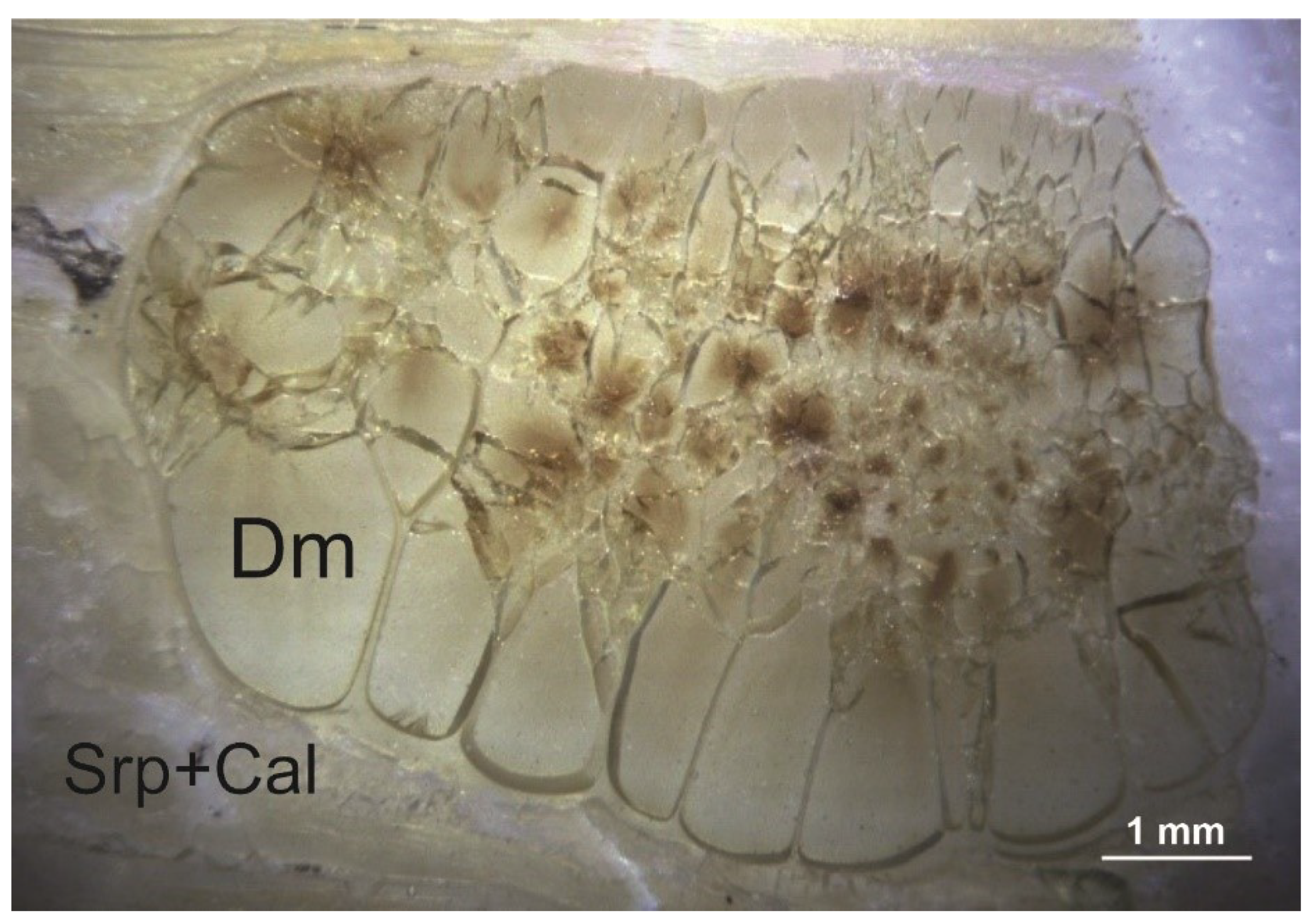
Vein serpentine (antigorite, clinochrysotile, and lizardite), carbonates (calcite, dolomite, magnesite, and aragonite), magnetite, brucite, and, sometimes, native copper, copper and nickel sulfides, cuprite, and other minerals are found in association with demantoids. Demantoids are rarely represented by poorly shaped rhombic dodecahedron crystals and, instead, are often composed of spherical aggregates made by of many individuals separated by veins of serpentine and calcite (Figure 3).
3. Materials and Methods
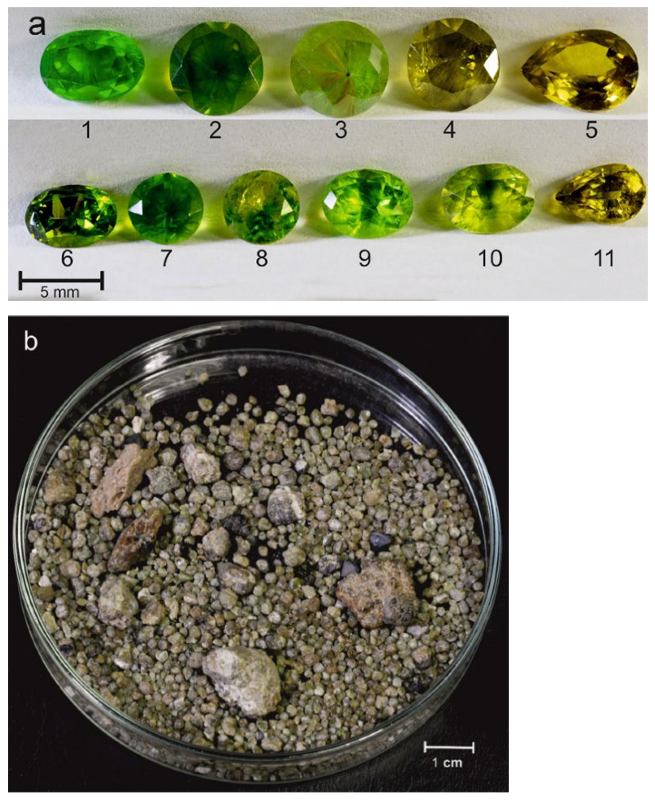
The faceted demantoids (Figure 4a) were borrowed from the collection of A. Kissin. Samples of untreated (rough) demantoid (Figure 4b) were obtained during the exploration work carried out by one of the authors (A.K.) in the Poldnevskoye deposit in 1978–1979. Additional materials were collected by the authors during visits to the Poldnevskoye and Korkodinskoye deposits in 2018–2020.
The investigations were carried out by optical methods using an Olympus microscope with DP2-TWAIN system at 10× to 400× magnification (Japan). The X-ray powder diffraction analysis of demantoid structure was performed at the Institute of Geology and Geochemistry of UrB RAS (Ekaterinburg, Russia).
Thin sections of the demantoid, as well as the demantoid grain surface morphology, were studied using scanning electron microscopy (SEM). The analyses were carried out using a Jeol JSM-6390LV scanning electron microscope (Jeol, Tokyo, Japan) with an INCA Energy 450 X-Max 80 (Oxford Instruments, Abingdon, UK) energy-dispersive spectrometer at the Geoanalitik Collective Use Center of the Institute of Geology and Geochemistry of the Ural Branch of the Russian Academy of Sciences (Ekaterinburg, Russia). The operating conditions included an acceleration voltage of 20 kV, a beam current 85 µA, and an operating distance of 10 mm.
Raman spectra in the region of 100–1000 cm−1 were obtained in back-scattering geometry using a Horiba LabRam HR800 Evolution (Horiba, Palaiseau, France) equipped with an Olympus BX-FM confocal microscope (Olympus, Tokyo, Japan), He-Ne laser (radiation wavelength 633 nm, with laser power of 2 mW) at the Geoanalitik Collective Use Center of the Institute of Geology and Geochemistry of the Ural Branch of the Russian Academy of Sciences (Ekaterinburg, Russia). There was no photoluminescence observed with this excitation. An Olympus 100× objective (Olympus, Tokyo, Japan) with numerical aperture = 0.9 was used. The acquisition time was 10 s, with two accumulations per spectral segment. A diffraction grating of 1800 gr/mm and an electrically cooled charge-coupled device detector were used to record the spectra. The spectrometer calibration was guided along the Rayleigh line and the emission lines of a neon lamp.
4. Results
4.1. Microscopic Characteristics
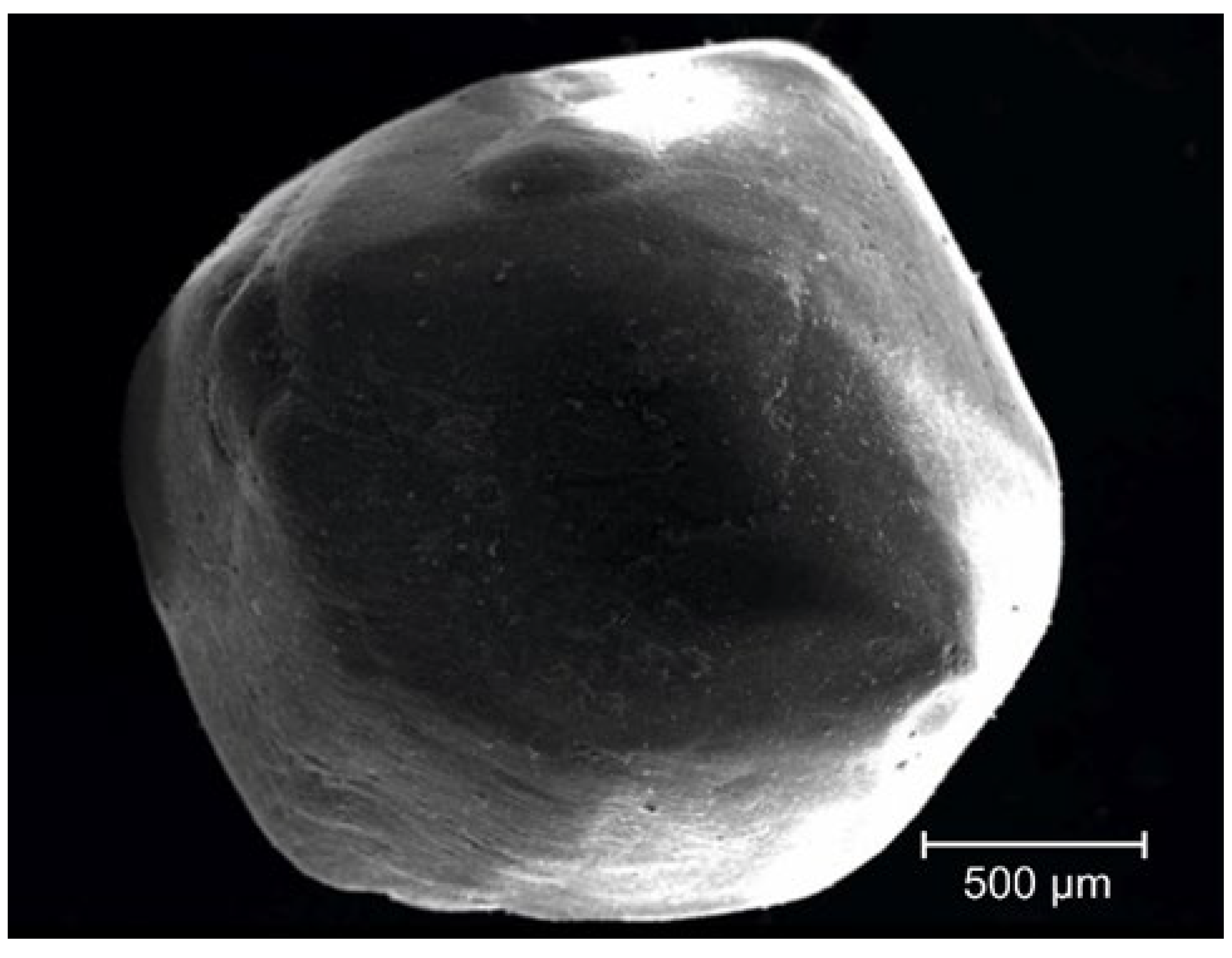
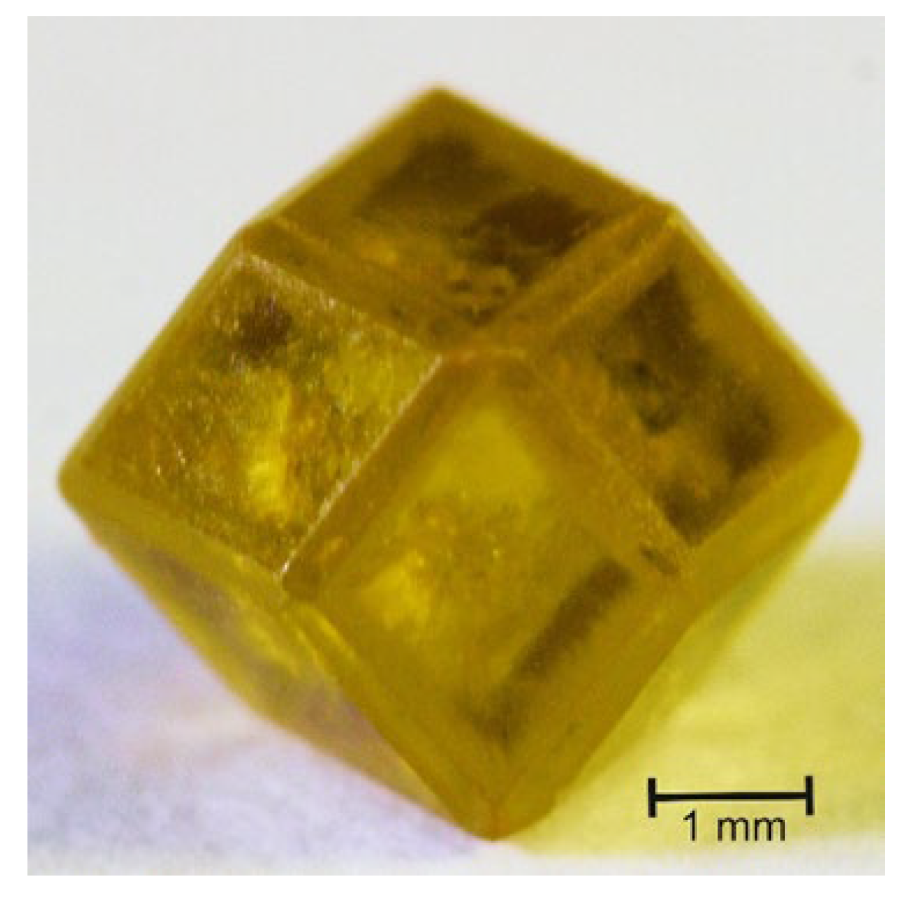
The Ural demantoid rarely has well-formed crystals and, instead, usually manifests as rhombic dodecahedron with a rounded shape and unexpressive ribs (Figure 5). It is very rare to find relatively well-faceted crystals (Figure 6). It is usually represented by grains with rounded irregular shape. Kidney-shaped formations consisting of many small grains with a sharply pronounced increase in size to the periphery of the aggregate are very typical (see Figure 3).
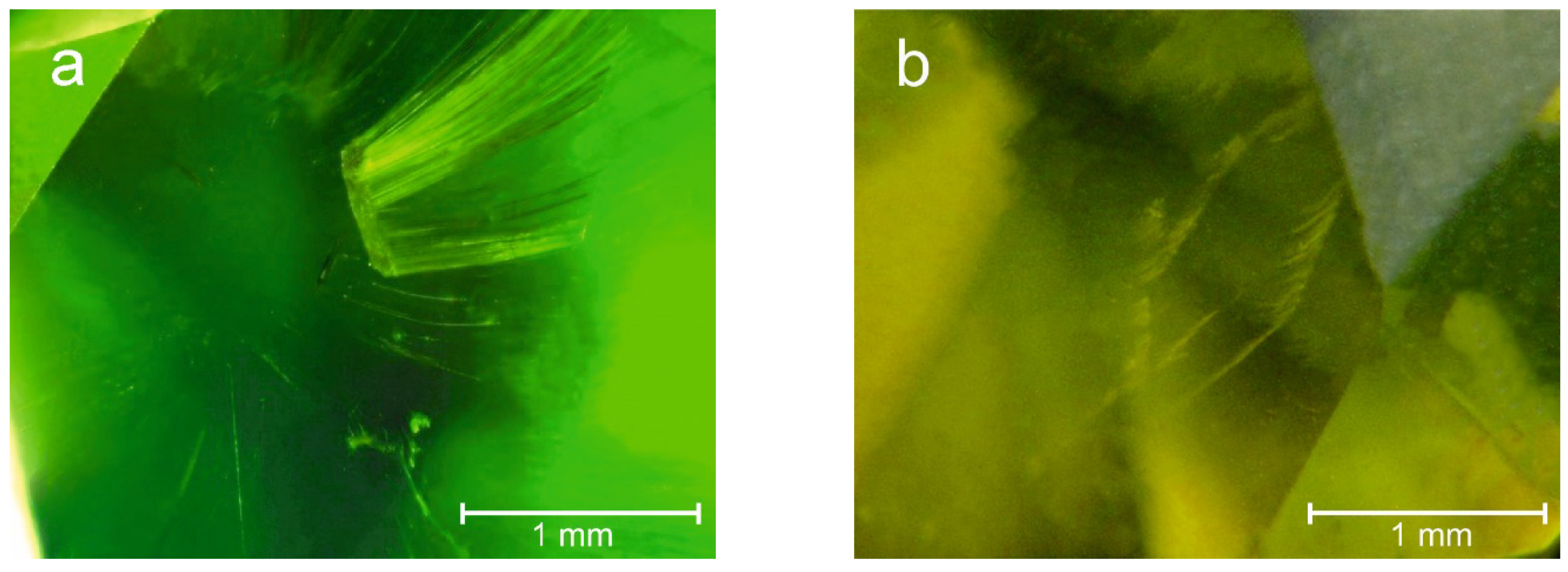
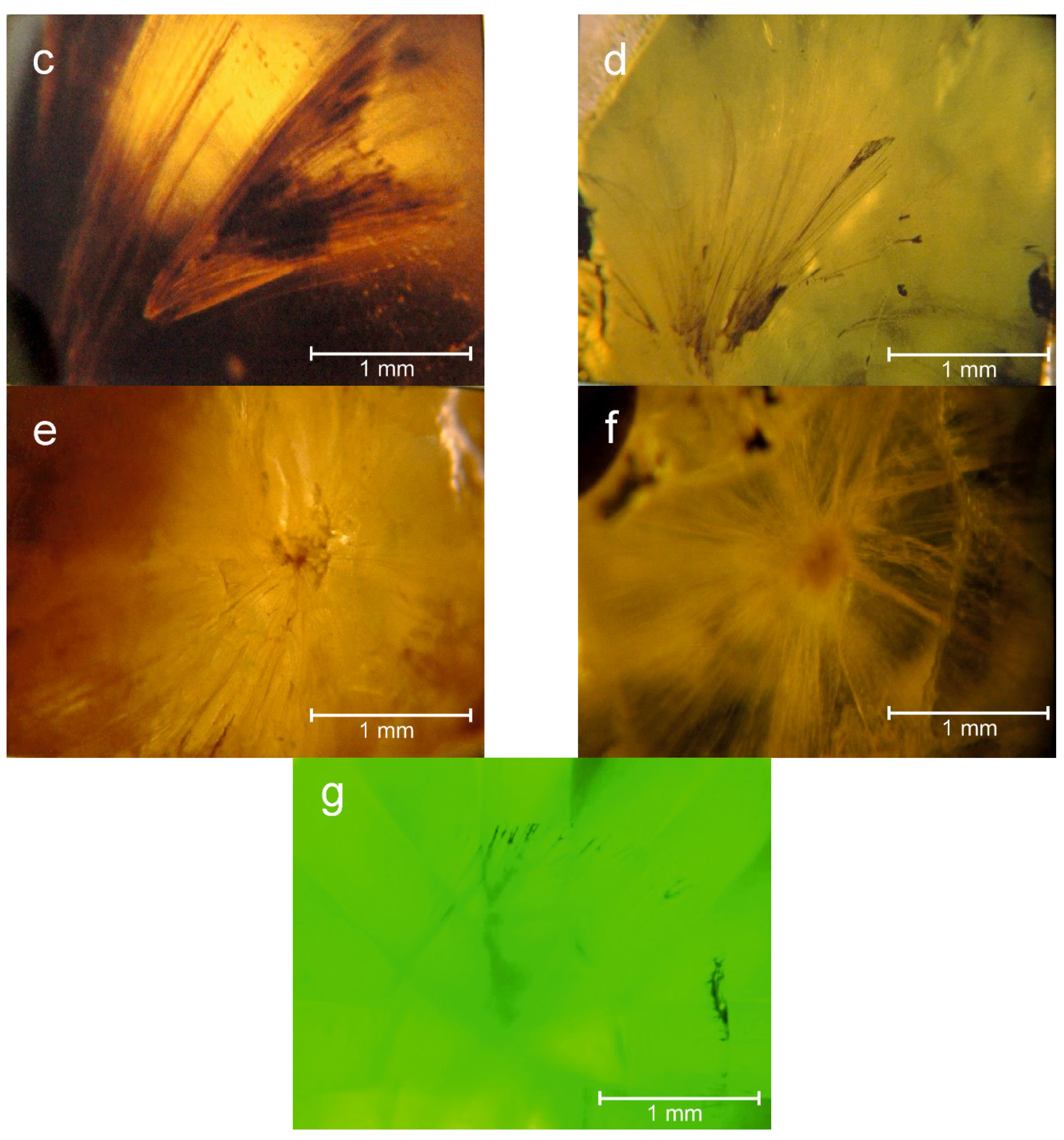
“Horsetail” inclusions are represented by hair-like formations of various sizes assembled in fan or sheaf-like bundles tapering toward the central part of the grain. Closely spaced inclusions form larger and denser formations. “Horsetail” inclusions may begin in the center of the grain from an inclusion of a dark ore mineral (chromospinel or magnetite, see Figure 2), or without a central inclusion. Sometimes “horsetail” inclusions start from a facet of the phantom, which can reflect (Figure 7a). “Horsetail” inclusions are often gathered not in a bundle (horsetail), but in a fan (Figure 7b). This can be seen especially well on the facets of the stone. They can start at any depth of the stone, with shallow “horsetail” inclusions often being v-shaped (Figure 7c). They are usually colorless and transparent, while in the demantoid from alluvial placers, horsetail inclusions are usually colored ochre-yellow (Figure 7e,f). In the stones that underwent high-temperature annealing (to improve the coloring), the inclusions sometimes acquire a dark brown coloring and lose transparency (Figure 7c,d). It is important to note that small disc-shaped cracks are not characteristic for annealed stones, although these cracks are a sign of heat treatment of the gemstones. The “horsetail” inclusions on the polished facets near the surface are dark colored and opaque (Figure 7g), which is due to process dirt.
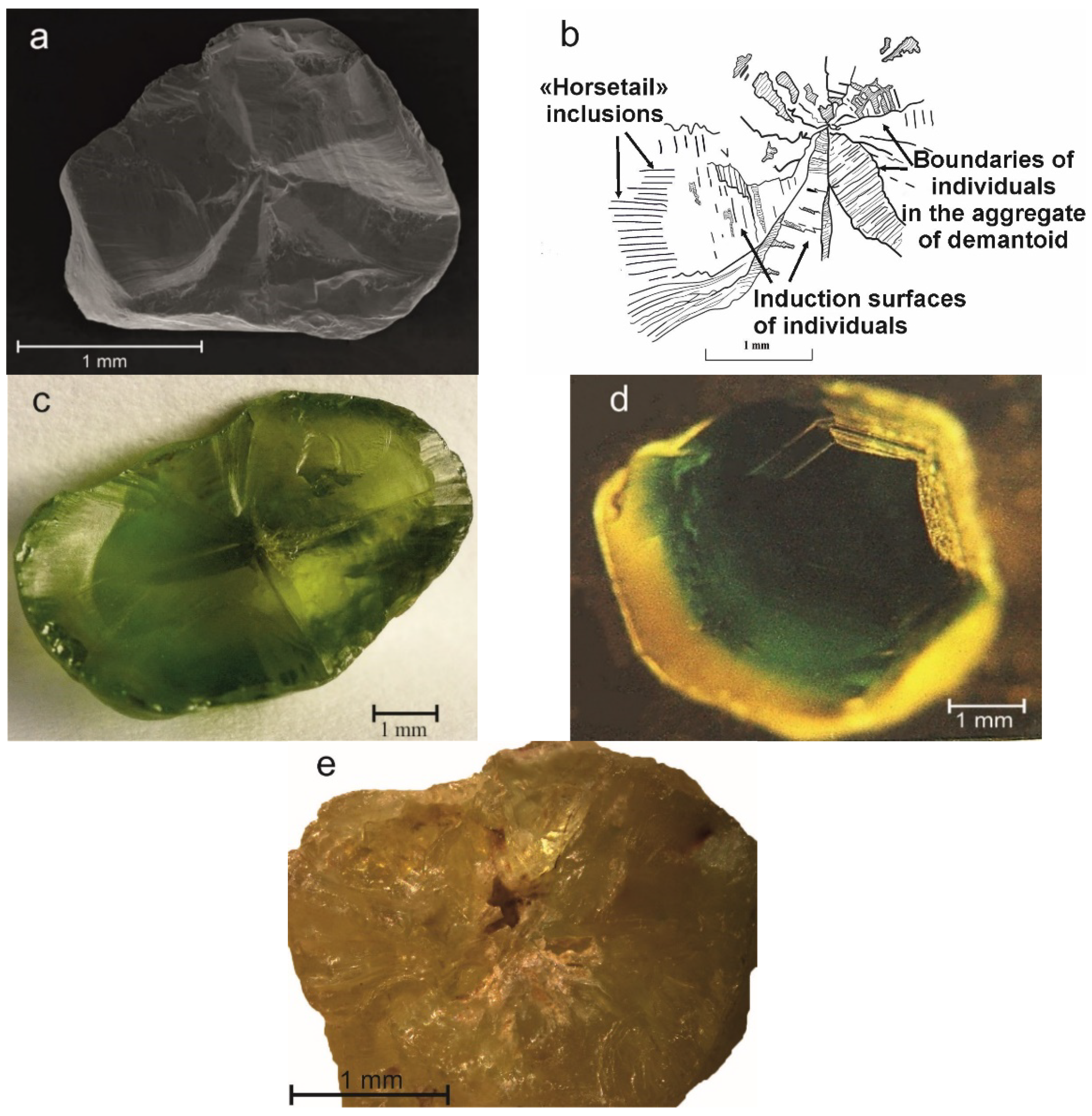
The study of “horsetail” inclusions in transparent, thin sections of the demantoid under a microscope, with and without an analyzer, did not reveal the presence of any mineral phases. When rotating the microscope table, only the reflection from the surface of the inclusions were observed. Sometimes, the chips made through the central part of the demantoid grain clearly showed its sectoral-radial structure (Figure 8a,b): the individuals of the demantoid diverged from the center along radians expanding rapidly. In some places, induction surfaces with coarse transverse striations were clearly visible. There were also longitudinal thin grooves, the number and size of which increased rapidly toward the periphery of the grain (see Figure 8b). These are the “horsetail” inclusions.
Figure 8c shows a similar chipping on the induction surfaces of another demantoid grain. Here, there is no coarse cross hatching, but fine hatching, creating inclusions of the “horsetail” type, is shown quite distinctly. Note the rounded shape of the grain. Figure 8d shows an individual polychrome demantoid with distinct induction surfaces on the dark green (interior) area, and abundant “horsetail” inclusions in its light yellow–green (exterior) area. These inclusions begin only when the induction surfaces with coarse transverse hatching disappear.
Sometimes, there is a cavern in the central part of the demantoid grain, from which “horsetail” inclusions start (Figure 8e). A drop of water placed in the cavern is instantly absorbed by the stone due to the presence of an effective capillary system caused by the “horsetail” inclusions.
4.2. X-ray Powder Diffraction and Thermal Analysis
X-ray powder diffraction of the demantoid samples with numerous “horsetail” inclusions (loss of transparency) detected only andradite. These results were confirmed by thermal research methods.
4.3. Electron Scanning Microscope
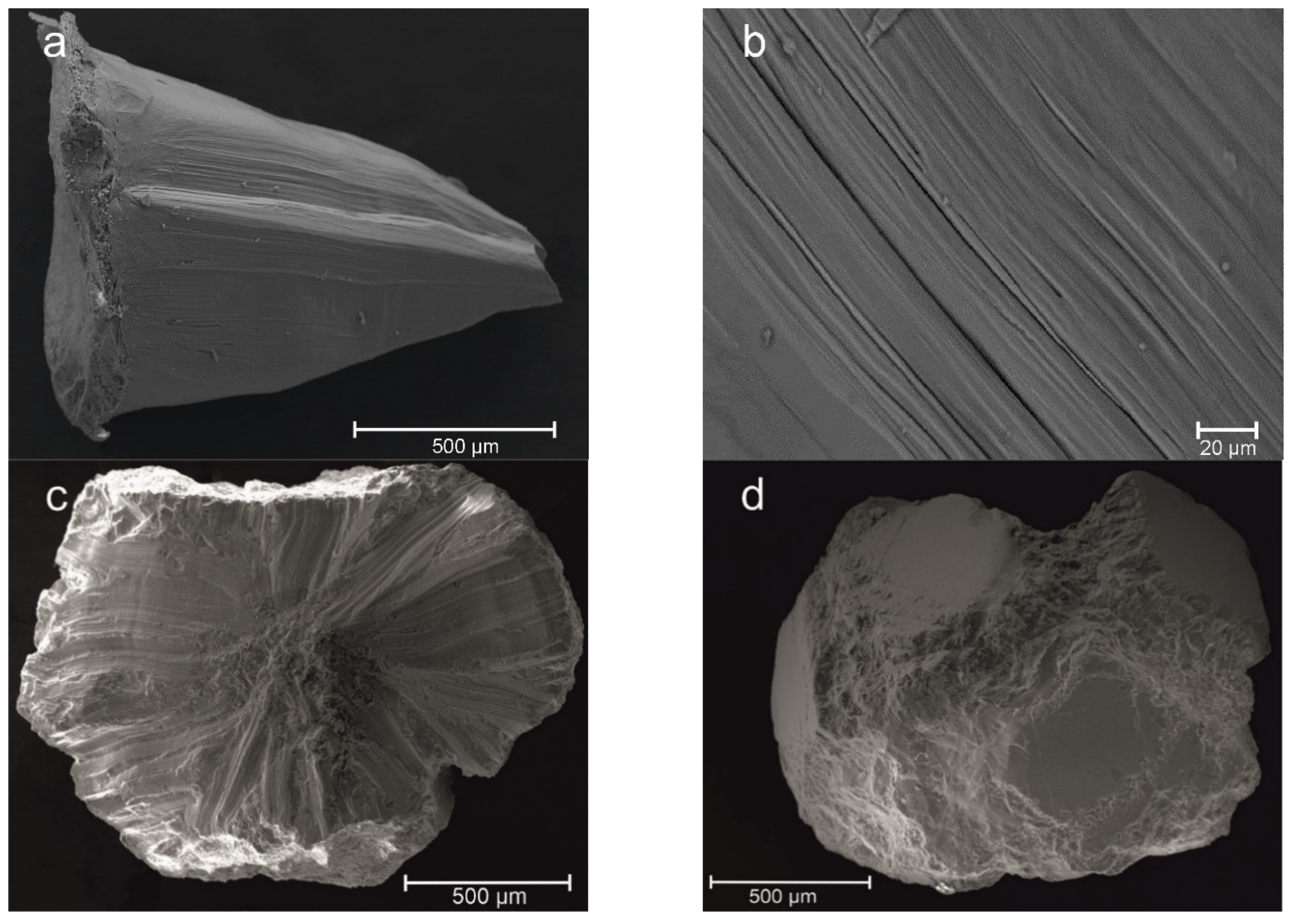
The study of hair-like inclusions on fresh chips of the Ural demantoid grains under an electron scanning microscope showed very interesting results. Even the seemingly monocrystal andradite (see Figure 5) had an aggregate structure. When crushing such a crystal, many fragments were cone-shaped (Figure 9a) with thin longitudinal grooves and knolls. Note also the transverse irregularities (steps) enveloping the cone, which are characteristic of the induction surfaces of crystals. The longitudinal section of “horsetail” inclusions is shown in Figure 9b: many subparallel grooves of different lengths and depths were observed; there were split and relatively short grooves which disappeared in both directions.
The appearance of a chip of brown andradite crystal (Figure 9c) from the Poldnevskoye deposit is very like a “trapiche” crystal of corundum or beryl: flat faces with deep grooves in place of the ribs of the crystal (Figure 9d). Here, too, “horsetail” inclusions were visible, but in some areas they were parallel and also v-shaped.
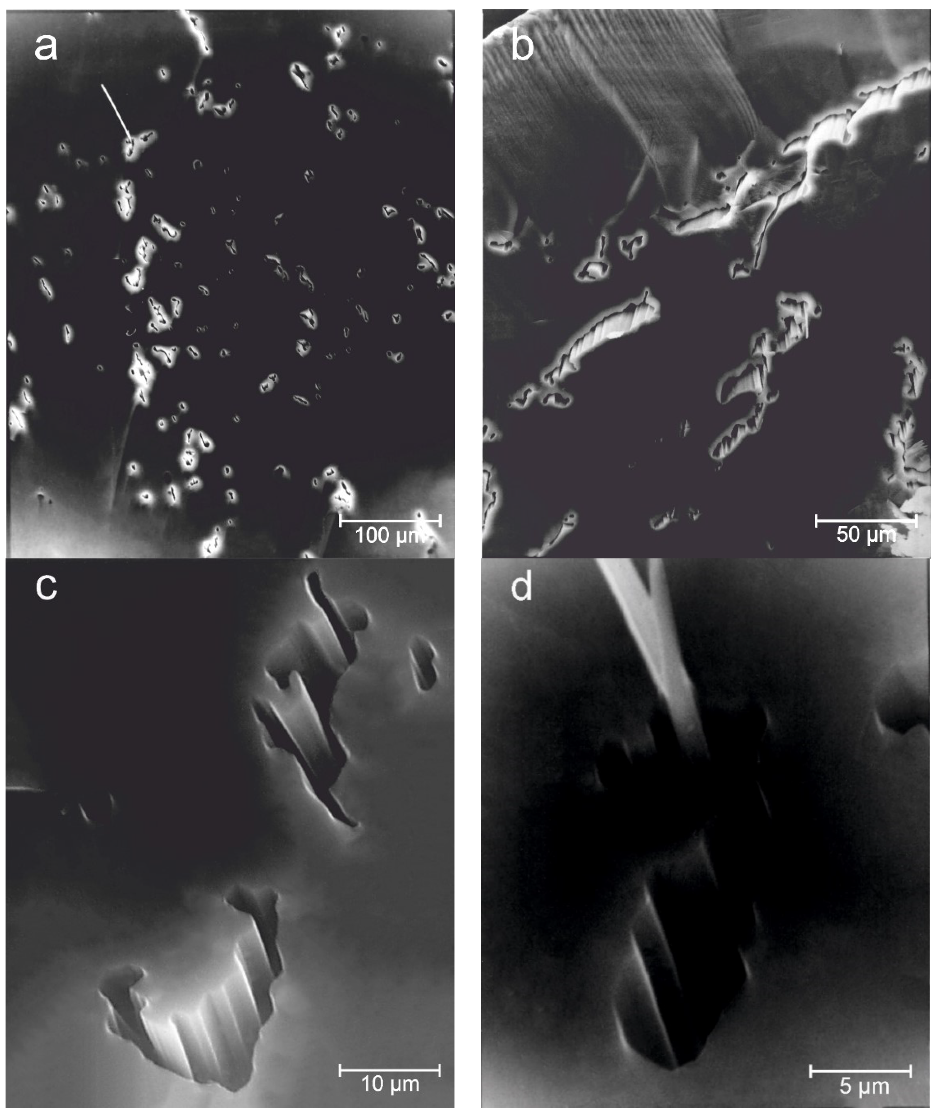
The mineral composition of the “horsetail” inclusions was not detected, or they were represented by single inclusions, which were not identified due to their small size (Figure 10a,d). The vast majority of the “horsetail” inclusions were hollow channels (Figure 10). The configuration of their sections was very bizarre, but closest to slit-like (Figure 10b). The walls of the channels were corrugated, resembling the faces of negative crystals (Figure 10c,d).
4.4. Raman Spectroscopy
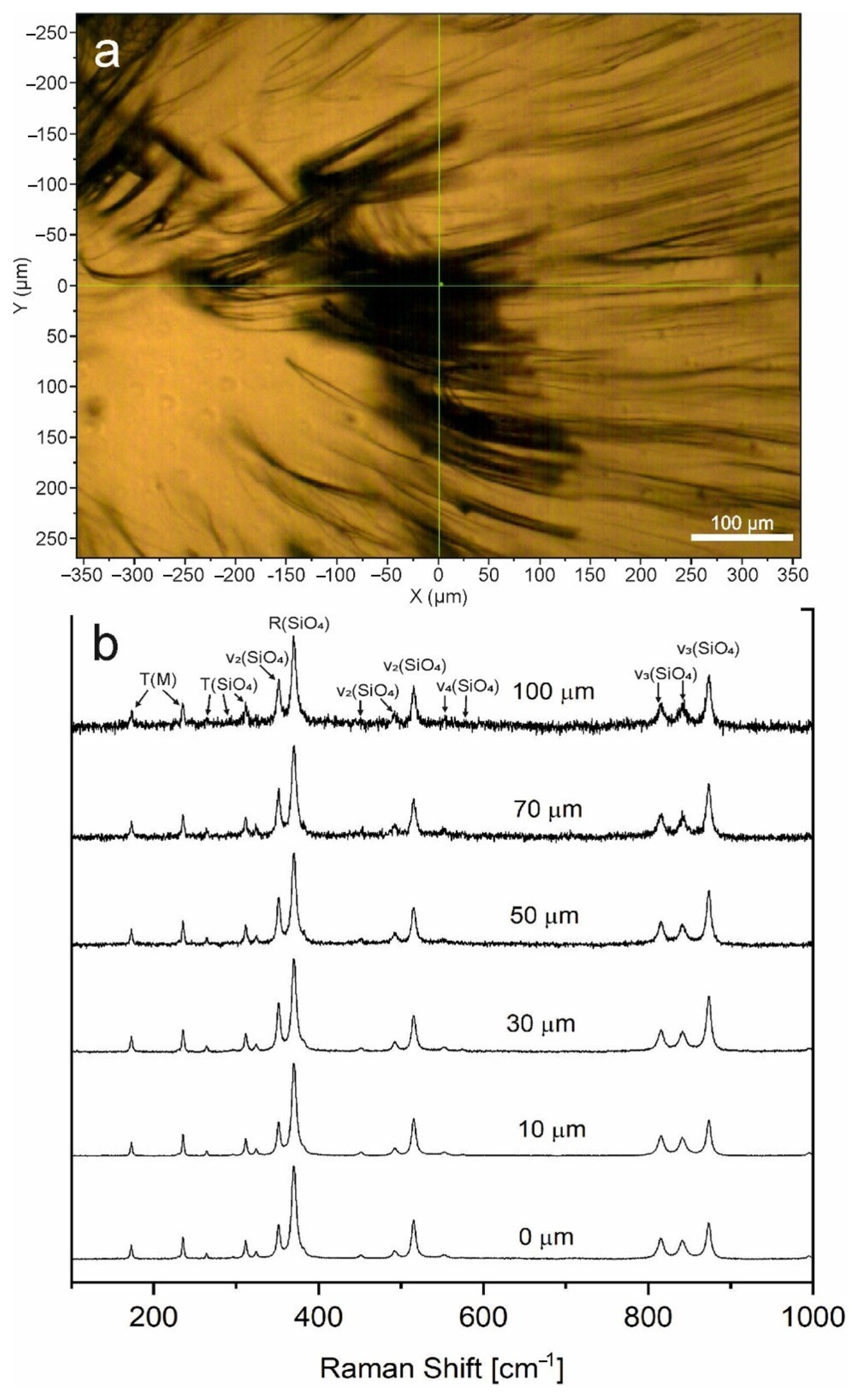
Raman spectroscopy of a mineral filling in the large “horsetail” inclusions (Figure 11a) did not detect in the depth ranges of 0–100 µm. The obtained Raman spectra corresponded to the reference data of the demantoid (Figure 11b). In the Ca3Fe2(SiO4)3 demantoid spectra (space group Ia3d), 15 out of the 25 Raman-active vibrational modes were collected, which were predicted by group-theoretical analysis [24]. No photoluminescence occurred at the selected excitation wavelength.
In the region 100–400 cm−1, modes were associated with the translation of SiO4—T(SiO4), the rotation of SiO4—R(SiO4), and the translation of YO6(Y = Fe3+, Cr3+)—T(M). The bending vibrations of SiO4 tetrahedra – ν2, in the region 493–516 cm−1, with bending vibrations of SiO4 tetrahedra – ν4 and ν2, while in the region 816–874 cm−1, were associated with stretching vibrations of the SiO4 tetrahedra – ν3 [11,24].
5. Discussion
This study on the Ural demantoids, via various methods, showed that “horsetail” inclusions are usually represented by hollow tubular formations. This can explain the problems associated with identifying these inclusions by optical microscopy, X-ray powder diffraction, thermal methods, and Raman spectroscopy. The presence of hollow channels should be already inferred from the analysis of the cause of coloring of “horsetail” inclusions in the demantoids from alluvial placers (see Figure 7e,f), those subjected to high-temperature annealing (see Figure 7c,d), or, sometimes, those exiting on the polished surface of a cut stone (see Figure 7g). In the first case, the staining is due to the penetration of iron hydroxides into the stone, while in the second and third cases it is due to combustion products and technological dirt, respectively. This would be impossible if the “horsetail” inclusions were filled with solid mineral matter. This is also indicated by the absence of disc-shaped cracks around the inclusions in the annealed demantoids, which are inevitable in the presence of solid mineral inclusions. It is noteworthy that similar conclusions were reached by Fritz et al. [25], who studied andradite garnet from Namibia.
The formation of hollow channels in the demantoid could not be the result of dissolution of the mineral inclusion under natural conditions, because the carbonate-serpentine vein masses that host the demantoid (see Figure 3) showed no signs of dissolution, even calcite. Moreover, serpentine filled the “horsetail” inclusions on the surface of the demantoid grain, as its envelopes (see Figure 3). The mouths of hollow channels can also be filled with other minerals. For example, we observed the junction of demantoid and a large magnetite crystal: the contact of the mouths of the “horsetail” inclusions were filled with magnetite.
“Horsetail” inclusions often have a fan-shape, in the plane of which induction surfaces are sometimes observed (see Figure 8a–d), and the demantoid grains themselves have an aggregate sectoral structure. It is the sectoral growth of the demantoid crystals that explains the appearance of “horsetail” inclusions. Even in well-formed crystals (see Figure 6), the sectoral structure is observed (Figure 8a,b).
The reason for the sectoral growth of the demantoid crystals is caused, in our opinion, by mineral formation under the conditions of decompression during the rise of the crust-mantle mixture, experiencing autometamorphism and autometasomatism [26,27]. At the Poldnevskoye deposit, rounded grains of calcite and, sometimes, magnetite, in association with demantoid in the carbonate-serpentine vein mass, had a similar structure. The mechanism of such crystal growth under decompression conditions, however, requires further study.
6. Conclusions
1. “Horsetail” inclusions in the Ural demantoids are represented by tubular hollow formations, sometimes containing serpentine;
2. The formation of “horsetail” inclusions is caused by the spherical growth of the demantoid crystals under conditions of decompression, autometamorphism, and autometasomatism of the up-moving ultrabasite massif;
3. The mechanism of split growth of the demantoid crystals and the formation of “horsetail” inclusions is not well understood and requires additional research.
Author Contributions
Conceptualization, A.Y.K.; methodology, V.V.M. and E.S.K.; software, E.S.K.; visualization, V.V.M. and E.S.K.; writing—original draft, A.Y.K.; writing—review and editing, A.Y.K. and V.V.M. All authors have read and agreed to the published version of the manuscript.
Funding
The work was performed within the framework of topic no. АААА-А18-118052590028-9 of the IGG UB RAS state assignment.
Data Availability Statement
Not applicable.
Acknowledgments
The editor of Minerals and guest editors are thanked for organizing the Special Issue on “Gems and Gem Minerals” and extending an invitation to submit a contribution for consideration. We thank the Reviewers for the important comments and constructive suggestions, which helped us to improve the quality of the manuscript.
Conflicts of Interest
The authors declare no conflict of interest. The funders had no role in the design of the study; in the collection, analyses, or interpretation of data; in the writing of the manuscript, or in the decision to publish the results.
References
- O’Donoghue, M. (Ed.) Gems, 6th ed.; Butterworth-Heinemann: Oxford, UK, 2006. [Google Scholar]
- Murzin, V.V.; Mamin, N.A.; Kissin, A.J.; Demchuk, I.G. Demantoid garnet mineralization of the Verh-Neivinskii alpinotype ultramafic intrusion (Urals). Intergems-95 Turnov 1995, 38–41. [Google Scholar]
- Phillips, W.R.; Talantsev, A.S. Russian demantoid, czar of the garnet family. Gems Gemol. 1996, 32, 100–111. [Google Scholar] [CrossRef]
- Kutyev, F.S.; Anikin, L.P.; Ivanov, B.V.; Kutyeva, G.V.; Lyapichev, I.G.; Pavshukov, V.V.; Samoilovich, M.I.; Sidorov, E.G.; Simonova, L.S.; Sugrobov, V.M.; et al. Finding of demantoids and topazolites in the Koryak Mountain Range. Doclady Acad. NAUK SSSR 1983, 269, 198–200. [Google Scholar]
- Kievlenko, E.Y. Geology of Gems; Ministry of Natural Resources of the Russian Federation, RGU “Quartzsamotsvety”, Earth, Association “Ecost”: Moscow, Russia, 2001; 584p, ISBN 5-900395-25-1. (In Russian)
- Adamo, I.; Bocchio, R.; Diella, V.; Pavese, A.; Vignola, P.; Prosperi, L.; Palanza, V. Demantoid from Val Malenco, Italy: Review and update. Gems Gemol. 2009, 45, 280–287. [Google Scholar] [CrossRef] [Green Version]
- Kashkai, M.-A. On demantoid from ultramafic rocks of Azerbaijan. Rep. USSR Acad. Sci. 1939, 22, 512–514. (In Russian) [Google Scholar]
- Du Toi, G.; Mayerson, W.; van Der Bogert, C.; Douman, M.; Befi, R.; Koivula, J.I.; Kiefert, L. Demantoid from Iran. Gems Gemol. 2006, 42, 131. [Google Scholar]
- Milisenda, C.C.; Henn, U.; Henn, J. Demantoide aus Pakistan. Gemmol. Z. Dtsch. Gemmol. Ges. 2001, 50, 51–56. [Google Scholar]
- Liu, G.; Kang, X.; Zhang, L. On the genesis of demantoid from Xinjiang, China. Chin. J. Geochem. 1986, 5, 381–390. [Google Scholar]
- Štubňa, J.; Bačík, P.; Fridrichová, J.; Hanus, R.; Illášová, L.; Milovská, S.; Škoda, R.; Vaculovič, T.; Čerňanský, S. Gem-Quality Green Cr-Bearing Andradite (var. Demantoid) from Dobšiná, Slovakia. Minerals 2019, 9, 164. [Google Scholar] [CrossRef] [Green Version]
- Giuliani, G.; Pignatelli, I.; Fallick, A.; Boyce, A.; Andriamamonjy, A.; Razafindratsimba, S.; Khan, T. Gem Andradite Garnet Deposits Demantoid Variety. InColor 2017, 36, 28–39. [Google Scholar]
- Danet, F. Gem News International: New discovery of demantoid from Ambanja, Madagascar. Gems Gemol. 2009, 45, 218–219. [Google Scholar]
- Rondeau, B.; Fritsch, E.; Mocquet, B.; Lulyac, Y. Ambanja (Madagascar)—New source of gem demantoid garnet. InColor 2009, 11, 16–20. [Google Scholar]
- Stockton, C.M.; Manson, D.V. Gem andradite garnets. Gems Gemol. 1983, 19, 202–208. [Google Scholar] [CrossRef]
- Kornilov, N.I.; Solodova, Y.P. Jewelry Stones; Nedra: Moscow, Russia, 1986. (In Russian) [Google Scholar]
- Gübelin, E.J.; Koivula, J.I. Photoatlas of Inclusions in Gemstones; ABC Edition: Zurich, Switzerland, 1992; 532p. [Google Scholar]
- Gübelin, E.J. Internal World of Gemstones; ABC Druckerei & Verlags AG: Zurich, Switzerland, 1974. [Google Scholar]
- Gübelin, E.J. Mineral inclusions in gemstones recently observed, analysed and identified. In Proceedings of the XXIV International Gemmological Conference, Paris, France, 19–24 July 1993; p. 85. [Google Scholar]
- Hoskin, P.W.O.; Grapes, R.H.; Catchpole, H.; Klaudius, J. Horsetail inclusions in demantoid from Val Malenco, Italy. J. Gemmol. 2003, 28, 333–336. [Google Scholar] [CrossRef]
- Adamo, I.; Bocchio, R.; Diella, V.; Caucia, F.; Schmetzer, K. Demantoid from Balochistan, Pakistan: Gemmological and Mineralogical Characterization. J. Gemmol. 2015, 34, 428–433. [Google Scholar] [CrossRef]
- Kissin, A.Y.; Murzin, V.V. Hair-like inclusions in demantoid: What is it? In Yearbook-96; IGG UB RAS: Ekaterinburg, Russia, 1997; pp. 113–115. (In Russian) [Google Scholar]
- Kissin, A.Y.; Murzin, V.V.; Karaseva, E.S.; Ogorodnikov, V.N.; Polenov, Y.A.; Seleznev, S.G.; Ozornin, D.A. Problems of structural control of demantoid mineralization at the Poldnevskoe deposit. News UGMU 2020, 2, 64–73. (In Russian) [Google Scholar] [CrossRef]
- Hofmeister, A.M.; Chopelas, A. Vibrational spectroscopy of end-member silicate garnets. Phys. Chem. Miner. 1991, 17, 503–526. [Google Scholar] [CrossRef]
- Fritz, E.A.; Laurs, B.M. Gem News International: Andradite from Balochistan, Pakistan. Gems Gemol. 2007, 43, 373. [Google Scholar]
- Kissin, A.Y.; Murzin, V.V.; Pritchin, M.E. Tectonic position of the gold mineralization of the Karabash Mountain (The Southern Urals): On the results of the study of small structural forms. Lithosphere 2016, 4, 79–91. (In Russian) [Google Scholar]
- Murzin, V.; Chudnenko, K.; Palyanova, G.; Kissin, A.; Varlamov, D. Physicochemical model of formation of gold-bearing magnetite-chlorite-carbonate rocks at the Karabash massif of ultramafic rocks (Southern Urals, Russia). Minerals 2018, 8, 306. [Google Scholar] [CrossRef] [Green Version]
Figure 1.
Geographic position of the demantoid deposits in the Central Urals.

Figure 2.
“Horsetail” inclusions in the demantoid from the Bobrovka river placer (Elizavetinskiy village, the Central Urals); image width 4 mm. Photo by E.S. Karaseva.
Figure 2.
“Horsetail” inclusions in the demantoid from the Bobrovka river placer (Elizavetinskiy village, the Central Urals); image width 4 mm. Photo by E.S. Karaseva.

Figure 3.
Slice of a rounded aggregate of demantoid in the carbonate-serpentine vein (Poldnevskoye deposit). Srp+Cal: serpentine and calcite, Dm: andradite (brown - Ti-containing, light yellow-green-demantoid). Photo by E.S. Karaseva.
Figure 3.
Slice of a rounded aggregate of demantoid in the carbonate-serpentine vein (Poldnevskoye deposit). Srp+Cal: serpentine and calcite, Dm: andradite (brown - Ti-containing, light yellow-green-demantoid). Photo by E.S. Karaseva.

Figure 4.
Research material: (a) collection of faceted demantoids with “horsetail” inclusions sourced from Poldnevskoye (1–2), Elizavetinskoye (3; alluvial), and Korkodinskoye (4–11); and (b) untreated demantoid (rough material) from the Poldnevskoye deposit.
Figure 4.
Research material: (a) collection of faceted demantoids with “horsetail” inclusions sourced from Poldnevskoye (1–2), Elizavetinskoye (3; alluvial), and Korkodinskoye (4–11); and (b) untreated demantoid (rough material) from the Poldnevskoye deposit.

Figure 5.
Rhombic dodecahedron shape of the demantoid crystal from the Poldnevskoye deposit. SE image.
Figure 5.
Rhombic dodecahedron shape of the demantoid crystal from the Poldnevskoye deposit. SE image.

Figure 6.
The demantoid crystal from the Ufaleiskiy manifestation.

Figure 7.
Features of “horsetail” inclusions revealed by optical methods: (a) “horsetail” inclusions start from the reflecting surface of the crystal phantom (stone 2 in Figure 4a); (b) curtains of threadlike inclusions; (c) v-shaped dark-brown inclusion in annealed stone; (d) inclusion in annealed stone; (e,f) colored inclusions in the demantoids from alluvial placers; and (g) dark, opaque ends of “horsetail” inclusions, exiting to the polished surface (stone 1 in Figure 4a).


Figure 8.
Features of the structure of the Ural demantoid grains: (a) photo of chipping through the central part of the demantoid grain, showing induction surfaces; (b) same, sketch from photo (a); (c) chipping through the central part of the demantoid grain (induction surfaces); (d) induction surfaces on the polychrome demantoid grain recovered from the aggregate; and (e) cavity in the central part of the demantoid grain.

Figure 9.
BSE-images of demantoids: (a) cone-shaped fragment; (b) surface of the chip in the “horsetail” inclusions plane; and (c) chip through the center of an opaque andradite crystal with negative-shaped ribs (d).
Figure 9.
BSE-images of demantoids: (a) cone-shaped fragment; (b) surface of the chip in the “horsetail” inclusions plane; and (c) chip through the center of an opaque andradite crystal with negative-shaped ribs (d).

Figure 10.
BSE-images of “horsetail” inclusions in fresh chipping of the demantoid: (a) point-like hollow channels (one contains a needle-like inclusion of an unknown mineral); (b) slit-like hollow channels; (c) morphology of cross sections of point and slit-like channels; and (d) mineral inclusion in a hollow channel (fragment of Figure 10a).

Figure 11.
Raman spectrum of the Demantoid from the Poldnevskoye deposit: (a) optical image of the “horsetail” inclusion in the demantoid (thin sections); and (b) Raman spectra of inclusions in the depth range of 0–100 µm.
Figure 11.
Raman spectrum of the Demantoid from the Poldnevskoye deposit: (a) optical image of the “horsetail” inclusion in the demantoid (thin sections); and (b) Raman spectra of inclusions in the depth range of 0–100 µm.

Publisher’s Note: MDPI stays neutral with regard to jurisdictional claims in published maps and institutional affiliations. |
© 2021 by the authors. Licensee MDPI, Basel, Switzerland. This article is an open access article distributed under the terms and conditions of the Creative Commons Attribution (CC BY) license (https://creativecommons.org/licenses/by/4.0/).
Share and Cite
MDPI and ACS Style
Kissin, A.Y.; Murzin, V.V.; Karaseva, E.S. “Horsetail” Inclusions in the Ural Demantoids: Growth Formations. Minerals 2021, 11, 825. https://doi.org/10.3390/min11080825
AMA Style
Kissin AY, Murzin VV, Karaseva ES. “Horsetail” Inclusions in the Ural Demantoids: Growth Formations. Minerals. 2021; 11(8):825. https://doi.org/10.3390/min11080825
Chicago/Turabian StyleKissin, Aleksander Yurevich, Valery Vasilevich Murzin, and Elizaveta Sergeevna Karaseva. 2021. "“Horsetail” Inclusions in the Ural Demantoids: Growth Formations" Minerals 11, no. 8: 825. https://doi.org/10.3390/min11080825
Note that from the first issue of 2016, this journal uses article numbers instead of page numbers. See further details here.