Article

The Effects of Temperature, Light, and Feeding on the Physiology of Pocillopora damicornis, Stylophora pistillata, and Turbinaria reniformis Corals

by
Kerri L. Dobson
1,*,
Christine Ferrier-Pagès
2,
Casey M. Saup
1 and
Andréa G. Grottoli
1,†
1
School of Earth Sciences, The Ohio State University, Columbus, OH 43210, USA
2
Centre Scientifique de Monaco, 98000 Monaco, Monaco
*
Author to whom correspondence should be addressed.
Co-correspondence and senior author.
Water 2021, 13(15), 2048; https://doi.org/10.3390/w13152048
Submission received: 28 May 2021 / Revised: 22 July 2021 / Accepted: 23 July 2021 / Published: 27 July 2021
(This article belongs to the Special Issue Climate Impact on Sustainability of Aquatic Organisms)

Abstract

:
Evidence has shown that individually feeding or reduced light can mitigate the negative effects of elevated temperature on coral physiology. We aimed to evaluate if simultaneous low light and feeding would mitigate, minimize, or exacerbate negative effects of elevated temperature on coral physiology and carbon budgets. Pocillopora damicornis, Stylophora pistillata, and Turbinaria reniformis were grown for 28 days under a fully factorial experiment including two seawater temperatures (ambient temperature of 25 °C, elevated temperature of 30 °C), two light levels (high light of 300 μmol photons m−2 s−1, low light of 150 μmol photons m−2 s−1), and either fed (Artemia nauplii) or unfed. Coral physiology was significantly affected by temperature in all species, but the way in which low light and feeding altered their physiological responses was species-specific. All three species photo-acclimated to low light by increasing chlorophyll a. Pocillopora damicornis required feeding to meet metabolic demand irrespective of temperature but was unable to maintain calcification under low light when fed. In T. reniformis, low light mitigated the negative effect of elevated temperature on total lipids, while feeding mitigated the negative effects of elevated temperature on metabolic demand. In S. pistillata, low light compounded the negative effects of elevated temperature on metabolic demand, while feeding minimized this negative effect but was not sufficient to provide 100% metabolic demand. Overall, low light and feeding did not act synergistically, nor additively, to mitigate the negative effects of elevated temperature on P. damicornis, S. pistillata, or T. reniformis. However, feeding alone was critical to the maintenance of metabolic demand at elevated temperature, suggesting that sufficient supply of heterotrophic food sources is likely essential for corals during thermal stress (bleaching) events.

1. Introduction

Under predicted future ocean conditions, mean global ocean temperature is estimated to increase 1–4 °C by 2100 [1]. As corals already live close to their upper thermal maximum, this baseline temperature shift is predicted to increase the frequency and severity of mass bleaching events [2,3]. Increasing sea surface temperature can cause the coral host-algal symbiosis to break down, resulting in coral bleaching as the endosymbiotic algae (Symbiodiniaceae) and/or algal pigments are lost from the coral tissue (e.g., [4,5,6]).
Within a healthy coral holobiont, endosymbiotic algae photosynthesize in excess of their daily metabolic needs and translocate fixed carbon to the coral host, often providing more than 100% of daily metabolic requirements (e.g., [7,8,9]), while heterotrophy can provide between ≈5 and 46% of daily metabolic requirements (e.g., [7,10,11]). However, under thermal stress, the loss of the endosymbiotic algae and/or photosynthetic pigments causes photosynthesis to decline (e.g., [12,13,14]), and corals are no longer able to meet metabolic demand through photosynthesis alone [7]. To compensate for this decrease in photosynthetic capacity, coral can undertake one or more of the following strategies to aid in the recovery and maintenance of metabolic demand: (1) increase heterotrophy (e.g., [7,15,16,17]), (2) catabolize energy reserves (e.g., [7,18,19]), (3) decrease respiration (e.g., [14,20,21]), and (4) decrease calcification rates (e.g., [22,23,24]). Heterotrophy can also mitigate bleaching damage to algal endosymbionts by stimulating photosynthesis and the re-establishment of host–algal symbioses [20,25,26], and by providing fixed carbon for tissue building and lipid synthesis [17,27,28].
Increases in photosynthetically active radiation (PAR) can inhibit the photosystems of endosymbiotic algae (photoinhibition) [29], which can be exacerbated with increased temperature (e.g., [30,31,32]). Therefore, reduced PAR may decrease damage from, or mitigate the negative effects of, elevated temperature stress on algal endosymbionts [33], so decreasing the thermal sensitivity of the holobiont [34,35,36,37,38]. Furthermore, Lesser [39] determined that the production of reactive oxygen species (ROS) by Symbiodiniaceae is greater under high light and thermal stress, and that low light could prevent, or decrease, the production of ROS that occurs under elevated temperature. Such evidence suggests that shading of corals (i.e., naturally on cloudy days or artificially via manipulation with shade-cloths) could provide a mechanism for protecting corals from elevated temperature stress. However, as photosynthetic capacity also decreases under low light, corals must photo-acclimate (i.e., increasing algal and/or pigment densities) [40,41,42] and/or increase heterotrophy to compensate [43,44,45,46].
Much work has been carried out on the individual effects of increased temperature, light, and feeding on coral physiology (e.g., [20,47,48]), but only one has tested the multi-stressor effects of all three simultaneously [49]. This study found feeding and low light levels to mitigate the negative effects of elevated temperature on chlorophyll a concentrations and photosynthetic rates in Turbinaria reniformis and Stylophora pistillata. However, no experiment to date has examined the interactive effects of all three variables on the overall coral holobiont (i.e., coral + endosymbiotic algae) physiology (i.e., calcification, biomass, lipids, chlorophyll a, and biogeochemistry) and carbon budgets of multiple species.
Here, we experimentally assess the individual and interactive multi-stressor effects of elevated temperature, reduced light, and feeding on the physiology, biogeochemistry, and carbon budget of three aquarium-cultured Red Sea coral: Pocillopora damicornis, Stylophora pistillata, and Turbinaria reniformis. These species were chosen as they represent a range of morphologies (fine branching, thick branching, and foliose, respectively), are ubiquitous throughout the tropical Pacific and Indian oceans, and have been well studied [9,16,49,50,51,52,53,54,55]. We hypothesize that reduced light and feeding will act synergistically to mitigate or minimize negative effects of elevated temperature on coral holobiont physiology and carbon budgets.

2. Materials and Methods

2.1. Experimental Design

Full metadata associated with this study is given in Table S1 [56]. Five colonies of S. pistillata and T. reniformis (originally sourced from the Red Sea) were collected from the Centre Scientifique de Monaco where they had been reared for >5 years in Mediterranean flow-through seawater tanks at 24.5 °C, 200 μmol photons m−2 s−1 with a 12:12 h day/night diurnal light cycle and fed two-day-old Artemia nauplii twice weekly. All colonies were assumed to be distinct genets. Additionally, in November 2012, five colonies of P. damicornis (originally sourced from the Red Sea) were acquired from five different mariculture facilities (Centre Scientifique de Monaco, Cap d’Agde, Oceanopolis, Nausicaa, and Marineland-Antibes), where they were reared under similar conditions for prolonged periods. Acquiring the P. damicornis from the 5 facilities increased the probability that colonies were distinct genets.
In March 2013, each colony of P. damicornis, S. pistillata, and T. reniformis was divided into 8 large and 8 small ramets, totaling 40 large (≈5 cm2) and 40 small (≈3 cm2) ramets per species. All ramets recovered under ambient seawater conditions (25 °C flow-through, filtered, oligotrophic Mediterranean seawater, 200 μmol photons m−2 s−1, and fed two-day-old Artemia nauplii twice weekly) for one month. On 12 April 2013, ramets were distributed randomly and evenly among eight 17 L experimental tanks such that each tank contained one large and one small ramet from each coral colony of each species (N = 30 per tank). P. damicornis and T. reniformis ramets, each with a clay epoxy base engraved with a unique identifier, were placed on the tank bottom. S. pistillata ramets were suspended mid-tank by nylon thread with unique identifiers. Ramets acclimated for two additional weeks under ambient conditions as described above.
On 2 May 2013, a fully factorial experiment with eight treatments was initiated (Figure 1), including two temperatures (ambient temperature of 25 °C, elevated temperature of 30 °C), two light levels (high light of 300 μmol photons m−2 s−1, low light of 150 μmol photons m−2 s−1), and either fed (ad libitum twice a week for approximately 70 min with two-day old Artemia nauplii at an average concentration of 12.3 Artemia nauplii mL−1) or unfed (Figure 1). While individual treatment replication was not possible due to equipment and space constraints within the experimental facility, there were four tanks of each treatment type rendering statistical analyses robust at that level (Figure 1). Any interpretation of effects between individual tanks was done with this caveat in mind. Coral ramets under ambient temperature, high light, and unfed served as the control within the experiment in order to test the effect of each treatment. Temperature conditions in each of the elevated temperature tanks was increased by 0.5 °C per day, until the target temperature of 30 °C was reached after 11 days. Temperature was continuously monitored and regulated within ±0.2 °C using temperature controllers, and it was recorded almost daily starting on 6 May 2013 (Figure 2). The ambient temperature of 25 °C is the long-term average temperature that these aquaria-reared corals have experienced for the past several years, while the average elevated temperature of 30 °C was employed because it has been shown that Red Sea species require a much larger temperature increase to experience temperature stress [16,57,58,59]. Previous studies have generally considered low light to be <300 μmol photons m−2 s−1 (e.g., [60,61,62,63,64]). The photosystem II of each species is saturated at 300 μmol photons m−2 s−1 (Stylophora pistillata [49], Pocillopora damicornis [64], and Turbinaria reniformis [65]). In addition, the Mediterranean seawater supplied to the tanks was filtered, but it still contained dissolved and particulate organic carbon, which is a known source of heterotrophic carbon to corals [66,67,68,69,70]. The fed corals were supplied Artemia nauplii twice a week at the same time and on the same days as they were accustomed to for the past several years, while the unfed corals were not. Coral ramets were subjected to experimental conditions (ramping plus target conditions) for 28 days, on a 12:12 h day/night diurnal light cycle, with an average flow rate of 14.4 L hr−1. Ramet positions within each tank were rotated three times per week to remove any positional effects within the tanks, and tanks were cleaned once a week to minimize filamentous algal growth.

2.2. Host and Symbiont Physiology

Each large ramet was buoyant weighed at the start (3–4 May 2013) and end (27–29 May 2013) of the experiment [71]. During the last 3 days of the experiment (27 May–29 May 2013), the following measurements were also made on a subset (N = 3) of the small ramets: maximum photosynthetic rate (Pmax) [72], light-enhanced dark respiration (R) [72] and feeding rate capacity [16]. Additionally, large and small ramets were photographed underwater. Once these measurements were made, all ramets were frozen on 30 May 2013, shipped, and stored at −80 °C at the Ohio State University until further analysis.
Surface area of large and small ramets were determined using the wax dipping technique [73] for P. damicornis and the foil technique [74] for S. pistillata and T. reniformis. Each small ramet of each species was airbrushed, and the tissue slurry was collected for isotopic analyses. Due to the delicate nature of P. damicornis ramets, air-brushed slurry was prepared for further downstream analyses of this species for small and large ramets. The large ramets of S. pistillata and T. reniformis were ground into a homogenous paste using a mortar and pestle. Chlorophyll a content was measured according to methods in Jeffrey and Humphrey [75], calcification using the buoyant weight technique of Jokiel et al. [71], and biomass according to methods in McLachlan et al. [76]. Photosynthesis (P), respiration (R), feeding rate, chlorophyll a, calcification, and biomass were standardized to surface area. Total lipids were extracted according to methods in McLachlan et al. [77], normalized to tissue biomass (g g−1 ash-free dry weight), and converted into Joules [78]. Total lipids were not measured on P. damicornis due to the limited sample available.
Carbon Budgets. PG and R were used to calculate the Contribution of Zooxanthellae (Symbiodiniaceae) to Animal Respiration (CZAR) [8], while feeding rate capacity was used to calculate Contribution of Heterotrophy to Animal Respiration (CHAR) from feeding on Artemia nauplii using equations modified from Grottoli et al. [7]:
CHAR = H C R C × 100 %
where RC is total daily respiration (gC day−1 gdw−1), and HC is the contribution of heterotrophy to daily metabolic demand (gC day−1 gdw−1). HC was modified and calculated as follows:
H C = ( F R × 3.65 ) × C Z × M i P i × 1.18
where FR is the feeding rate capacity (Artemia nauplii hour−1 gdw−1), CZ is the average proportion of carbon in Artemia nauplii (30%), Mi is the average ash-free dry weight of a single Artemia nauplius (3.04 × 10⁻⁶ g), Pi is the proportion of the diet that the plankton type contributes, 1.18 is the number of hours corals were allowed to feed on feeding days, and 3.65 is the proportionality constant that equates measured feeding rate capacity at the concentration of food available during feeding trials to the concentration of food available during the experiment, as the feeding capacity is related to food concentration [79,80,81]. The constant 3.65 was determined as follows:
B E B F = 3.65
where BE is the average concentration of Artemia nauplii per ml available for consumption during the experiment, considering tank volume and flow rate (12.25 Artemia nauplii mL−1), and BF is the average concentration of Artemia nauplii per ml available for consumption during feeding trials (3.36 Artemia nauplii mL−1). As coral were fed Artemia nauplii only two days per week, we assumed that on days they were not fed, the CHAR contribution was 0%. In addition, we did not measure the DOC and POC fluxes of the corals and so were unable to calculate CHAR from those potential heterotrophic sources. Thus, our CHAR calculations are very conservative as they only account for feeding on Artemia two days a week and do not include contributions from DOC or POC. To assess total carbon budget, CZAR and CHAR were summed to yield Contribution of Total fixed carbon to Animal Respiration (CTAR) [23]. Thus, the calculated CTAR values of fed coral only consider feeding days.
Host δ13C (δ13Ch) isotopes.Stylophora pistillata were prepared for isotopic analyses according to methods modified from Hughes and Grottoli [17]. A subsample of coral tissue slurry was homogenized, sonicated (probe sonicator: 20% amplitude for 60 s total, 1:1 cycle), and 35 mg mL−1 NaCl added. The subsample was filtered through 20 µm nitex mesh to remove skeletal material, the algal endosymbiont isolated onto GF/F filters, and the host tissue filtrate dried down in 9 mm × 10 mm tin capsules on a heat plate (60 °C) with ultra-pure nitrogen gas (this step does not affect sample nitrogen isotope values [82]. Pocillopora damicornis and T. reniformis were prepared for isotopic analyses according to methods modified from Hughes and Grottoli [17] and described in Price et al. [83]. All P. damicornis and T. reniformis host fractions were combusted in an Elementar Vario EL Cube/Micro Cube elemental analyzer interfaced to a PDZ Europa 20–20 isotope ratio mass spectrometer at the stable isotope facility at University of California—Davis. Repeated measurements of an internal standard have produced the long-term standard deviation of ±0.02‰ for δ13C. Stylophora pistillata host fractions were combusted in a Costech elemental analyzer stable isotope ratio mass spectrometer (EA-IRMS), and the resulting C gas was automatically analyzed with a Thermo Finnigan Delta Plus isotope ratio mass spectrometer via a ConFlow open split interface in the Grottoli Stable Isotope Biogeochemistry Lab at Ohio State University. Repeated measurements of internal standards had a standard deviation of ±0.10‰ for δ13C. All δ13C values are reported as the permil deviation of the ratio of stable carbon isotopes 13C:12C relative to Vienna PeeDee Belemnite (v-PDB). Approximately 10% of samples were run in duplicate, with an average standard deviation of ±0.16‰.
Artemia nauplii fed to the corals during this experiment have previously been measured to have δ13C values of −12‰ [54].

2.3. Statistical Analyses

A Euclidean distance-based resemblance matrix was constructed to test the effect of treatment on overall coral holobiont physiological profile using normalized data of the primary physiological variables: chlorophyll a, δ13Ch, calcification, biomass, and (for S. pistillata and T. reniformis only) total lipids. Photosynthesis, respiration, and feeding rate were not included, as these variables were only measured on a subset of ramets within each species (N = 3). Collinearity amongst physiological response variables was investigated using Draftsman’s plots and Pearson’s correlation coefficient. No variables were strongly correlated (i.e., |r| < 0.70 [84]) and thus were treated as independent. Homogeneity of variance in the multivariate dispersions (PERMDISP) was tested, before three-way permutational multivariate analysis of variance (PERMANOVA) was used to evaluate the effects of temperature, light, and feeding on the overall physiology of each species [85]. Multivariate analyses were conducted using Primer v6 [85].
ANOVA analyses were also performed on each variable independently. Each variable dataset was tested for normality using Shapiro–Wilk’s test, and homogeneity of variance was assessed with plots of expected vs. residual values. Calcification in P. damicornis, total lipids in S. pistillata, and calcification and chlorophyll a in T. reniformis were log-transformed to meet assumptions of normal distribution prior to further analysis. Normal distribution was not achievable for CZAR, CTAR, and δ13Ch in P. damicornis, even with multiple transformation attempts. Cook’s Distance was used to identify outliers [86], but outlier removal did not improve normality. Since ANOVA is robust to data with a non-normal distribution [87], ANOVA were still performed on the non-normal datasets, and any interpretations were made with this caveat in mind. Univariate Type III four-way analysis of variance (ANOVA) was used to test the effects of temperature, light, feeding, and genet on each measured variable for each species. Temperature was fixed with 2 levels (25 °C, 30 °C), light was fixed with 2 levels (high light at 300 μmol photons m−2 s−1, low light at 150 μmol photons m−2 s−1), feeding was fixed with 2 levels (fed Artemia nauplii, unfed), and genotype was included as a random effect. As no genotypes were found to be systematically different from all others, we concluded that the colonies represented natural variation in the population, genotype was removed from the models, and three-way type III ANOVAs were run (temperature, light, feeding) and used in interpretation of the data. The lack of any consistent genotype effect agrees with findings of previous similarly designed experiments [14,24,88]. Bonferroni corrections were not employed so as to not enhance Type II error [89]. For significant single effects, post-hoc Tukey tests were run to determine which means were significantly different. Finally, post-hoc slice tests were run to test the effect of the temperature within each light and feeding treatment for each variable. The use of replicate genotypes across all treatments reduced the overall variation between treatments. Since all coral ramets were reared under the same conditions except treatment, any differences between treatments and controls for any variable were due to the treatment effects alone. Univariate parametric statistics were generated using SAS® software (SAS Institute, Cary, NC, USA) version 9.3 for Windows. p ≤ 0.05 were considered significantly different.

3. Results

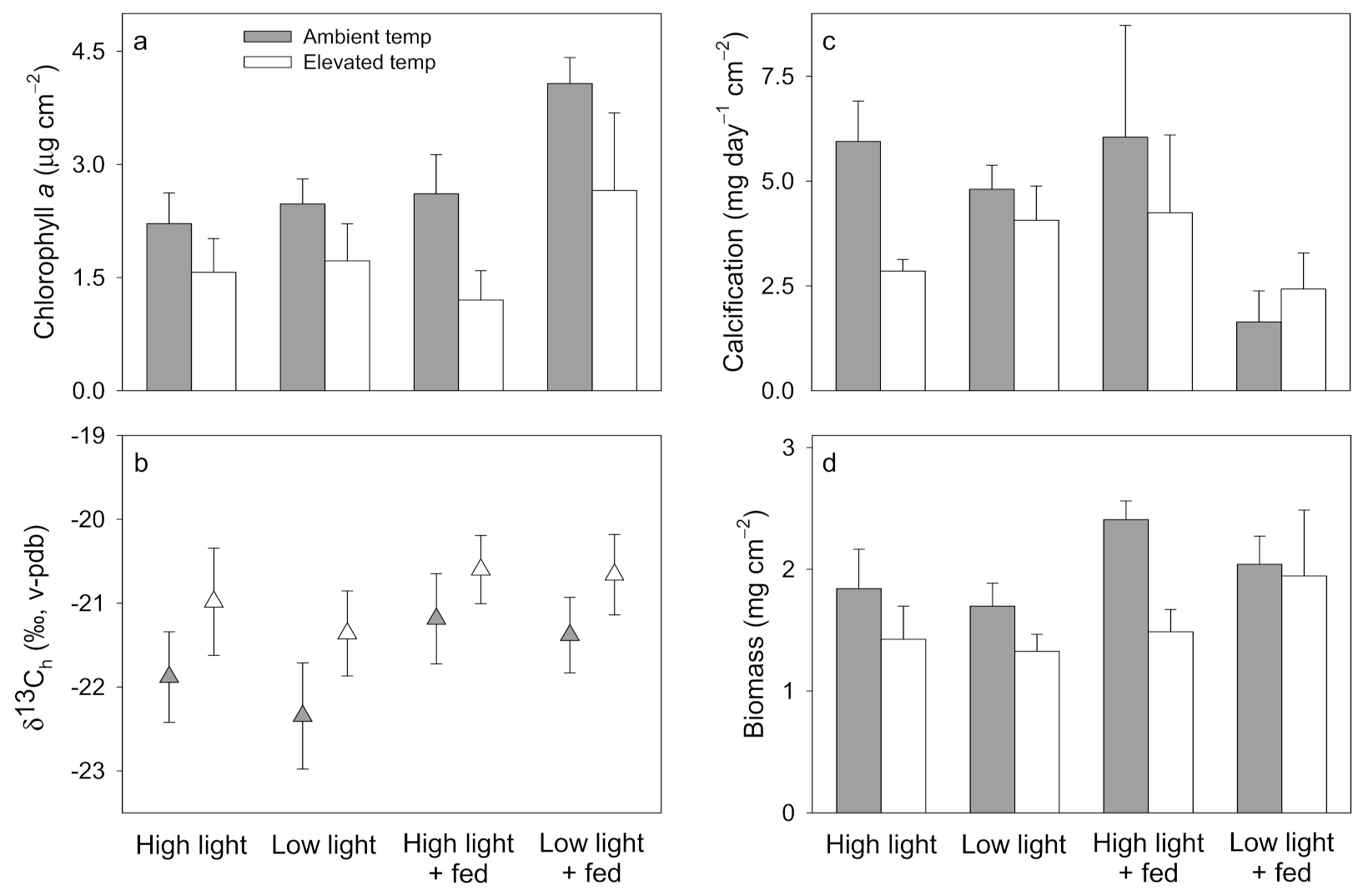
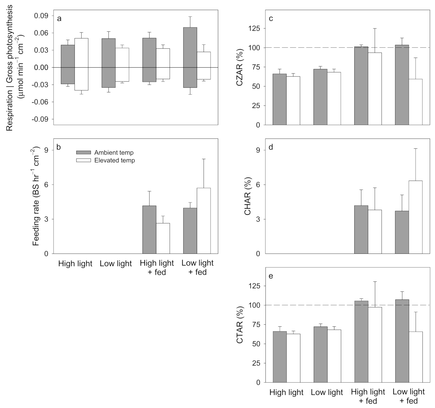
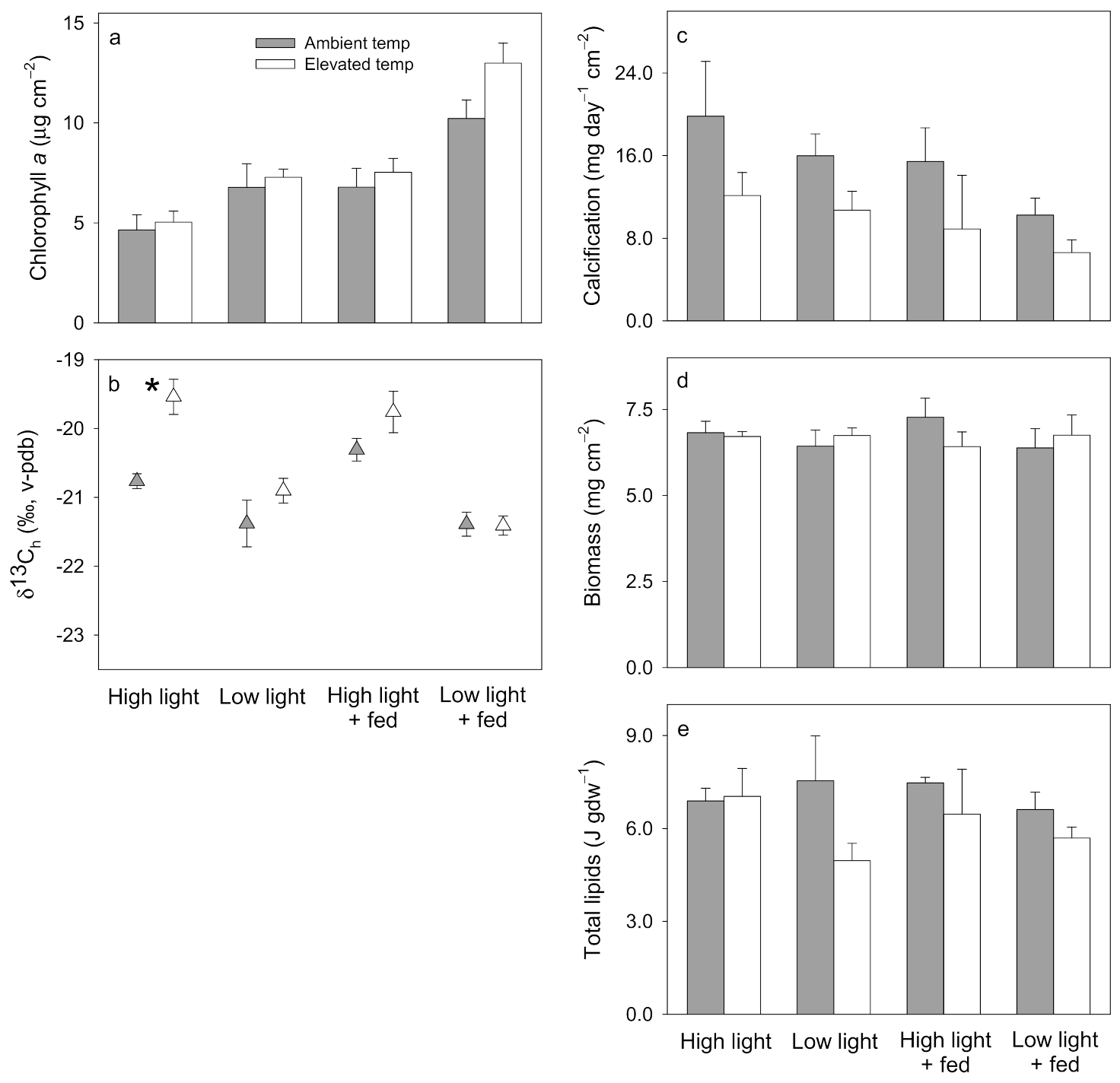
3.1. Pocillopora damicornis

All P. damicornis ramets survived the experiment. The P. damicornis holobiont physiological profile was significantly affected by temperature and feeding (Table S2). Chlorophyll a content was lower under elevated temperature compared to ambient temperature and under high light compared to low light (Figure 3a; Table S3). No significant differences were observed in δ13Ch (Figure 3b; Table S3). Calcification was lower in the low light + fed corals compared to all other treatments, irrespective of temperature (Figure 3c; Table S3), while no significant differences were observed in biomass (Figure 3d; Table S3).
No significant differences were observed in photosynthesis, respiration, feeding rate, CZAR, CHAR, or CTAR (Figure 4a–e; Table S4). However, corals were only able to meet 100% CTAR when fed at ambient temperature (Figure 4e).

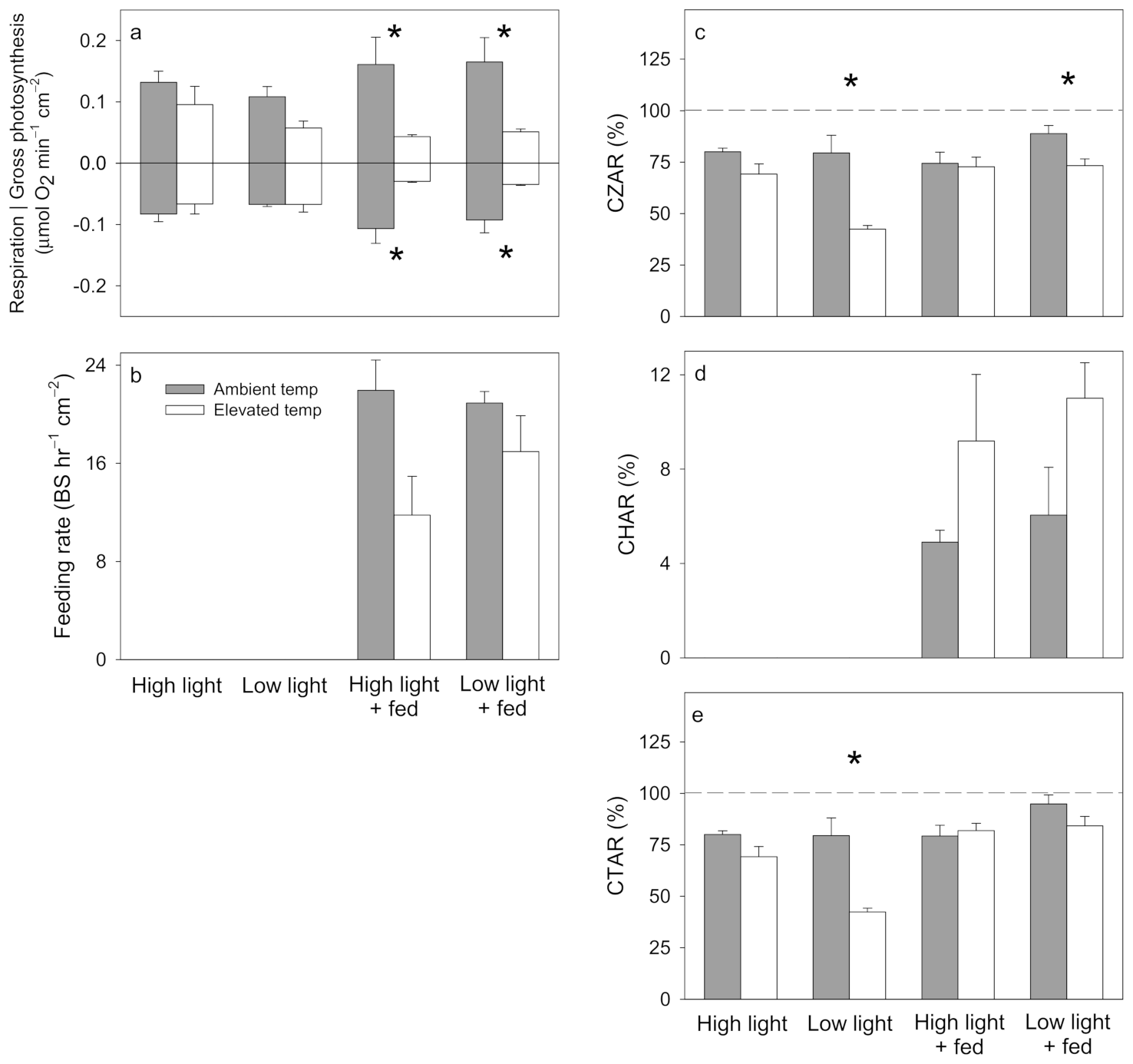
3.2. Stylophora pistillata

All S. pistillata ramets survived the experiment. The S. pistillata holobiont physiological profile was affected by temperature, light, and feeding (Table S5). Chlorophyll a content was higher in corals under low light than in corals under high light and in fed corals compared to unfed corals (Figure 5a; Table S6). Under high light, δ13Ch was more enriched in corals at elevated temperature than at ambient temperature (Figure 5b; Table S6). No significant effects were observed in calcification, biomass, or total lipids (Figure 5c–e; Table S6). However, while overall ANOVA models were not significant, a significant temperature effect in calcification (p = 0.0162) and total lipids (p = 0.0321) suggest a trend of lower calcification and total lipids in corals at elevated temperature compared to those at ambient temperature (Figure 5c,e; Table S6).
Photosynthesis and respiration were lower in corals under elevated temperature compared to ambient temperature when fed (Figure 6a; Table S7). Under low light, CZAR was lower in corals under elevated temperature compared to those under ambient temperature (Figure 6c; Table S7). No significant effects were detected in feeding rate or CHAR (Figure 6b,d; Table S7). Unfed corals in low light had lower CTAR at elevated temperature than at ambient temperature (Figure 6e; Table S7). Interestingly, S. pistillata was not able to meet 100% CTAR under any of the treatment conditions.

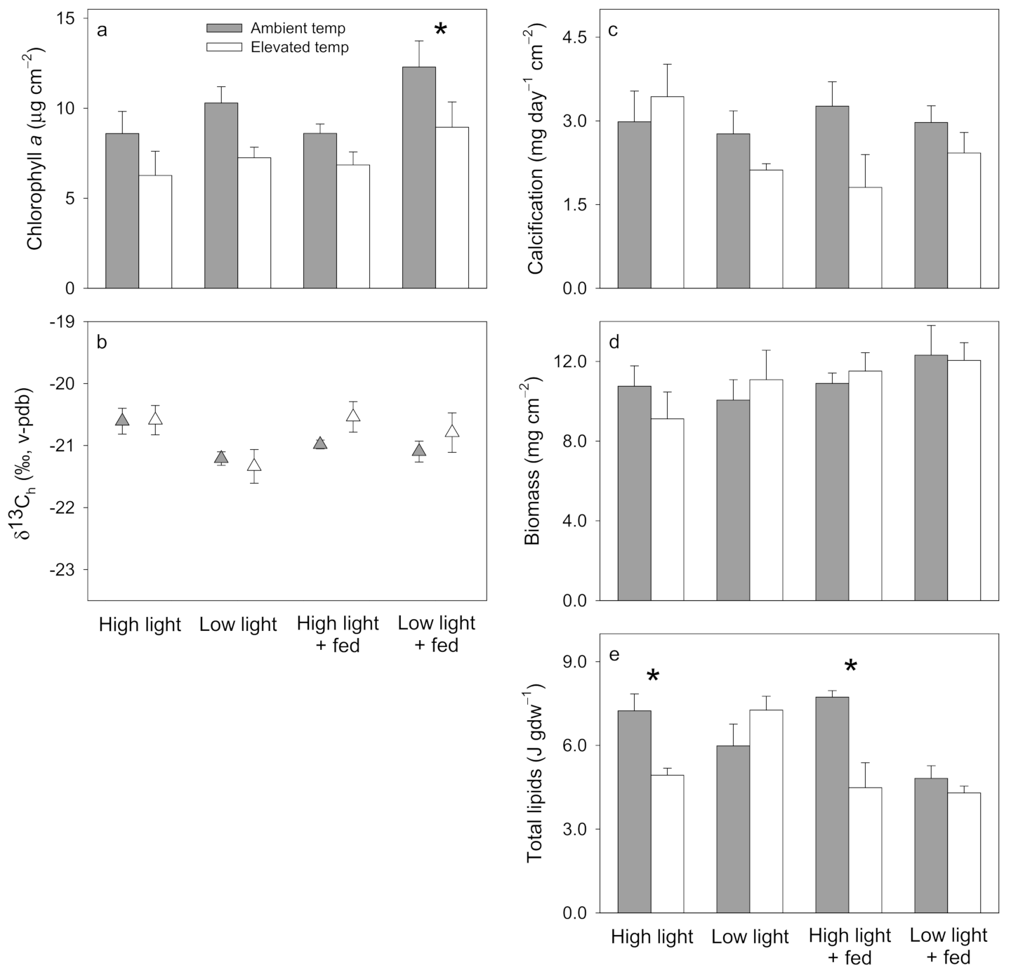
3.3. Turbinaria reniformis

All T. reniformis ramets survived the experiment. The T. reniformis holobiont physiological profile was significantly affected by both temperature and light (Table S8). Chlorophyll a content was lower in corals at elevated temperature than at ambient temperature, and it was also lower under high light compared to low light (Figure 7a; Table S9). No significant differences were observed in δ13Ch, calcification, or biomass across all treatment conditions (Figure 7b–d; Table S9). Under high light, total lipids were lower in corals at elevated temperature than at ambient temperature (Figure 7e; Table S9). Furthermore, fed corals had lower total lipids under low light than under high light (Figure 7e; Table S9).
Photosynthesis and respiration were lower in fed corals under elevated temperature (Figure 8a; Table S10). No significant differences were observed in feeding rate, but CHAR was higher in corals under elevated temperature (Figure 8d; Table S10). While the overall ANOVA models were not significant for CZAR or CTAR, a significant temperature by feeding effect on CZAR (p = 0.0347) and CTAR (p = 0.0052) suggest a trend of higher CTAR in corals at ambient temperature compared to elevated temperature when not fed (Figure 8c, e; Table S10). Only fed corals under elevated temperature were able to meet 100% CTAR (Figure 8e).

4. Discussion

Temperature had a significant effect on the overall coral physiology of each species; light influenced the overall physiology of P. damicornis and S. pistillata, and feeding influenced the overall physiology of S. pistillata and T. reniformis (Tables S2, S3 and S8). However, the way in which low light and feeding mitigated or minimized the negative effects of elevated temperature varied by species.

4.1. Pocillopora damicornis

Low light minimized the negative effect of elevated temperature on algal endosymbionts (Figure 3a; Table S3) and facilitated the maintenance of photosynthesis across treatments (Figure 4a). While unfed corals were not able to meet 100% of total carbon budget (CTAR), feeding on Artemia nauplii was insufficient to fully mitigate the negative effects of elevated temperature on the total carbon budget (CTAR) in low light conditions (Figure 4e).
Photo-acclimation of chlorophyll a content in Pocilloporid corals to low light conditions has previously been observed [90,91], but this process was not sufficient to maintain calcification here. While calcification typically decreases with light (e.g., [54,92,93]) and increases with feeding in P. damicornis (e.g., [65,81,94]), the combination of low light and feeding actually resulted in the lowest calcification rates (Figure 3c). It is possible that less carbon was translocated from the endosymbiont to the host under these conditions (similar to previous observations in S. pistillata; [48]). This, coupled with the lack of detectable incorporation of the Artemia’s carbon into the host tissue (Figure 3b), suggests that heterotrophically derived carbon was metabolized to aid in maintaining the total carbon budget (CTAR), rather than calcification. Calcification is independent of feeding in P. damicornis in the Gulf of Panama [95] and bleached Montipora capitata also metabolize heterotrophically derived fixed carbon to maintain energy reserves and endosymbiont population at the expense of calcification [7,28,96]. Feeding rates and contribution of heterotrophy to the total carbon budget (CHAR) were the lowest of the three species and did not significantly change under elevated temperature (Figure 4d). This lack of heterotrophic plasticity in response to temperature stress is consistent with previous findings in Red Sea P. damicornis [58,91]. At the same time, P. damicornis can linearly increase feeding on suspended particulate organic matter (POM) over a full range of concentrations [79], suggesting that feeding rate and the contribution of heterotrophy to the total carbon budget (CHAR) may have been greater in this study if higher concentrations of Artemia nauplii had been employed. Alternatively, this species may prefer to feed on suspended POM rather than Artemia nauplii. However, our results are consistent with previous findings that feeding is necessary to maintain metabolic demand by stimulating the contribution of photoautotrophic carbon [97]. While we found no evidence that low light or feeding mitigated the negative effects of elevated temperature on the various coral holobiont physiological variables measured, P. damicornis was only able to meet 100% of the total carbon budget (CTAR) when fed, highlighting the fundamental role of heterotrophy for this coral.

4.2. Stylophora pistillata

Individually, low light and feeding stimulated chlorophyll a content in S. pistillata (Figure 5a). At the holobiont scale, the response patterns of total lipids, the contribution of photosynthesis to the total carbon budget (CZAR), and the total carbon budget (CTAR) were similar, suggesting that feeding on Artemia nauplii mitigated the compounded negative effects of elevated temperature and low light on total lipids and metabolic demand (Figure 5e and Figure 6c,e). However, the additional fixed carbon available from feeding (Figure 6d) was not sufficient to allow S. pistillata to meet 100% of the total carbon budget (CTAR) across treatments (Figure 6e) nor mitigate the negative effect of elevated temperature on calcification (Figure 5c).
Photo-acclimation by increasing chlorophyll a content under low light (e.g., [40,90,98]) and when fed (e.g., [49,94,99]) is well documented for S. pistillata. However, while a coral may not visibly exhibit paling in response to temperature stress, it can still be physiologically compromised [16,23]. Despite the benefits of low light for algal endosymbiont pigmentation, low light compounded negative effects of elevated temperature on the contribution of photosynthesis to the total carbon budget (CZAR) (Figure 6c), which was minimized in the total carbon budget (CTAR) by feeding (Figure 6e). Similar to findings by Tremblay et al. [26], feeding was not enough to mitigate or minimize the negative effect of elevated temperature on calcification. This species is a voracious feeder [80,81,94], so its inability to meet 100% of the total carbon budget (CTAR) across treatments, or restore calcification, may be an artefact of only being fed twice weekly. However, as feeding minimized the negative effect of elevated temperature stress on total lipids, perhaps feeding more frequently, for longer durations, or with a more nutritious food source (i.e., freshly caught zooplankton) would have resulted in the full mitigation of this coral’s total carbon budget (CTAR) and/or calcification under elevated temperature conditions.

4.3. Turbinaria reniformis

Low light stimulated T. reniformis chlorophyll a content, but not enough to avoid bleaching (Figure 7a). However, at the holobiont scale, feeding on Artemia nauplii did mitigate the negative effects of elevated temperature on the total carbon budget (CTAR), but at the expense of total lipids (Figure 7e and Figure 8d,e). T. reniformis was the only species to meet 100% of the total carbon budget (CTAR) and maintain calcification under elevated temperature, provided it was fed (Figure 8e).
Photo-acclimation to low light by increasing chlorophyll a content (Figure 7a) has been previously documented for this genus (Hoogenboom et al. 2009). While T. reniformis has previously been observed to catabolize lipids under low light, or when starved [46], our findings suggest that heat-stressed corals under high light may have catabolized lipids to aid in the maintenance of calcification and biomass (Figure 7c,e). In addition, when T. reniformis was fed under elevated temperature, photosynthesis and respiration were depressed and the contribution of heterotrophy to the total carbon budget (CHAR) increased, thus allowing the coral to meet 100% of the total carbon budget (CTAR). Therefore, feeding mitigated the negative effects of elevated temperature on the total carbon budget (CTAR) (Figure 8e), which is consistent with previous findings of feeding helping to sustain carbon budgets in bleached T. reniformis [16,19].

4.4. Implications

Reduced light and feeding on Artemia nauplii did not act synergistically, nor additively, to mitigate nor minimize the negative effects of elevated temperature on coral holobiont physiology and carbon budgets in any of the three coral species examined. The physiological responses of each species to elevated temperature, and the minimizing and/or mitigating effects of low light or feeding were unique to each species. While each species photo-acclimated to the low light conditions by increasing chlorophyll a content, this response did not always translate into the maintenance of daily carbon budgets and thus the minimization or mitigation of the negative effects of elevated temperature on holobiont physiology. This may be due to their different morphologies [100], associated Symbiodiniaceae species (P. damicornis in symbiosis with Cladocopium spp., formerly clade C [101,102]; S. pistillata in symbiosis with Symbiodinium spp., formerly clade A [101,102,103]; and T. reniformis in symbiosis with Cladocopium spp. and Durusdinium spp., formerly clades C and D respectively [102,103]), and/or energetic strategies employed, and agrees with previous studies which have shown that chlorophyll a content is not a reliable predictor of overall holobiont health [14]. Low light did minimize the negative effects of elevated temperature on total lipids in unfed T. reniformis but compounded the negative effects on the total carbon budget (CTAR) in S. pistillata. While feeding did not mitigate the negative effects of elevated temperature on the total carbon budget (CTAR) in P. damicornis, it did for T. reniformis, and it minimized the negative effects of elevated temperature on S. pistillata total carbon budget (CTAR) under low light conditions. Furthermore, feeding was required for P. damicornis to meet 100% of the total carbon budget (CTAR), irrespective of temperature. The role of feeding in the mitigation of elevated temperature stress within this study is likely conservative, as corals in situ would receive a more nutritious array of natural zooplankton and be exposed to zooplankton daily (rather than Artemia nauplii twice weekly). Additional research is needed to quantify dissolved and small particulate organic carbon contributions to the total carbon budget (CTAR), as these heterotrophic sources may bridge the gap for corals to meet 100% of the total carbon budget (CTAR) when heat-stressed [43,66].
Future research with higher tank replication and reef-sourced corals would be desirable to verify these results. Nevertheless, these results suggest that the physiological stress experienced by P. damicornis during bleaching events would not be reduced by low light, even in zooplankton-rich reef environments, due to its inability to maintain growth and lack of heterotrophic plasticity. It remains unclear whether low light would benefit S. pistillata and T. reniformis experiencing temperature stress. However, zooplankton-rich reef environments would likely provide refuge to S. pistillata and T. reniformis at elevated temperature stress, such as occurs during bleaching events. Therefore, these findings add to the growing body of research highlighting the importance of zooplankton availability on the physiological maintenance and persistence of many coral species in a warming ocean.

Supplementary Materials

The following are available online at https://www.mdpi.com/article/10.3390/w13152048/s1, Figure S1. Representative images of (A-H) Pocillopora damicornis, (I-P) Stylophora pistillata, and (Q-X) Turbinaria reniformis from each treatment: high light (300 μmol photons m−2 s−1, unfed), low light (150 μmol photons m−2 s−1, unfed), high light + fed (300 μmol photons m−2 s−1, fed), and low light + fed (150 μmol photons m−2 s−1, fed), under ambient temperature (25 °C) or elevated temperature (30 °C). These images were recorded on the last day of the experiment, 30 May 2013, prior to removal and preservation. Table S1. Project metadata. Modified from Table 2 in Grottoli et al. [56]. Table S2. Pocillopora damicornis results from three-way PERMANOVA, testing for the effects of temperature (Temp), light, and feeding on the coral holobiont physiological profile based on chlorophyll a, δ13Ch, calcification, and biomass. Significant effects are in bold. Table S3. Pocillopora damicornis results from three-way ANOVAs testing the effect of temperature (Temp), light, and feeding on chlorophyll a, δ13Ch, calcification, and biomass. Effects of temperature, light, and feeding were fixed and fully crossed. Significant effects are in bold. Df = degrees of freedom, Mean Sq = mean squares. Tukey test indicates which mean is significantly different from another, for ambient temperature (ambT), elevated temperature (elevT), high light (hiL), low light (lowL), fed (F), and unfed (UF). Table S4. Pocillopora damicornis results from three-way ANOVAs testing the effect of temperature (Temp), light, and feeding on photosynthesis, respiration, feeding rate, Contribution of Zooxanthellae (Symbiodiniaceae) to Animal Respiration (CZAR), Contribution of Heterotrophy to Animal Respiration (CHAR), and Contribution of Total acquired fixed carbon relative to Animal Respiration (CTAR). Effects of temperature, light, and feeding were fixed and fully crossed. Significant effects are in bold. Df = degrees of freedom, Mean Sq = mean squares. Table S5. Stylophora pistillata results from three-way PERMANOVA, testing for the effect of temperature (Temp), light, and feeding on the coral holobiont physiological profile based on chlorophyll a, δ13Ch, calcification, biomass, and total lipids. Significant effects are in bold. Table S6. Stylophora pistillata results from three-way ANOVAs testing the effect of temperature (Temp), light, and feeding on chlorophyll a, δ13Ch, calcification, biomass, and total lipids. Effects of temperature, light, and feeding were fixed and fully crossed. Significant effects are in bold. Df = degrees of freedom, Mean Sq = mean squares. Tukey test indicates which mean is significantly different from another, for ambient temperature (ambT), elevated temperature (elevT), high light (hiL), low light (lowL), fed (F), and unfed (UF). Table S7. Stylophora pistillata results from three-way ANOVAs testing the effect of temperature (Temp), light, and feeding on photosynthesis, respiration, feeding rate, Contribution of Zooxanthellae (Symbiodiniaceae) to Animal Respiration (CZAR), Contribution of Heterotrophy to Animal Respiration (CHAR), and Contribution of Total acquired fixed carbon relative to Animal Respiration (CTAR). Effects of temperature, light, and feeding were fixed and fully crossed. Significant effects are in bold. Df = degrees of freedom, Mean Sq = mean squares. Tukey test indicates which mean is significantly different from another, for ambient temperature (ambT), elevated temperature (elevT), high light (hiL), low light (lowL), fed (F), and unfed (UF). Table S8. Turbinaria reniformis results from three-way PERMANOVA, testing for the effect of temperature (Temp), light, and feeding on the coral holobiont physiological profile based on chlorophyll a, δ13Ch, calcification, biomass, and total lipid. Significant effects are in bold. Table S9. Turbinaria reniformis results from three-way ANOVAs testing the effect of temperature (Temp), light, and feeding on chlorophyll a, δ13Ch, calcification, biomass, and total lipids. Effects of temperature, light, and feeding were fixed and fully crossed. Significant effects are in bold. Df = degrees of freedom, Mean Sq = mean squares. Tukey test indicates which mean is significantly different from another, for ambient temperature (ambT), elevated temperature (elevT), high light (hiL), low light (lowL), fed (F), and unfed (UF). Table S10. Turbinaria reniformis results from three-way ANOVAs testing the effect of temperature (Temp), light, and feeding on photosynthesis, respiration, feeding rate, Contribution of Zooxanthellae (Symbiodiniaceae) to Animal Respiration (CZAR), Contribution of Heterotrophy to Animal Respiration (CHAR), and Contribution of Total acquired fixed carbon relative to Animal Respiration (CTAR). Effects of temperature, light, and feeding were fixed and fully crossed. Significant effects are in bold. Df = degrees of freedom, Mean Sq = mean squares. Tukey test indicates which mean is significantly different from another, for ambient temperature (ambT), elevated temperature (elevT), high light (hiL), low light (lowL), fed (F), and unfed (UF). Table S11. Raw physiology and biochemistry data: chlorophyll a (Chl a), carbon isotope data for host tissue (δ13Ch), calcification (calc), biomass, photosynthesis (P), respiration (R), feeding rate (FR), Contribution of Zooxanthellae to Animal Respiration (CZAR), Contribution of Heterotrophy to Animal Repsiration (CHAR), and Contribution of Total carbon to Animal Respiration (CTAR) for Pocillopora damicornis (P), Stylophora pistillata (S), and Turbinaria reniformis (T). Coral ID consists of species (P, S, or T), genotype (A–E), and treatment (1 = 25 °C, 150 μmol photons m−2 s−1, fed; 2 = 30 °C, 150 μmol photons m−2 s−1, fed; 3 = 30 °C, 150 μmol photons m−2 s−1, unfed; 4 = 25 °C, 150 μmol photons m−2 s−1, unfed; 5 = 30 °C, 300 μmol photons m−2 s−1, unfed; 6 = 25 °C, 300 μmol photons m−2 s−1, unfed; 7 = 30 °C, 300 μmol photons m−2 s−1, fed; 8 = 25 °C, 300 μmol photons m−2 s−1, fed). Dots indicate missing measurements due to insufficient sample material.

Author Contributions

A.G.G. secured the funding; A.G.G. and C.F.-P. designed and coordinated the study and participated in the experiment; K.L.D. and C.M.S. carried out laboratory analyses; K.L.D. carried out all laboratory analyses and drafted the manuscript; all authors contributed to revising the manuscript and gave final approval for publication. All authors have read and agreed to the published version of the manuscript.

Funding

Major funding for this work was provided to AGG by the PADI Foundation grant #6314. Additional funding from the Herbert W. Hoover Foundation and the National Science Foundation (OCE 1459536, 1514859, 1838667) to AGG supported students on this project.

Institutional Review Board Statement

Corals used in this study have been cultured at the Centre Scientifique de Monaco and were not sampled from the wild. Nevertheless, the study was conducted according to the guidelines of the Declaration of Helsinki.

Informed Consent Statement

Not applicable.

Data Availability Statement

Project metadata can be found in Table S1. The datasets supporting this manuscript can be found in Table S11.

Acknowledgments

Thank you to Severine Sikorski, Cecile Rottier, and Miriam Geronimus for running this experiment in Monaco, as well as the Centre Scientifique de Monaco for providing necessary tools and equipment. Thank you to Yohei Matsui for laboratory analysis training, and John Armstrong, Alec Moore, Margaret Otto, Jamie Price, Kristen Rockwell, and Katiri Snyder for laboratory assistance.

Conflicts of Interest

The authors declare no conflict of interest exist.

References

  1. Pachauri, R.K.; Allen, M.R.; Barros, V.R.; Broome, J.; Cramer, W.; Christ, R.; Church, J.A.; Clarke, L.; Dahe, Q.; Dasgupta, P.; et al. Climate Change 2014: Synthesis Report. Contribution of Working Groups I, II, and III to the Fifth Assessment Report of the Intergovernmental Panel on Climate Change; The Core Writing Team, Pachauri, R.K., Meyer, L.A., Eds.; IPCC: Geneva, Switzerland, 2014; p. 151. [Google Scholar]
  2. Donner, S.D.; Skirving, W.J.; Little, C.M.; Oppenheimer, M.; Hoegh-Guldberg, O. Global assessment of coral bleaching and required rates of adaptation under climate change. Glob. Chang. Biol. 2005, 11, 2251–2265. [Google Scholar] [CrossRef]
  3. Hughes, T.P.; Anderson, K.D.; Connolly, S.R.; Heron, S.F.; Kerry, J.T.; Lough, J.M.; Baird, A.H.; Baum, J.K.; Berumen, M.L.; Bridge, T.C.; et al. Spatial and temporal patterns of mass bleaching of corals in the Anthropocene. Science 2018, 359, 80–83. [Google Scholar] [CrossRef] [PubMed] [Green Version]
  4. Brown, B.E. Coral bleaching: Causes and consequences. Coral Reefs 1997, 16, S129–S138. [Google Scholar] [CrossRef]
  5. Gates, R.D.; Baghdasarian, G.; Muscatine, L. Temperature stress causes host-cell detachment in symbiotic Cnidarians—Implications for coral bleaching. Biol. Bull. 1992, 182, 324–332. [Google Scholar] [CrossRef] [PubMed]
  6. Glynn, P.W. Coral reef bleaching: Facts, hypotheses and implications. Glob. Chang. Biol. 1996, 2, 495–509. [Google Scholar] [CrossRef]
  7. Grottoli, A.G.; Rodrigues, L.J.; Palardy, J.E. Heterotrophic plasticity and resilience in bleached corals. Nature 2006, 440, 1186–1189. [Google Scholar] [CrossRef]
  8. Muscatine, L.; McCloskey, L.R.; Marian, R.E. Estimating the daily contribution of carbon from zooxanthellae to coral animal respiration. Limnol. Oceanogr. 1981, 26, 601–611. [Google Scholar] [CrossRef] [Green Version]
  9. Tremblay, P.; Grover, R.; Maguer, J.F.; Legendre, L.; Ferrier-Pages, C. Autotrophic carbon budget in coral tissue: A new C-13-based model of photosynthate translocation. J. Exp. Biol. 2012, 215, 1384–1393. [Google Scholar] [CrossRef] [Green Version]
  10. Ferrier-Pages, C.; Hoogenboom, M.; Houlbreque, F. The role of plankton in coral trophodynamics. In Coral Reefs: An Ecosystem in Transition; Dubinsky, Z., Stambler, N., Eds.; Springer: Dordrecht, The Netherlands, 2011; pp. 215–229. [Google Scholar] [CrossRef]
  11. Palardy, J.E.; Rodrigues, L.J.; Grottoli, A.G. The importance of zooplankton to the daily metabolic carbon requirements of healthy and bleached corals at two depths. J. Exp. Mar. Biol. Ecol. 2008, 367, 180–188. [Google Scholar] [CrossRef]
  12. Iglesias-Prieto, R.; Matta, J.L.; Robins, W.A.; Trench, R.K. Photosynthetic response to elevated temperature in the symbiotic dinoflagellate Symbiodinum microadriaticum in culture. Proc. Natl. Acad. Sci. USA 1992, 89, 10302–10305. [Google Scholar] [CrossRef] [Green Version]
  13. Levas, S.J.; Grottoli, A.G.; Hughes, A.; Osburn, C.L.; Matsui, Y. Physiological and biogeochemical traits of bleaching and recovery in the mounding species of coral Porites lobata: Implications for resilience in mouding corals. PLoS ONE 2013, 8, e63267. [Google Scholar] [CrossRef] [Green Version]
  14. Rodrigues, L.J.; Grottoli, A.G. Energy reserves and metabolism as indicators of coral recovery from bleaching. Limnol. Oceanogr. 2007, 52, 1874–1882. [Google Scholar] [CrossRef]
  15. Connolly, S.R.; Lopez-Yglesias, M.A.; Anthony, K.R.N. Food availability promotes rapid recovery from thermal stress in a scleractinian coral. Coral Reefs 2012, 31, 951–960. [Google Scholar] [CrossRef]
  16. Ferrier-Pages, C.; Rottier, C.; Beraud, E.; Levy, O. Experimental assessment of the feeding effort of three scleractinian coral species during a thermal stress: Effect on the rates of photosynthesis. J. Exp. Mar. Biol. Ecol. 2010, 390, 118–124. [Google Scholar] [CrossRef]
  17. Hughes, A.D.; Grottoli, A.G. Heterotrophic compensation: A possible mechanism for resilience of coral reefs to global warming or a sign of prolonged stress? PLoS ONE 2013, 8, e81172. [Google Scholar] [CrossRef]
  18. Grottoli, A.G.; Rodrigues, L.J. Bleached Porites compressa and Montipora capitata corals catabolize delta C-13-enriched lipids. Coral Reefs 2011, 30, 687–692. [Google Scholar] [CrossRef]
  19. Tolosa, I.; Treignier, C.; Grover, R.; Ferrier-Pages, C. Impact of feeding and short-term temperature stress on the content and isotopic signature of fatty acids, sterols, and alcohols in the scleractinian coral Turbinaria reniformis. Coral Reefs 2011, 30, 763. [Google Scholar] [CrossRef]
  20. Borell, E.M.; Yuliantri, A.R.; Bischof, K.; Richter, C. The effect of heterotrophy on photosynthesis and tissue composition of two scleractinian corals under elevated temperature. J. Exp. Mar. Biol. Ecol. 2008, 364, 116–123. [Google Scholar] [CrossRef]
  21. Levas, S. Biogeochemistry and Physiology of Bleached and Recovering Hawaiian and Caribbean Corals. Ph.D. Thesis, The Ohio State University, Columbus, OH, USA, 2012. [Google Scholar]
  22. Cohen, A.L.; Holcomb, M. Why corals care about ocean acidification: Uncovering the mechanism. Oceanography 2009, 22, 118–127. [Google Scholar] [CrossRef]
  23. Grottoli, A.G.; Warner, M.E.; Levas, S.J.; Aschaffenburg, M.D.; Schoepf, V.; McGinley, M.; Baumann, J.; Matsui, Y. The cumulative impact of annual coral bleaching can turn some coral species winners into losers. Glob. Chang. Biol. 2014, 20, 3823–3833. [Google Scholar] [CrossRef]
  24. Schoepf, V.; Grottoli, A.G.; Levas, S.J.; Aschaffenburg, M.D.; Baumann, J.H.; Matsui, Y.; Warner, M.E. Annual coral bleaching and the long-term recovery capacity of coral. Proc. R. Soc. B Biol. Sci. 2015, 282, 20151887. [Google Scholar] [CrossRef] [Green Version]
  25. Borell, E.M.; Bischof, K. Feeding sustains photosynthetic quantum yield of a scleractinian coral during thermal stress. Oecologia 2008, 157, 593–601. [Google Scholar] [CrossRef]
  26. Tremblay, P.; Gori, A.; Maguer, J.F.; Hoogenboom, M.; Ferrier-Pages, C. Heterotrophy promotes the re-establishment of photosynthate translocation in a symbiotic coral after heat stress. Sci. Rep. 2016, 6, 38112. [Google Scholar] [CrossRef] [Green Version]
  27. Baumann, J.; Grottoli, A.G.; Hughes, A.D.; Matsui, Y. Photoautotrophic and heterotrophic carbon in bleached and non-bleached coral lipid acquisition and storage. J. Exp. Mar. Biol. Ecol. 2014, 461, 469–478. [Google Scholar] [CrossRef]
  28. Hughes, A.D.; Grottoli, A.G.; Pease, T.K.; Matsui, Y. Acquisition and assimilation of carbon in non-bleached and bleached corals. Mar. Ecol. Prog. Ser. 2010, 420, 91–101. [Google Scholar] [CrossRef]
  29. Brown, B.E.; Ambarsari, I.; Warner, M.E.; Fitt, W.K.; Dunne, R.P.; Gibb, S.W.; Cummings, D.G. Diurnal changes in photochemical efficiency and xanthophyll concentrations in shallow water reef corals: Evidence for photoinhibition and photoprotection. Coral Reefs 1999, 18, 99–105. [Google Scholar]
  30. Bhagooli, R.; Hidaka, M. Photoinhibition, bleaching susceptibility and mortality in two scleractinian corals, Platygyra ryukyuensis and Stylophora pistillata, in response to thermal and light stresses. Comp. Biochem. Physiol. A-Mol. Integr. Physiol. 2004, 137, 547–555. [Google Scholar] [CrossRef]
  31. Coles, S.L.; Jokiel, P.L. Synergistic effects of temperature, salinity and light on hermatypic coral Montipora verrucosa. Mar. Biol. 1978, 49, 187–195. [Google Scholar] [CrossRef]
  32. Lesser, M.P.; Stochaj, W.R.; Tapley, D.W.; Shick, J.M. Bleaching in coral reef Anthozoans—Effects of irradiance, ultraviolet-radiation, and temperature on the activities of protective enzymens against active oxygen. Coral Reefs 1990, 8, 225–232. [Google Scholar] [CrossRef]
  33. Browne, N.; Braoun, C.; McIlwain, J.; Nagarajan, R.; Zinke, J. Borneo coral reefs subject to high sediment loads show evidence of resilience to various environmental stressors. PeerJ 2019, 7, e7382. [Google Scholar] [CrossRef] [Green Version]
  34. Cacciapaglia, C.; van Woesik, R. Climate-change refugia: Shading reef corals by turbidity. Glob. Chang. Biol. 2016, 22, 1145–1154. [Google Scholar] [CrossRef] [PubMed]
  35. Morgan, K.M.; Perry, C.T.; Johnson, J.A.; Smithers, S.G. Nearshore turbid-zone corals exhibit high bleaching tolerance on the Great Barrier Reef following the 2016 ocean warming event. Front. Mar. Sci. 2017, 4, 224. [Google Scholar] [CrossRef]
  36. Nitschke, M.R.; Gardner, S.G.; Goyen, S.; Fujise, L.; Camp, E.F.; Ralph, P.J.; Suggett, D.J. Utility of photochemical traits as diagnostics of thermal tolerance amongst Great Barrier Reef corals. Front. Mar. Sci. 2018, 5, 460. [Google Scholar] [CrossRef] [Green Version]
  37. Sully, S.; van Woesik, R. Turbid reefs moderate coral bleaching under climate-related temperature stress. Glob. Chang. Biol. 2020, 26, 1367–1373. [Google Scholar] [CrossRef] [Green Version]
  38. Courtial, L.; Roberty, S.; Shick, J.M.; Houlbreque, F.; Ferrier-Pages, C. Interactive effects of ultraviolet radiation and thermal stress on two reef-building corals. Limnol. Oceanogr. 2017, 62, 1000–1013. [Google Scholar] [CrossRef]
  39. Lesser, M.P. Phylogenetic signature of light and thermal stress for the endosymbiotic dinoflagellates of corals (Family Symbiodiniaceae). Limnol. Oceanogr. 2019, 64, 1852–1863. [Google Scholar] [CrossRef]
  40. Falkowski, P.G.; Dubinsky, Z. Light-shade adaptation of Stylophora pistillata, a hermatypic coral from the Gulf of Eilat. Nature 1981, 289, 172–174. [Google Scholar] [CrossRef]
  41. Porter, J.W.; Muscatine, L.; Dubinsky, Z.; Falkowski, P.G. Primary production and photoadaptation in light-adapted and shade-adapted colonies of the symbiotic coral, Stylophora pistillata. Proc. R. Soc. Ser. B Biol. Sci. 1984, 222, 161–180. [Google Scholar] [CrossRef]
  42. Titlyanov, E.A.; Titlyanova, T.V.; Yamazato, K.; van Woesik, R. Photo-acclimation dynamics of the coral Stylophora pistillata to low and extremely low light. J. Exp. Mar. Biol. Ecol. 2001, 263, 211–225. [Google Scholar] [CrossRef]
  43. Anthony, K.R.N.; Fabricius, K.E. Shifting roles of heterotrophy and autotrophy in coral energetics under varying turbidity. J. Exp. Mar. Biol. Ecol. 2000, 252, 221–253. [Google Scholar] [CrossRef]
  44. Falkowski, P.G.; Dubinsky, Z.; Muscatine, L.; Porter, J.W. Light and the bioenergetics of a symbiotic coral. Bioscience 1984, 34, 705–709. [Google Scholar] [CrossRef]
  45. Levy, O.; Karako-Lampert, S.; Ben-Asher, H.W.; Zoccola, D.; Pages, G.; Ferrier-Pages, C. Molecular assessment of the effect of light and heterotrophy in the scleractinian coral Stylophora pistillata. Proc. R. Soc. B Biol. Sci. 2016, 283, 20153025. [Google Scholar] [CrossRef] [Green Version]
  46. Treignier, C.; Grover, R.; Ferrier-Pages, C.; Tolosa, I. Effect of light and feeding on the fatty acid and sterol composition of zooxanthellae and host tissue isolated from the scleractinian coral Turbinaria reniformis. Limnol. Oceanogr. 2008, 53, 2702–2710. [Google Scholar] [CrossRef] [Green Version]
  47. Tilstra, A.; Wijgerde, T.; Dini-Andreote, F.; Eriksson, B.K.; Salles, J.F.; Pen, I.; Osinga, R.; Wild, C. Light induced intraspecific variability in response to thermal stress in the hard coral Stylophora pistillata. PeerJ 2017, 5, e3802. [Google Scholar] [CrossRef] [Green Version]
  48. Tremblay, P.; Grover, R.; Maguer, J.F.; Hoogenboom, M.; Ferrier-Pages, C. Carbon translocation from symbiont to host depends on irradiance and food availability in the tropical coral Stylophora pistillata. Coral Reefs 2014, 33, 1–13. [Google Scholar] [CrossRef]
  49. Hoogenboom, M.O.; Campbell, D.A.; Beraud, E.; DeZeeuw, K.; Ferrier-Pages, C. Effects of light, food availability and temperature stress on the function of photosystem II and photosystem I of coral symbionts. PLoS ONE 2012, 7, e30167. [Google Scholar] [CrossRef] [Green Version]
  50. Ferrier-Pages, C.; Gattuso, J.P.; Dallot, S.; Jaubert, J. Effect of nutrient enrichment on growth and photosynthesis of the zooxanthellate coral Stylophora pistillata. Coral Reefs 2000, 19, 103–113. [Google Scholar] [CrossRef]
  51. Maor-Landaw, K.; Karako-Lampert, S.; Ben-Asher, H.W.; Goffredo, S.; Falini, G.; Dubinsky, Z.; Levy, O. Gene expression profiles during short-term heat stress in the red sea coral Stylophora pistillata. Glob. Chang. Biol. 2014, 20, 3026–3035. [Google Scholar] [CrossRef] [Green Version]
  52. Reynaud, S.; Ferrier-Pages, C.; Meibom, A.; Mostefaoui, S.; Mortlock, R.; Fairbanks, R.; Allemand, D. Light and temperature effects on Sr/Ca and Mg/Ca ratios in the scleractinian coral Acropora sp. Geochim. Cosmochim. Acta 2007, 71, 354–362. [Google Scholar] [CrossRef]
  53. Reynaud, S.; Martinez, P.; Houlbreque, F.; Billy, I.; Allemand, D.; Ferrier-Pages, C. Effect of light and feeding on the nitrogen isotopic composition of a zooxanthellate coral: Role of nitrogen recycling. Mar. Ecol. Prog. Ser. 2009, 392, 103–110. [Google Scholar] [CrossRef] [Green Version]
  54. Reynaud-Vaganay, S.; Ferrier, C.; Sambrotto, R.; Juillet-Leclerc, A.; Gattuso, J.P. Effect of feeding on the carbon isotopic composition of the zooxanthellate coral Stylophora pistillata. Geochim. Cosmochim. Acta 2002, 66, A636. [Google Scholar]
  55. Hoogenboom, M.; Rottier, C.; Sikorski, S.; Ferrier-Pages, C. Among-species variation in the energy budgets of reef-building corals: Scaling from coral polyps to communities. J. Exp. Biol. 2015, 218, 3866–3877. [Google Scholar] [CrossRef] [PubMed] [Green Version]
  56. Grottoli, A.G.; Toonen, R.J.; van Woesik, R.; Vega Thurber, R.; Warner, M.E.; McLachlan, R.H.; Price, J.T.; Bahr, K.D.; Baums, I.B.; Castillo, K.; et al. Increasing comparability among coral bleaching experiments. Ecol. Appl. 2020, 31, e02262. [Google Scholar] [CrossRef]
  57. Fine, M.; Gildor, H.; Genin, A. A coral reef refuge in the Red Sea. Glob. Chang. Biol. 2013, 19, 3640–3647. [Google Scholar] [CrossRef]
  58. Grottoli, A.G.; Tchernov, D.; Winters, G. Physiological and biogeochemical responses of super-corals to thermal stress from the Northern Gulf of Aqaba, Red Sea. Front. Mar. Sci. 2017, 4, 215. [Google Scholar] [CrossRef]
  59. Osman, E.O.; Smith, D.J.; Ziegler, M.; Kurten, B.; Conrad, C.; El-Haddad, K.M.; Voolstra, C.R.; Suggett, D.J. Thermal refugia against coral bleaching throughout the northern Red Sea. Glob. Chang. Biol. 2018, 24, E474–E484. [Google Scholar] [CrossRef]
  60. Lesser, M.P. Oxidative stress causes coral bleaching during exposure to elevated temperatures. Coral Reefs 1997, 16, 187–192. [Google Scholar] [CrossRef]
  61. Marubini, F.; Barnett, H.; Langdon, C.; Atkinson, M.J. Dependence of calcification on light and carbonate ion concentration for the hermatypic coral Porites compressa. Mar. Ecol. Prog. Ser. 2001, 220, 153–162. [Google Scholar] [CrossRef]
  62. Nakamura, T.; Yamasaki, H. Flicker light effects on photosynthesis of symbiotic algae in the reef-building coral Acropora digitifera (Cnidaria: Anthozoa: Scleractinia). Pac. Sci. 2008, 62, 341–350. [Google Scholar] [CrossRef] [Green Version]
  63. Schutter, M.; Kranenbarg, S.; Wijffels, R.H.; Verreth, J.; Osinga, R. Modification of light utilization for skeletal growth by water flow in the scleractinian coral Galaxea fascicularis. Mar. Biol. 2011, 158, 769–777. [Google Scholar] [CrossRef]
  64. Ulstrup, K.E.; Ralph, P.J.; Larkum, A.W.D.; Kuhl, M. Intra-colonial variability in light acclimation of zooxanthellae in coral tissues of Pocillopora damicornis. Mar. Biol. 2006, 149, 1325–1335. [Google Scholar] [CrossRef]
  65. Houlbreque, F.; Tambutte, E.; Allemand, D.; Ferrier-Pages, C. Interactions between zooplankton feeding, photosynthesis and skeletal growth in the scleractinian coral Stylophora pistillata. J. Exp. Biol. 2004, 207, 1461–1469. [Google Scholar] [CrossRef] [Green Version]
  66. Levas, S.; Grottoli, A.G.; Schoepf, V.; Aschaffenburg, M.; Baumann, J.; Bauer, J.E.; Warner, M.E. Can heterotrophic uptake of dissolved organic carbon and zooplankton mitigate carbon budget deficits in annually bleached corals? Coral Reefs 2016, 35, 495–506. [Google Scholar] [CrossRef]
  67. Ferrier-Pages, C.; Peirano, A.; Abbate, M.; Cocito, S.; Negri, A.; Rottier, C.; Riera, P.; Rodolfo-Metalpa, R.; Reynaud, S. Summer autotrophy and winter heterotrophy in the temperate symbiotic coral Cladocora caespitosa. Limnol. Oceanogr. 2011, 56, 1429–1438. [Google Scholar] [CrossRef]
  68. Leal, M.C.; Ferrier-Pages, C.; Calado, R.; Brandes, J.A.; Frischer, M.E.; Nejstgaard, J.C. Trophic ecology of the facultative symbiotic coral Oculina arbuscula. Mar. Ecol. Prog. Ser. 2014, 504, 171–179. [Google Scholar] [CrossRef]
  69. Tremblay, P.; Naumann, M.S.; Sikorski, S.; Grover, R.; Ferrier-Pages, C. Experimental assessment of organic carbon fluxes in the scleractinian coral Stylophora pistillata during a thermal and photo stress event. Mar. Ecol. Prog. Ser. 2012, 453, 63–77. [Google Scholar] [CrossRef]
  70. Levas, S.; Grottoli, A.G.; Warner, M.E.; Cai, W.-J.; Bauer, J.; Schoepf, V.; Baumann, J.H.; Matsui, Y.; Gearing, C.; Melman, T.F.; et al. Organic carbon fluxes mediated by corals at elevated pCO2 and temperature. Mar. Ecol. Prog. Ser. 2015, 519, 153–164. [Google Scholar] [CrossRef]
  71. Jokiel, P.L.; Maragos, J.E.; Franzisket, L. Coral growth: Buoyant weight technique. In Coral Reef: Research Methods; Stoddart, D., Johannes, R.E., Eds.; UNESCO: Paris, France, 1978; Volume 78, pp. 379–396. [Google Scholar]
  72. Ferrier-Pages, C.; Gevaert, F.; Reynaud, S.; Beraud, E.; Menu, D.; Janquin, M.A.; Cocito, S.; Peirano, A. In situ assessment of the daily primary production of the temperate symbiotic coral Cladocora caespitosa. Limnol. Oceanogr. 2013, 58, 1409–1418. [Google Scholar] [CrossRef] [Green Version]
  73. Veal, C.J.; Carmi, M.; Fine, M.; Hoegh-Guldberg, O. Increasing the accuracy of surface area estimation using single wax dipping of coral fragments. Coral Reefs 2010, 29, 893–897. [Google Scholar] [CrossRef]
  74. Marsh, J.A. Primary productivity of reef building calcareous red algae. Ecology 1970, 51, 255–263. [Google Scholar] [CrossRef]
  75. Jeffrey, S.W.; Humphrey, G.F. New spectrophotometric equations for determining chlorophylls a, b, c1, and c2 in higher-plants, algae and natural phytoplankton. Biochem. Und Physiol. Der Pflanz. 1975, 167, 191–194. [Google Scholar] [CrossRef]
  76. McLachlan, R.H.; Dobson, K.L.; Grottoli, A.G. Quantification of Total Biomass in Ground Coral Samples. Available online: http://www.protocols.io/view/quantification-of-biomass-in-ground-coral-samples-bdyai7se (accessed on 23 April 2020).
  77. McLachlan, R.H.; Munoz-Garcia, A.; Grottoli, A.G. Extraction of Total Soluble Lipid from Ground Coral Samples. Available online: https://www.protocols.io/view/extraction-of-total-soluble-lipid-from-ground-cora-bc4qiyvw (accessed on 14 March 2020).
  78. Gnaiger, E.; Bitterlich, G. Proximate biochemical composition and caloric content calculated from elemental CHN analysis—A stochiometric concept. Oecologia 1984, 62, 289–298. [Google Scholar] [CrossRef] [PubMed]
  79. Anthony, K.R.N. Coral suspension feeding on fine particulate matter. J. Exp. Mar. Biol. Ecol. 1999, 232, 85–106. [Google Scholar] [CrossRef]
  80. Ferrier-Pages, C.; Allemand, D.; Gattuso, J.P.; Jaubert, J.; Rassoulzadegan, R. Microheterotrophy in the zooxanthellate coral Stylophora pistillata: Effects of light and ciliate density. Limnol. Oceanogr. 1998, 43, 1639–1648. [Google Scholar] [CrossRef]
  81. Ferrier-Pages, C.; Witting, J.; Tambutte, E.; Sebens, K.P. Effect of natural zooplankton feeding on the tissue and skeletal growth of the scleractinian coral Stylophora pistillata. Coral Reefs 2003, 22, 229–240. [Google Scholar] [CrossRef]
  82. Sturaro, N.; Hsieh, Y.E.; Chen, Q.; Wang, P.-L.; Denis, V. Toward a standardised protocol for the stable isotope analysis of scleractinian corals. Rapid Commun. Mass Spectrom. 2020, 34, e8663. [Google Scholar] [CrossRef]
  83. Price, J.; Smith, A.; Dobson, K.D.; Grottoli, A.G. Airbrushed Coral Sample Preparation for Organic Stable Carbon and Nitrogen Isotope Analyses. Available online: https://www.protocols.io/view/airbrushed-coral-sample-preparation-for-organic-st-bgi7juhn (accessed on 10 July 2020).
  84. Dormann, C.F.; Elith, J.; Bacher, S.; Buchmann, C.; Carl, G.; Carre, G.; Garcia Marquez, J.R.; Gruber, B.; Lafourcade, B.; Leitao, P.J.; et al. Collinearity: A review of methods to deal with it and a simulation study evaluating their performance. Ecography 2013, 36, 27–46. [Google Scholar] [CrossRef]
  85. Clarke, K.; Gorley, R. Primer v6: User Manual/Tutorial; PRIMER-E Ltd.: Plymouth, UK, 2006. [Google Scholar]
  86. Cook, R.D. Detection of influential observation in linear regression. Technometrics 1977, 19, 15–18. [Google Scholar] [CrossRef]
  87. Kikvidze, Z.; Moya-Laraño, J. Unexpected failures of recommended tests in basic statistical analyses of ecological data. Web Ecol. 2008, 8, 67–73. [Google Scholar] [CrossRef] [Green Version]
  88. Grover, R.; Maguer, J.F.; Reynaud-Vaganay, S.; Ferrier-Pages, C. Uptake of ammonium by the scleractinian coral Stylophora pistillata: Effect of feeding, light, and ammonium concentrations. Limnol. Oceanogr. 2002, 47, 782–790. [Google Scholar] [CrossRef] [Green Version]
  89. Moran, M.D. Arguments for rejecting the sequential Bonferroni in ecological studies. Oikos 2003, 100, 403–405. [Google Scholar] [CrossRef]
  90. Titlyanov, E.A.; Titlyanova, T.V.; Yamazato, K. Acclimation of symbiotic reef-building corals to extremely low light. Symbiosis 2002, 33, 125–143. [Google Scholar]
  91. Ziegler, M.; Roder, C.M.; Buechel, C.; Voolstra, C.R. Limits to physiological plasticity of the coral Pocillopora verrucosa from the central Red Sea. Coral Reefs 2014, 33, 1115–1129. [Google Scholar] [CrossRef] [Green Version]
  92. Cohen, I.; Dubinsky, Z.; Erez, J. Light enhanced calcification in hermatypic corals: New insights from light spectral responses. Front. Mar. Sci. 2016, 2, 122. [Google Scholar] [CrossRef] [Green Version]
  93. Kawaguti, S.; Sakumoto, D. The effect of light on the calcium deposition of corals. Bull. Oceanogr. Inst. Taiwan 1948, 4, 65–70. [Google Scholar]
  94. Houlbreque, F.; Tambutte, E.; Ferrier-Pages, C. Effect of zooplankton availability on the rates of photosynthesis, and tissue and skeletal growth in the scleractinian coral Stylophora pistillata. J. Exp. Mar. Biol. Ecol. 2003, 296, 145–166. [Google Scholar] [CrossRef]
  95. Wellington, G.M. An experimental analysis of the effects of light and zooplankton on coral zonation. Oecologia 1982, 52, 311–320. [Google Scholar] [CrossRef]
  96. Rodrigues, L.J.; Grottoli, A.G. Calcification rate and the stable carbon, oxygen, and nitrogen isotopes in the skeleton, host tissue, and zooxanthellae of bleached and recovering Hawaiian corals. Geochim. Cosmochim. Acta 2006, 70, 2781–2789. [Google Scholar] [CrossRef]
  97. Lyndby, N.H.; Holm, J.B.; Wangpraseurt, D.; Grover, R.; Rottier, C.; Kuhl, M.; Ferrier-Pages, C. Effect of temperature and feeding on carbon budgets and O2 dynamics in Pocillopora damicornis. Mar. Ecol. Prog. Ser. 2020, 652, 49–62. [Google Scholar] [CrossRef]
  98. Mass, T.; Einbinder, S.; Brokovich, E.; Shashar, N.; Vago, R.; Erez, J.; Dubinsky, Z. Photoacclimation of Stylophora pistillata to light extremes: Metabolism and calcification. Mar. Ecol. Prog. Ser. 2007, 334, 93–102. [Google Scholar] [CrossRef] [Green Version]
  99. Titlyanov, E.A.; Titlyanova, T.V.; Yamazato, K.; van Woesik, R. Photo-acclimation of the hermatypic coral Stylophora pistillata while subjected to either starvation or food provisioning. J. Exp. Mar. Biol. Ecol. 2001, 257, 163–181. [Google Scholar] [CrossRef]
  100. Loya, Y.; Sakai, K.; Yamazato, K.; Nakano, Y.; Sambali, H.; van Woesik, R. Coral bleaching: The winners and the losers. Ecol. Lett. 2001, 4, 122–131. [Google Scholar] [CrossRef]
  101. Godinot, C.; Ferrier-Pages, C.; Sikorski, S.; Grover, R. Alkaline phosphatase activity of reef-building corals. Limnol. Oceanogr. 2013, 58, 227–234. [Google Scholar] [CrossRef]
  102. LaJeunesse, T.C.; Parkinson, J.E.; Gabrielson, P.W.; Jeong, H.J.; Reimer, J.D.; Voolstra, C.R.; Santos, S.R. Systematic revision of Symbiodiniaceae highlights the antiquity and diversity of coral endosymbionts. Curr. Biol. 2018, 28, 2570–2580.e6. [Google Scholar] [CrossRef] [Green Version]
  103. Tremblay, P.; Maguer, J.F.; Grover, R.; Ferrier-Pages, C. Trophic dynamics of scleractinian corals: Stable isotope evidence. J. Exp. Biol. 2015, 218, 1223–1234. [Google Scholar] [CrossRef] [Green Version]
Figure 1. Experimental design schematic. Five parent colonies of Pocillopora damicornis Water 13 02048 i001, Stylophora pistillata Water 13 02048 i002, and Turbinaria reniformis Water 13 02048 i003 
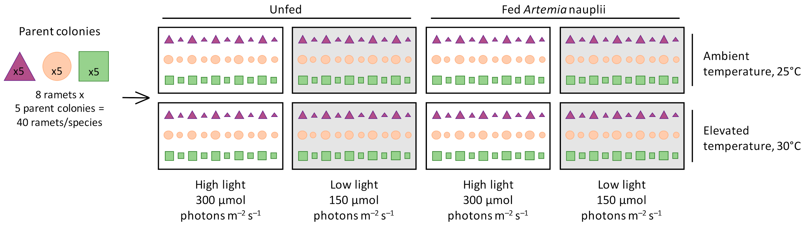
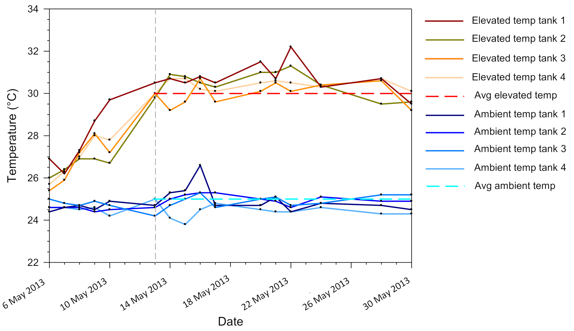
were fragmented into eight large and eight small ramets and distributed randomly and evenly among eight treatments of a fully factorial experimental design: two seawater temperatures (ambient temperature of 25 °C, elevated temperature of 30 °C), two light levels (high light of 300 μmol photons m−2 s−1, low light of 150 μmol photons m−2 s−1), and either fed (Artemia nauplii) or unfed.
Water 13 02048 g001
Figure 2. Temperature (°C) in each of the eight tanks over the duration of the study. The temperature ramp ended on 13 May 2013 as indicated by the vertical dashed gray line. Average temperature of the four ambient temperature tanks was 25 °C ± 0.5 (1SD), while average temperature for the elevated temperature tanks was 30 °C ± 0.6 (1SD). Average temperatures were calculated from the end of the ramping period on 13 May 2013 until the end of the experiment on 30 May 2013.
Water 13 02048 g002
Figure 3. Pocillopora damicornis average (±1 SE) (a) chlorophyll a, (b) δ13Ch, (c) calcification, and (d) biomass, under ambient temperature (25 °C, solid bars and symbols) or elevated temperature (30 °C, open bars and symbols) within high light (300 μmol photons m−2 s−1, unfed), low light (150 μmol photons m−2 s−1, unfed), high light + fed (300 μmol photons m−2 s−1, fed), and low light + fed (150 μmol photons m−2 s−1, fed) treatments. N = 3–4 for all averages. Significant effects from corresponding ANOVAs indicated in Table S3.
Water 13 02048 g003
Figure 4. Pocillopora damicornis average (±1 SE) (a) photosynthesis and respiration, (b) feeding rate, (c) Contribution of Zooxanthellae (Symbiodiniaceae) to Animal Respiration (CZAR), (d) Contribution of Heterotrophy to Animal Respiration (CHAR) from feeding on Artemia nauplii, and (e) Contribution of Total acquired fixed carbon relative to Animal Respiration (CTAR = CZAR + CHAR), under ambient temperature (25 °C, solid bars) and elevated temperature (30 °C, open bars) within high light (300 μmol photons m−2 s−1, unfed), low light (150 μmol photons m−2 s−1, unfed), high light + fed (300 μmol photons m−2 s−1, fed), and low light + fed (150 μmol photons m−2 s−1, fed) treatments. N = 3 for all averages. Significant effects from corresponding ANOVAs in Table S4.
Water 13 02048 g004
Figure 5. Stylophora pistillata average (±1 SE) (a) chlorophyll a, (b) δ13Ch, (c) calcification, (d) biomass, and (e) total lipids, under ambient temperature (25 °C, solid bars) and elevated temperature (30 °C, open bars) within high light (300 μmol photons m−2 s−1, unfed), low light (150 μmol photons m−2 s−1, unfed), high light + fed (300 μmol photons m−2 s−1, fed), and low light + fed (150 μmol photons m−2 s−1, fed) treatments. Asterisks indicate significant differences between ambient and elevated temperature treatments within each light and feeding treatment combination. N = 5 for all averages. Significant effects from corresponding ANOVAs in Table S6.
Water 13 02048 g005
Figure 6. Stylophora pistillata average (±1 SE) (a) photosynthesis and respiration, (b) feeding rate, (c) Contribution of Zooxanthellae (Symbiodiniaceae) to Animal Respiration (CZAR), (d) Contribution of Heterotrophy to Animal Respiration (CHAR) from feeding on Artemia nauplii, and (e) Contribution of Total acquired fixed carbon relative to Animal Respiration (CTAR), under ambient temperature (25 °C, solid bars) and elevated temperature (30 °C, open bars) within high light (300 μmol photons m−2 s−1, unfed), low light (150 μmol photons m−2 s−1, unfed), high light + fed (300 μmol photons m−2 s−1, fed), and low light + fed (150 μmol photons m−2 s−1, fed) treatments. Asterisks indicate significant differences between ambient and elevated temperature treatments within each light and feeding treatment combination. N = 3 for all averages. Significant effects from the corresponding ANOVAs in Table S7.
Water 13 02048 g006
Figure 7. Turbinaria reniformis average (±1 SE) (a) chlorophyll a, (b) δ13Ch, (c) calcification, (d) biomass, and (e) total lipids, under ambient temperature (25 °C, solid bars) and elevated temperature (30 °C, open bars) within high light (300 μmol photons m−2 s−1, unfed), low light (150 μmol photons m−2 s−1, unfed), high light + fed (300 μmol photons m−2 s−1, fed), and low light + fed (150 μmol photons m−2 s−1, fed) treatments. Asterisks indicate significant differences between ambient and elevated temperature treatments within each light and feeding treatment combination. N = 4–5 for all averages. Significant effects from corresponding ANOVAs in Table S9.
Water 13 02048 g007
Figure 8. Turbinaria reniformis average (±1 SE) (a) photosynthesis and respiration, (b) feeding rate, (c) Contribution of Zooxanthellae (Symbiodiniaceae) to Animal Respiration (CZAR), (d) Contribution of Heterotrophy to Animal Respiration (CHAR) from feeding on Artemia nauplii, and (e) Contribution of Total acquired fixed carbon relative to Animal Respiration (CTAR), under ambient temperature (25 °C, solid bars) and elevated temperature (30 °C, open bars) within high light (300 μmol photons m−2 s−1, unfed), low light (150 μmol photons m−2 s−1, unfed), high light + fed (300 μmol photons m−2 s−1, fed), and low light + fed (150 μmol photons m−2 s−1, fed) treatments. Asterisks indicate significant differences between the ambient and elevated temperature treatments within each light and feeding treatment combination. N = 3 for all averages. Significant effects from corresponding ANOVAs in Table S10.
Water 13 02048 g008
Publisher’s Note: MDPI stays neutral with regard to jurisdictional claims in published maps and institutional affiliations.

