
The Efficiency of Biocidal Silica Nanosystems for the Conservation of Stone Monuments: Comparative In Vitro Tests against Epilithic Green Algae


Abstract
:1. Introduction
2. Materials and Methods
2.1. Biocidal Description
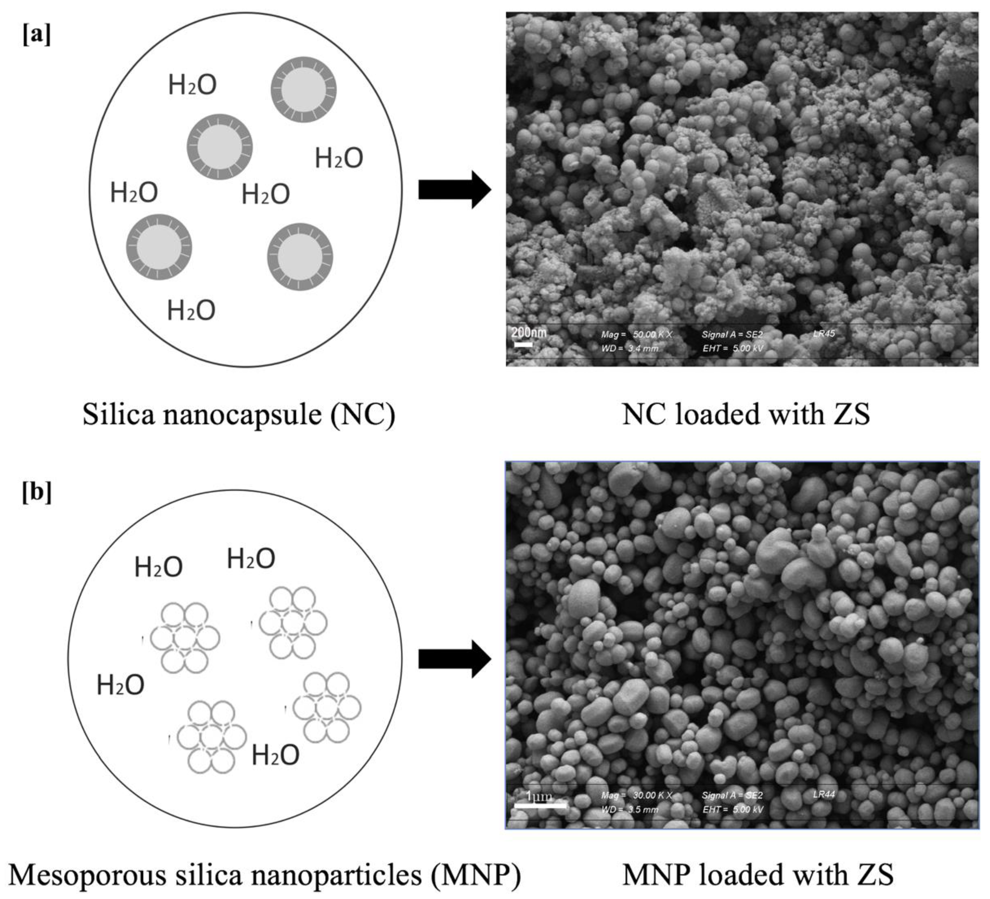
2.2. Encapsulation Step
2.3. In Vitro Tests
3. Results
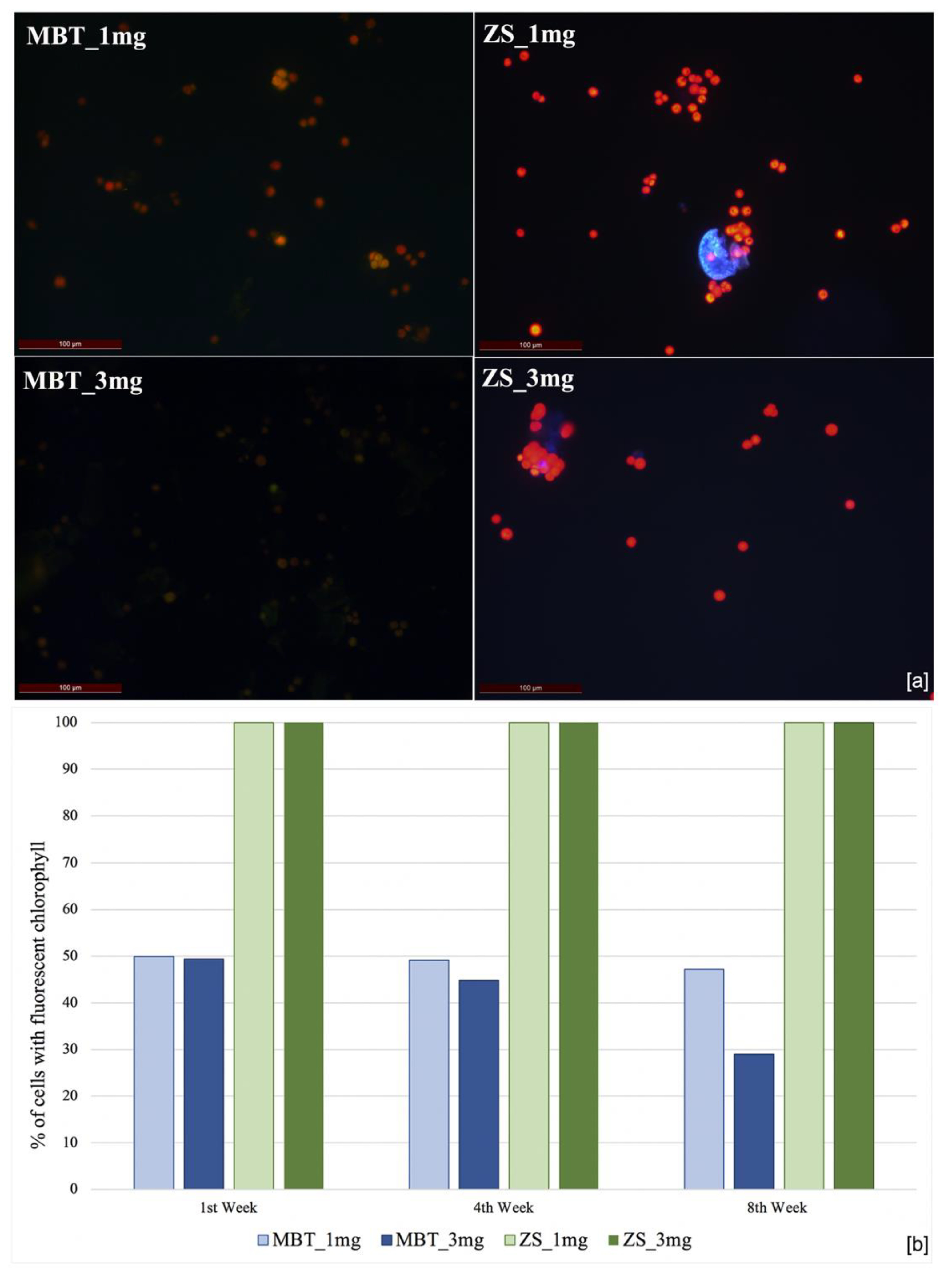
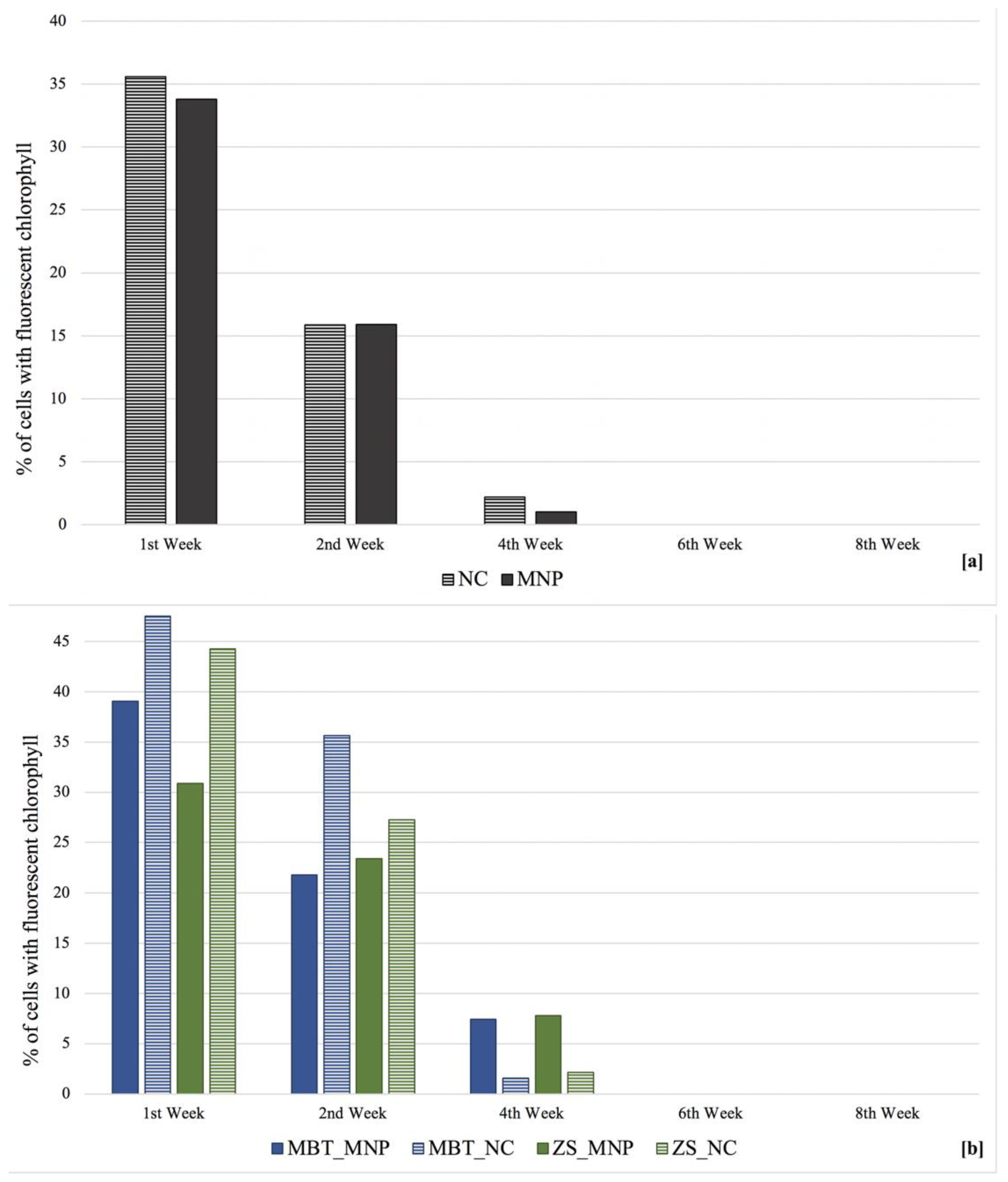
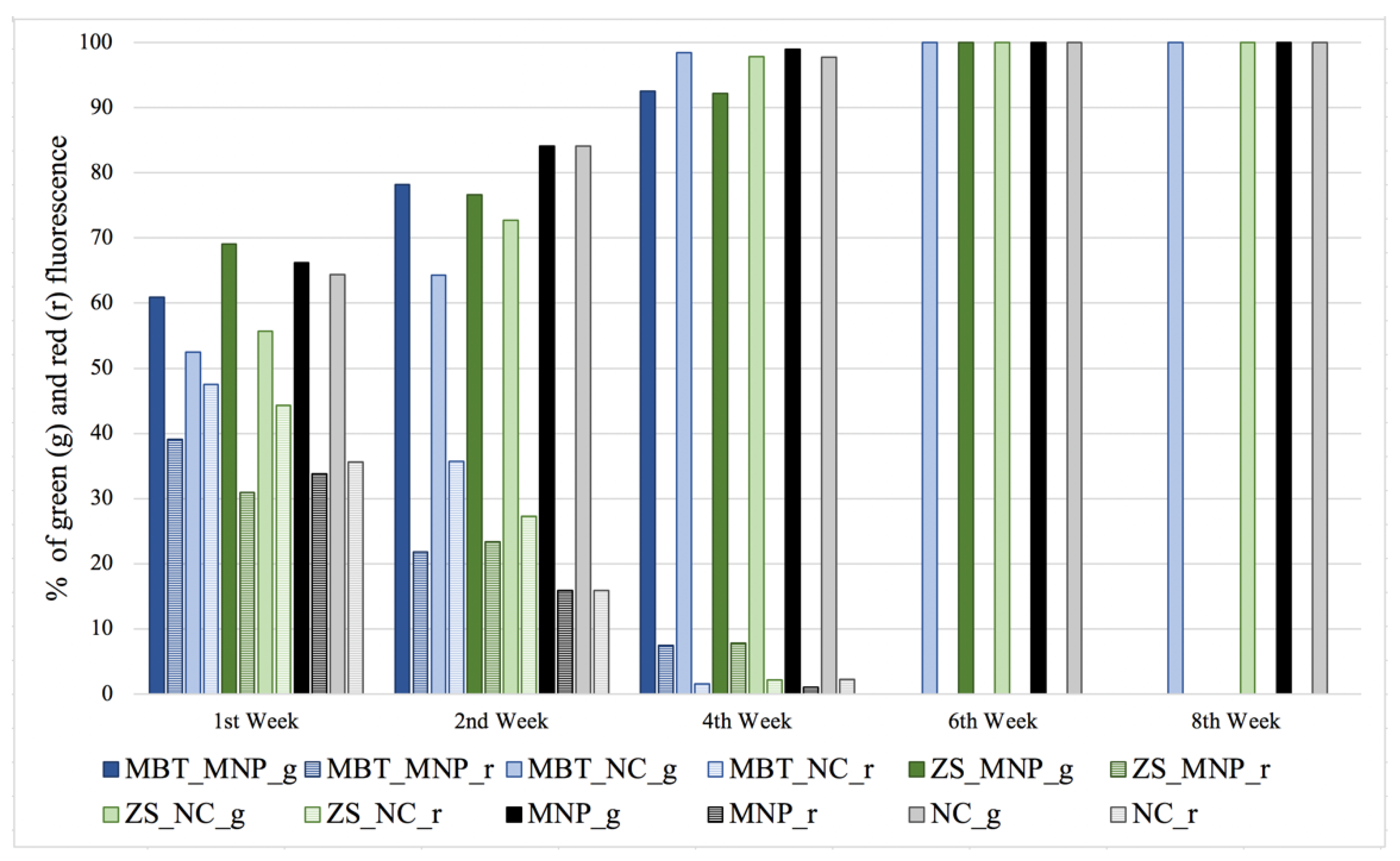
Biocidal Efficiency
4. Discussion
5. Conclusions
Author Contributions
Funding
Institutional Review Board Statement
Informed Consent Statement
Acknowledgments
Conflicts of Interest
References
- Videla, H.A.; Herrera, L.K. Biodeterioration and weathering effects on rock decay. Corros. Rev. 2004, 22, 341–364. [Google Scholar] [CrossRef]
- Caneva, G.; Nugari, M.P.; Salvadori, O. Plant. Biology for Cultural Heritage: Biodeterioration and Conservation; The Getty Conservation Institute: Los Angeles, CA, USA, 2008. [Google Scholar]
- Camuffo, D. Microclimate for Cultural Heritage: Measurement, Risk Assessment, Conservation, Restoration, and Maintenance of Indoor and Outdoor Monuments, 3rd ed.; Elsevier: Amsterdam, The Netherlands, 2019. [Google Scholar]
- Cappitelli, F.; Villa, F.; Sorlini, C. New environmentally friendly approaches against biodeterioration of outdoor cultural heritage. In Biocolonization of Stone: Middle Missouri Plains Control and Preventive Village Sites Methods, Proceedings of the MCI Workshop Series; Elena Charola, A., Ed.; Smithsonian Contributions to Museum Conservation; Smithsonian Institution Scholarly Press: Washington, DC, USA, 2011; pp. 51–58. [Google Scholar]
- Baglioni, P.; Chelazzi, D.; Giorgi, R. Nanotechnologies in the Conservation of Cultural Heritage: A Compendium of Materials and Techniques; Springer: Dordrecht, The Netherlands; Berlin/Heidelberg, Germany; New York, NY, USA; London, UK, 2014. [Google Scholar] [CrossRef]
- Serafini, I.; Ciccola, A. Nanotechnologies and Nanomaterials. Nanotechnologies and Nanomaterials for Diagnostic. Conserv. Restor. Cult. Herit. 2019, 325–380. [Google Scholar] [CrossRef]
- Tortora, L.; Di Carlo, G.; Mosquera, M.J.; Ingo, G.M. Nanoscience and Nanomaterials for the Knowledge and Conservation of Cultural Heritage. Front. Mater. Sci. 2020, 7, 372–374. [Google Scholar] [CrossRef]
- Fidanza, M.R.; Caneva, G. Natural biocides for the conservation of stone cultural heritage: A review. J. Cult. Herit. 2019, 38, 271–286. [Google Scholar] [CrossRef]
- Caneva, G.; Nugari, M.P.; Pinna, D.; Salvadori, O. Il Controllo del Degrado Biologico: I Biocidi nel Restauro dei Materiali Lapidei; Nardini Editore: Fiesole, Italy, 1996; p. 151. [Google Scholar]
- Pinna, D. Coping with Biological Growth on Stone Heritage Objects: Methods, Products, Applications, and Perspectives; Apple Academic Press: Waretown, NJ, USA, 2017. [Google Scholar]
- Kakakhel, M.A.; Wu, F.; Gu, J.D.; Feng, H.; Shah, K.; Wang, W. Controlling biodeterioration of cultural heritage objects with biocides: A review. Int. Biodeterior. Biodegrad. 2019, 143, 104721. [Google Scholar] [CrossRef]
- Nugari, M.P.; Pallecchi, P.; Pinna, D. Methodological Evaluation of Biocidal Interference with Stone Minerals—Preliminary Laboratory Tests. In Proceedings of the International RILEM/UNESCO Congress on Conservation of Stone and Other Materials, Paris, France, 29 June–1 July 1993; Thiel, M.J., Ed.; E. & F. N. Spon: London, UK, 1993; pp. 295–302. [Google Scholar]
- Ruggiero, L.; Bartoli, F.; Fidanza, M.R.; Zurlo, F.; Marconi, E.; Gasperi, T.; Tuti, S.; Crociani, L.; Di Bartolomeo, E.; Caneva, G.; et al. Encapsulation of environmentally-friendly biocides in silica nanosystems for multifunctional coatings. Appl. Surf. Sci. 2020, 514, 145908. [Google Scholar] [CrossRef]
- Ruggiero, L.; Fidanza, M.R.; Iorio, M.; Tortora, L.; Caneva, G.; Ricci, M.A.; Sodo, A. Synthesis and characterization of TEOS coating added with innovative antifouling silica nanocontainers and TiO2 nanoparticles. Front. Mater. 2020, 7, 185. [Google Scholar] [CrossRef]
- Silva, M.; Rosado, T.; Teixeira, D.; Candeias, A.; Caldeira, A.T. Production of green biocides for cultural heritage. Novel biotechnological solutions. Int. J. Conserv. Sci. 2015, 6, 519–530. [Google Scholar]
- Silva, M.; Salvador, C.; Candeias, M.F.; Teixeira, D.; Candeias, A.; Caldeira, A.T. Toxicological assessment of novel green biocides for cultural heritage. Int. J. Conserv. Sci. 2016, 7, 265–272. [Google Scholar]
- Rotolo, V.; Barresi, G.; Di Carlo, E.; Giordano, A.; Lombardo, G.; Crimi, E.; Costa, E.; Bruno, M.; Palla, F. Plant extracts as green potential strategies to control the biodeterioration of cultural heritage. Int. J. Conserv. Sci. 2016, 7, 839–846. [Google Scholar]
- Dresler, C.; Saladino, M.; Demirbag, C.; Caponetti, E.; Martino, D.F.C.; Alduina, R. Development of controlled release systems of biocides for the conservation of cultural heritage. Int. Biodeterior. Biodegrad. 2017, 125, 150–156. [Google Scholar] [CrossRef]
- Kuznetsova, A.; Domingues, P.M.; Silva, T.; Almeida, A.; Zheludkevich, M.L.; Tedim, J.; Ferreira, M.G.S.; Cunha, A. Antimicrobial activity of 2-mercaptobenzothiazole released from environmentally friendly nanostructured layered double hydroxides. J. Appl. Microbiol. 2017, 122, 1207–1218. [Google Scholar] [CrossRef] [PubMed]
- Ruggiero, L.; Crociani, L.; Zendri, E.; El Habra, N.; Guerriero, P. Incorporation of the zosteric sodium salt in silica nanocapsules: Synthesis and characterization of new fillers for antifouling coatings. Appl. Surf. Sci. 2018, 439, 705–711. [Google Scholar] [CrossRef]
- Ruggiero, L.; Di Bartolomeo, E.; Gasperi, T.; Luisetto, I.; Talone, A.; Zurlo, F.; Peddis, D.; Ricci, M.A.; Sodo, A. Silica nanosystems for active antifouling protection: Nanocapsules and mesoporous nanoparticles in controlled release applications. J. Alloys Compd. 2019, 798, 144–148. [Google Scholar] [CrossRef]
- Ruggiero, L.; Sodo, A.; Cestelli-Guidi, M.; Romani, M.; Sarra, A.; Postorino, P.; Ricci, M.A. Raman and ATR FT-IR investigations of innovative silica nanocontainers loaded with a biocide for stone conservation treatments. Microchem. J. 2020, 155, 104766. [Google Scholar] [CrossRef]
- Arreche, R.; Vázquez, P. Green biocides to control biodeterioration in materials science and the example of preserving World Heritage Monuments. Curr. Opin. Green Sustain. Chem. 2020, 100359. [Google Scholar] [CrossRef]
- Palla, F. Biotechnology and Cultural Heritage Conservation. In Heritage; IntechOpen: London, UK, 2020. [Google Scholar] [CrossRef] [Green Version]
- McCusker, L.; Liebau, F.; Engelhardt, G. Nomenclature of structural and compositional characteristics of ordered microporous and mesoporous materials with inorganic hosts (IUPAC Recommendations 2001). Pure Appl. Chem. 2009, 73, 381e394. [Google Scholar] [CrossRef] [Green Version]
- Mattos, B.D.; Rojas, O.J.; Magalhães, W.L. Biogenic silica nanoparticles loaded with neem bark extract as green, slow-release biocide. J. Clean. Prod. 2017, 142, 4206–4213. [Google Scholar] [CrossRef]
- Campanella, L.; Angeloni, R.; Cibin, F.; Dell’Aglio, E.; Grimaldi, F.; Reale, R.; Vitali, M. Capsulated essential oil in gel spheres for the protection of cellulosic cultural heritage. Nat. Prod. Res. 2021, 35, 116–123. [Google Scholar] [CrossRef] [PubMed]
- Chen, H.; He, J.; Tang, H.; Yan, C. Porous silica nanocapsules and nanospheres: Dynamic self-assembly synthesis and application in controlled release. Chem. Mater. 2008, 20, 5894–5900. [Google Scholar] [CrossRef]
- Popat, A.; Liu, J.; Hu, Q.; Kennedy, M.; Peters, B.; Lu, G.Q.; Qiao, S.Z. Adsorption and release of biocides with mesoporous silica nanoparticles. Nanoscale 2012, 4, 970e975. [Google Scholar] [CrossRef]
- Chan, A.C.; Bravo Cadena, M.; Townley, H.E.; Fricker, M.D.; Thompson, I.P. Effective delivery of volatile biocides employing mesoporous silicates for treating biofilms. J. R. Soc. Interface 2017, 14, 20160650. [Google Scholar] [CrossRef] [Green Version]
- European Parliament. Directive of the European Parliament and of the council of 16 February 1998 concerning the placing of biocidal products on the market. Off. J. Eur. Commun. 1998, L123, 1–63. [Google Scholar]
- Orlita, A. Microbial biodeterioration of leather and its control: A review. Int. Biodeterior. Biodegrad. 2004, 53, 157–163. [Google Scholar] [CrossRef]
- Nascimbene, J.; Salvadori, O. Lichen recolonization on restored calcareous statues of three Venetian villas. Int. Biodeterior. Biodegrad. 2008, 62, 313–318. [Google Scholar] [CrossRef]
- Jendresen, C.B.; Nielsen, A.T. Production of zosteric acid and other sulfated phenolic biochemicals in microbial cell factories. Nat. Commun. 2019, 10, 1–10. [Google Scholar] [CrossRef] [Green Version]
- Daidone, G.; Maggio, B.; Schillaci, D. Salicylanilide and its heterocyclic analogues. A comparative study of their antimicrobial activity. Pharmazie 1990, 45, 441–442. [Google Scholar] [PubMed]
- Franchini, C.; Muraglia, M.; Corbo, F.; Florio, M.A.; Di Mola, A.; Rosato, A.; Matucci, R.; Nesi, M.; van Bambeke, F.; Vitali, C. Synthesis and Biological Evaluation of 2-Mercapto-1, 3-benzothiazole Derivatives with Potential Antimicrobial Activity. Arch. Pharm. 2009, 342, 605–613. [Google Scholar] [CrossRef]
- Zhang Newby, B.; Cutright, T.; Barrios, C.A.; Xu, Q. Zosteric acid—An effective antifoulant for reducing fresh water bacterial attachment on coatings. JCT Res. 2006, 3, 69–70. [Google Scholar]
- Caneva, G.; De Marco, G.; Dinelli, A.; Vinci, M. The wall vegetation of the roman archaeological areas. Sci. Technol. Cult. Herit. 1992, 1, 217–226. [Google Scholar]
- Cappitelli, F.; Villa, F. Novel Antibiofilm Non-Biocidal Strategies. In Microorganisms in the Deterioration and Preservation of Cultural Heritage; Edith, J., Ed.; Springer: Berlin, Germany, 2021; p. 117. [Google Scholar] [CrossRef]
- UNI 10923. Beni culturali: Materiali lapidei naturali ed artificiali. In Allestimento di Preparati Biologici per L’osservazione al Microscopio Ottico; UNI: Milano, Italy, 2001. [Google Scholar]
- Guiry, M.D.; Guiry, G.M. AlgaeBase Version 4.2. World-Wide Electronic Publication; National University of Ireland: Maynooth, Ireland, 2007; Available online: http://www.algaebase.org (accessed on 2 February 2020).
- Ramírez, M.; Hernández-Mariné, M.; Novelo, E.; Roldán, M. Cyanobacteria-containing biofilms from a Mayan monument in Palenque, Mexico. Biofouling 2010, 26, 399–409. [Google Scholar] [CrossRef]
- Hsieh, P.; Pedersen, J.Z.; Bruno, L. Photoinhibition of cyanobacteria and its application in cultural heritage conservation. Photochem. Photobiol. 2014, 90, 533–543. [Google Scholar] [CrossRef]
- García-Plazaola, J.I.; Fernández-Marín, B.; Duke, S.O.; Hernández, A.; López-Arbeloa, F.; Becerril, J.M. Autofluorescence: Biological functions and technical applications. Plant Sci. 2015, 236, 136–145. [Google Scholar] [CrossRef]
- Donaldson, L. Autofluorescence in plants. Molecules 2020, 25, 2393. [Google Scholar] [CrossRef] [PubMed]
- Lamb, J.J.; Røkke, G.; Hohmann-Marriott, M.F. Chlorophyll fluorescence emission spectroscopy of oxygenic organisms at 77 K. Photosynthetica 2018, 56, 105–124. [Google Scholar] [CrossRef] [Green Version]
- Abràmoff, M.D.; Magalhães, P.J.; Ram, S.J. Image processing with ImageJ. Biophotonics Int. 2004, 11, 36–42. [Google Scholar]
- Rasband, W.S. ImageJ; US National Institutes of Health: Bethesda, MD, USA, 1997.
- Colangiuli, D.; Calia, A.; Bianco, N. Novel multifunctional coatings with photocatalytic and hydrophobic properties for the preservation of the stone building heritage. Constr. Build. Mater. 2015, 93, 189–196. [Google Scholar] [CrossRef]
- La Russa, M.F.; Ruffolo, S.A.; Rovella, N.; Belfiore, C.M.; Palermo, A.M.; Guzzi, M.T.; Crisci, G.M. Multifunctional TiO2 coatings for cultural heritage. Prog. Org. Coat. 2012, 74, 186–191. [Google Scholar] [CrossRef]
- Ruffolo, S.A.; La Russa, M.F. Nanostructured coatings for stone protection: An overview. Front. Mater. Sci. 2019, 6, 147. [Google Scholar] [CrossRef]
- Zuena, M.; Ruggiero, L.; Della Ventura, G.; Bemporad, E.; Ricci, M.A.; Sodo, A. Effectiveness and Compatibility of Nanoparticle Based Multifunctional Coatings on Natural and Man-Made Stones. Coatings 2021, 11, 480. [Google Scholar] [CrossRef]
- Becerra, J.; Mateo, M.; Ortiz, P.; Nicolás, G.; Zaderenko, A.P. Evaluation of the applicability of nano-biocide treatments on limestones used in cultural heritage. J. Cult. Herit. 2019, 38, 126–135. [Google Scholar] [CrossRef]
- Castaldo, R.; de Luna, M.S.; Siviello, C.; Gentile, G.; Lavorgna, M.; Amendola, E.; Cocca, M. On the acid-responsive release of benzotriazole from engineered mesoporous silica nanoparticles for corrosion protection of metal surfaces. J. Cult. Herit. 2020, 44, 317–324. [Google Scholar] [CrossRef]
- Rogers, S.O.; Bendich, A.J. Extraction of DNA from plant tissues. In Plant Molecular Biology Manual; Springer: Dordrecht, The Netherlands, 1989; pp. 73–83. [Google Scholar] [CrossRef]
- Ray, P.C.; Yu, H.; Fu, P.P. Toxicity and environmental risks of nanomaterials: Challenges and future needs. J. Environ. Sci. Health C 2009, 27, 1–35. [Google Scholar] [CrossRef] [PubMed] [Green Version]
- EU Regulation 1907/2006: Registration, Evaluation, Authorisation and Restriction of Chemicals (REACH). Available online: https://osha.europa.eu/en/legislation/directives/regulation-ec-no-1907-2006-of-the-european-parliament-and-of-the-council (accessed on 23 July 2021).
- Pastoriza-Santos, I.; Pérez-Juste, J.; Liz-Marzán, L.M. Silica-coating and hydrophobation of CTAB-stabilized gold nanorods. Chem. Mater. 2006, 18, 2465–2467. [Google Scholar] [CrossRef]
- Todd, J.S.; Zimmerman, R.C.; Crews, P.; Alberte, R.S. The AF activity of natural and synthetic phenolic-acid sulfate esters. Phytochemistry 1993, 34, 401–404. [Google Scholar] [CrossRef]
- Villa, F.; Remelli, W.; Forlani, F.; Vitali, A.; Cappitelli, F. Altered expression level of Escherichia coli proteins in response to treatment with the antifouling agent zosteric acid sodium salt. Environ. Microbiol. 2012, 14, 1753–1761. [Google Scholar] [CrossRef]
- Vilas-Boas, C.; Sousa, E.; Pinto, M.; Correia-da-Silva, M. An antifouling model from the sea: A review of 25 years of zosteric acid studies. Biofouling 2017, 33, 927–942. [Google Scholar] [CrossRef]
- Villa, F.; Pitts, B.; Stewart, P.S.; Giussani, B.; Roncoroni, S.; Albanese, D.; Giordano, C.; Tunesi, M.; Cappitelli, F. Efficacy of zosteric acid sodium salt on the yeast biofilm model Candida albicans. Microb. Ecol. 2011, 62, 584. [Google Scholar] [CrossRef] [Green Version]
- Geiger, T.; Delavy, P.; Hany, R.; Schleuniger, J.; Zinn, M. Encapsulated zosteric acid embedded in poly [3-hydroxyalkanoate] coatings—Protection against biofouling. Polym. Bull. 2004, 52, 65–72. [Google Scholar] [CrossRef] [Green Version]
- Boopalan, M.; Sasikumar, A. Studies on biocide free and biocide loaded zeolite hybrid polymer coatings on zinc phosphated mild steel for the protection of ships hulls from biofouling and corrosion. Silicon 2011, 3, 207–214. [Google Scholar] [CrossRef]
- Cattò, C.; Dell’Orto, S.; Villa, F.; Villa, S.; Gelain, A.; Vitali, A.; Marzano, V.; Baroni, S.; Forlani, F.; CAppitelli, F. Unravelling the Structural and Molecular Basis Responsible for the Anti-Biofilm Activity of Zosteric Acid. PLoS ONE 2015, 10, e0131519. [Google Scholar] [CrossRef] [PubMed] [Green Version]
- Caneva, G.; Salvadori, O.; Ricci, S.; Ceschin, S. Ecological analysis and biodeterioration processes over time at the Hieroglyphic Stairway in the Copán (Honduras) archaeological site. Plant Biosyst. 2005, 139, 295–310. [Google Scholar] [CrossRef]
- Caneva, G.; Bartoli, F.; Savo, V.; Futagami, Y.; Strona, G. Combining statistical tools and ecological assessments in the study of biodeterioration patterns of stone temples in Angkor (Cambodia). Sci. Rep. 2016, 6, 1–8. [Google Scholar] [CrossRef] [PubMed] [Green Version]
- Municchia, A.C.; Bartoli, F.; Taniguchi, Y.; Giordani, P.; Caneva, G. Evaluation of the biodeterioration activity of lichens in the Cave Church of Üzümlü (Cappadocia, Turkey). Int. Biodeterior. Biodegrad. 2018, 127, 160–169. [Google Scholar] [CrossRef]
- Valentini, F.; Diamanti, A.; Carbone, M.; Bauer, E.M.; Palleschi, G. New cleaning strategies based on carbon nanomaterials applied to the deteriorated marble surfaces: A comparative study with enzyme-based treatments. App. Surf. Sci. 2012, 258, 5965–5980. [Google Scholar] [CrossRef]


Publisher’s Note: MDPI stays neutral with regard to jurisdictional claims in published maps and institutional affiliations. |
© 2021 by the authors. Licensee MDPI, Basel, Switzerland. This article is an open access article distributed under the terms and conditions of the Creative Commons Attribution (CC BY) license (https://creativecommons.org/licenses/by/4.0/).
Share and Cite
Bartoli, F.; Zuena, M.; Sodo, A.; Caneva, G. The Efficiency of Biocidal Silica Nanosystems for the Conservation of Stone Monuments: Comparative In Vitro Tests against Epilithic Green Algae. Appl. Sci. 2021, 11, 6804. https://doi.org/10.3390/app11156804
Bartoli F, Zuena M, Sodo A, Caneva G. The Efficiency of Biocidal Silica Nanosystems for the Conservation of Stone Monuments: Comparative In Vitro Tests against Epilithic Green Algae. Applied Sciences. 2021; 11(15):6804. https://doi.org/10.3390/app11156804
Chicago/Turabian StyleBartoli, Flavia, Martina Zuena, Armida Sodo, and Giulia Caneva. 2021. "The Efficiency of Biocidal Silica Nanosystems for the Conservation of Stone Monuments: Comparative In Vitro Tests against Epilithic Green Algae" Applied Sciences 11, no. 15: 6804. https://doi.org/10.3390/app11156804