Article

Microbiological Changes during Orthodontic Aligner Therapy: A Prospective Clinical Trial

1
Unit of Orthodontics and Pediatric Dentistry, Section of Dentistry, Department of Clinical, Surgical, Diagnostic and Pediatric Sciences, University of Pavia, 27100 Pavia, Italy
2
Unit of Dental Hygiene, Section of Dentistry, Department of Clinical, Surgical, Diagnostic and Pediatric Sciences, University of Pavia, 27100 Pavia, Italy
3
UOC di Odontostomatologia e Chirurgia Orale Az. USL di Modena, 41121 Modena, Italy
*
Authors to whom correspondence should be addressed.
Appl. Sci. 2021, 11(15), 6758; https://doi.org/10.3390/app11156758
Submission received: 9 June 2021 / Revised: 7 July 2021 / Accepted: 21 July 2021 / Published: 23 July 2021
(This article belongs to the Section Environmental Sciences)

Abstract

:
The purpose of this study is to assess the variations induced by Clear Aligner Treatment (CAT) on the periodontal status and microbiological composition of oral microbiota. A total of 20 orthodontic patients were submitted to professional oral hygiene and, subsequently, underwent CAT (Group one, trials). A total of 20 non orthodontic patients (Group two, controls) remained untreated after oral hygiene. At T0 (baseline) and T1 (after 2 months), the participants were clinically examined for Probing Pocket Depth (PPD), Bleeding on Probing (BOP) and Plaque Index (PI), and microbiological tests were performed to detect bacteria of the red and orange complexes as well as the presence of Aggregatibacter actinomycetemcomitans. No significant inter- or intra-group differences were shown neither for PPD, PI and BOP (p > 0.05), nor for bacteria expressed in copies/microlitre or in percentages (p > 0.05). The only significant difference was assessed from T0 (baseline) to T1 (2 months) in both groups and is related to the total bacteria count increase. However, this parameter encompasses all the bacteria of the common oral microbiota, thus, not representing a significant result from a clinical point of view. Despite the limitations of this study, CAT does not significantly affect periodontal and microbiological parameters with respect to untreated patients for the first two months of therapy.

1. Introduction

Oral microbiota consists of more than 700 different kinds of microorganisms, including bacteria, viruses, mycoplasmas, fungi and protozoa, which inhabits the human mouth establishing an equilibrium with consequent benefits both for the microorganisms themselves and for the guest [1]. One of the major advantages brought by the resident microbiota is represented by the prevention of colonisation by pathogenetic microorganisms, which, conversely, would lead to a state of “dysbiosis”; this event is favoured by different conditions, such as plaque accumulation, the alteration of immune defences of the subject and antibiotics therapies [2].
Bacterial plaque accumulation is the leading cause of gingivitis, an inflammatory process of the gums that, if untreated, might evolve into periodontitis affecting the soft and hard tooth-supporting tissues. The latest classification of periodontal diseases dates back to 2017 [3] and it proposes the diagnosis of periodontitis according to stages (from stage I to stage IV, based on the severity and complexity of management) and grades (from grade A to grade C, according to the evidence or risk of rapid progression).
The bacteria most related to periodontitis development have traditionally been considered to be Porphyromonas gingivalis, Troponema denticola and Tannerella forsythensys, constituting the so-called “red complex”, despite the fact that this classification is quite no more actual [4].
The insertion of fixed orthodontic appliances in the mouth, aimed at the replacement of teeth in a correct position, actually provides the presence of more sites for bacterial adhesion and biofilm formation (plaque accumulation) [5], with an eventual progression toward the abovementioned inflammatory processes [6]. In addition to that, the increase in bacteria is also referable to the difficulty in realising proper oral hygiene caused by the presence of the appliances [7]. Accordingly, the use of removable clear aligners has also been proposed with the pros of facilitating teeth brushing in orthodontic patients avoiding the risk of white spot lesions as well as gingivitis and periodontitis [8,9]. Despite contrasting results, studies have generally shown a reduced plaque accumulation and periodontal risk in patients who underwent CAT (Clear Aligner Treatment), if compared to fixed appliances therapies [10,11,12]. However, it has not been widely investigated if the risk of the abovementioned factors is similar in patients undergoing CAT with respect to patients not under treatment.
Therefore, the purpose of this clinical trial is to compare the variations induced after two months of CAT on periodontal status and microbiological composition, compared to people not under orthodontic treatment. The first null hypothesis is that there is no statistically significant difference, neither for periodontal nor for microbiological parameters, between T0 and T1. The second null hypothesis is that no significant difference occurs between the trials and the controls considering the abovementioned parameters.

2. Materials and Methods
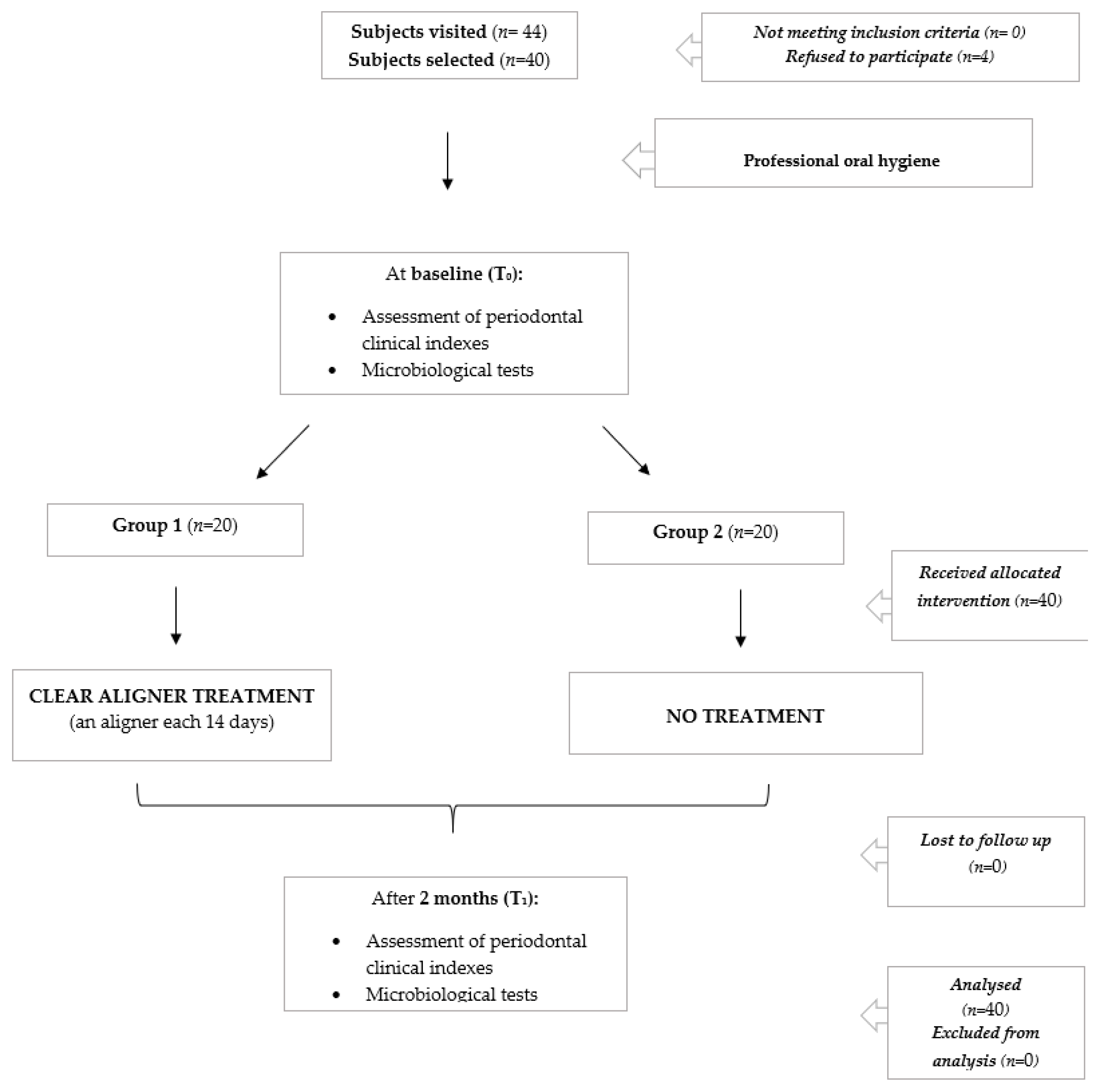
2.1. Trial Design

This is a parallel group, randomised, active controlled trial with a 1:1 allocation ratio. Unit Internal Review Board approved the study (IRB 2020-0129).

2.2. Participants

A total of 40 patients referring to the Unit of Orthodontics and Paediatric Dentistry, Section of Dentistry, Department of Clinical, Surgical, Diagnostic and Paediatric Sciences, University of Pavia, Pavia, Italy and undergoing clear aligner orthodontic therapy were recruited from June 2019 to October 2020 and followed for 2 months. The study lasted until December 2020. The consent of participants, or that of parents in the case of underage patients, was required.
The participant inclusion criteria were the following: aged more than 12 years old, good health status, good periodontal status, skeletal Class I, molar Class I, dental malocclusion, the absence of previous periodontal treatments on the teeth considered. Conversely, the exclusion criteria were the following: smoking any number of cigarettes/day; contraindications for the orthodontic treatment; the presence of dental restorations near the marginal gum of the teeth considered; the presence of fixed prostheses; the use of antibiotics, steroids or a nonsteroidal anti-inflammatory drug in the last six months; previous non-surgical periodontal treatments performed in the last year. As regards this last factor, all the patients underwent a professional oral hygiene before the beginning of the orthodontic treatment; some of them effectively needed to be subdued to a subsequent professional oral hygiene during the treatment, but this was performed at least one year before the beginning of the trial.

2.3. Interventions and Outcomes

Patients selected for the study underwent a professional supragingival and subgingival oral hygiene using a piezoelectric and Gracey curettes (Hu-Friedy, Chicago, IL, USA) with a final periodontal pockets decontamination by means of Air-flow Plus (EMS SA). Participants were then instructed to a correct domiciliary oral hygiene consisting of the use of an electric toothbrush for 2 min three times a day, as well as of the use of a dental floss once/day with abstention from mouthwashes in order to avoid an alteration of the resident bacterial flora. After 14 days from the professional hygiene treatment, patients were recalled. At this time (T0), periodontal indexes were assessed, and microbiological tests were carried out. The former encompassed Probing Pocket Depth (PPD), Bleeding on Probing (BOP) and Plaque Index (PI) (Table 1) [4,13,14], which were assessed by means of a probe (UNC probe 15; Hu-Friedy, Chicago, IL, USA) in correspondence of the “Ramfjord teeth” (maxillary right first molar, maxillary left central incisor, maxillary left first premolar, mandibular left first molar, mandibular right central incisor and mandibular right first premolar), as commonly considered in the literature [15].
Conversely, microbiological tests were performed by means of a Real Time PCR-based test (BPA Basic Lite, Biomolecular Diagnostic Srl, Firenze, Italy), considering, respectively, the mesio vestibular crevicular sulcus of maxillary right first molar, maxillary left central incisor, mandibular left first molar and mandibular right central incisor. The specific kit used allowed for the detection of the following bacteria: Aggregatibacter actinomycetemcomitans, Tannerella forsythensys, Porphyromonas gingivalis, Troponema denticola, Prevotella intermedia and Fusobacterium nucleatum. Microbiological samples were collected with sterile papers, inserted for 30 s in periodontal pockets and then stored in a sterile test tube to be sent to the laboratory (storage at −20 °C). Patients were not asked to avoid food or beverage intake before sampling procedures.
After outcomes assessment, participants were divided into two groups according to the succeeding orthodontic treatment. To those of group 1 (trials), a first clear aligner (Arc Angel, Modena, Italy) was delivered. Participants of group 2 were considered as controls, as they did not immediately undergo orthodontic therapy (the beginning of the therapy was delayed by 2 months). Patients in the trial group were recommended to wear the clear aligner for at least 20 h/day (as generally recommended by clear aligners manufacturers) and to respect a periodical follow up every 2 weeks to receive the subsequent appliance.
At T1, after 2 months from T0, periodontal parameters assessment and microbiological tests were performed again, as previously described.
The protocol of the study is shown in Table 2. The descriptions of the interventions performed are according to the TIDieR (Template for Intervention Description and Replication) Checklist.

2.4. Sample Size

The sample size calculation was conducted. Concerning the variable plaque index (primary outcome), an expected mean of 4.8 was hypothesised, with a standard deviation of 1.48 [16]. The expected difference between the means was supposed to be 1.35; therefore, 20 patients were requested for each group. Loss to follow-up and incomplete compliance with therapy were excluded.
A total of 44 participants, of which 21 were in the trial group and 23 were controls, were recruited. After the first visit, 1 trial and 3 controls refused to participate. A total of 40 final subjects when then selected, 20 trials (mean age: 33 years; standard deviation: 10 years and 4 months) and 20 controls (mean age: 36 years; standard deviation: 9 years) as requested by the sample size calculation.
The flow chart of the study is shown in Figure 1.

2.5. Randomisation and Blinding

Since the patients of the two groups differ for the need or the consent to undergo an orthodontic treatment using clear aligners, randomisation was not technically possible in this study. Since it was not possible to remove attachments, blinding the operator who performed the periodontal assessment was not feasible. The microbiological tests were conducted by an external centre that received samples without references to clinical data. Finally, the data analyst was blinded, too.

2.6. Statistical Methods

Data were submitted for statistical analysis with R Software (R version 3.1.3, R Development Core Team, R Foundation for Statistical Computing, Wien, Austria). Significance for all statistical tests was predetermined at p < 0.05.
For each group and variable, descriptive statistics (mean, standard deviation, median, minimum and maximum value) were calculated.
PPD was calculated in millimetres, whereas PI and BOP with the relative score. The presence of each specific microorganism tested was expressed both as the number of copies/microlitre and as a percentage of the total bacterial count.
For each variable, a repeated measures ANOVA was applied for each variable to detect differences among the groups.

3. Results

3.1. Periodontal Clinical Indexes

No significant inter- or intra-group differences were shown for PPD, PI and BOP (p > 0.05), as shown in Table 3.
In the control group, only PPD decreased from T0 to T1, whereas BOP and PI increased during this period. Conversely, the exact opposite pattern was assessed in the trial group. However, any statistically significant difference was found in all these variations.
The table shows that PPD, BOP and PI did not significantly change, neither in the control group (no treatment) nor in the trial group (clear aligner treatment) from the beginning to the end of the study.

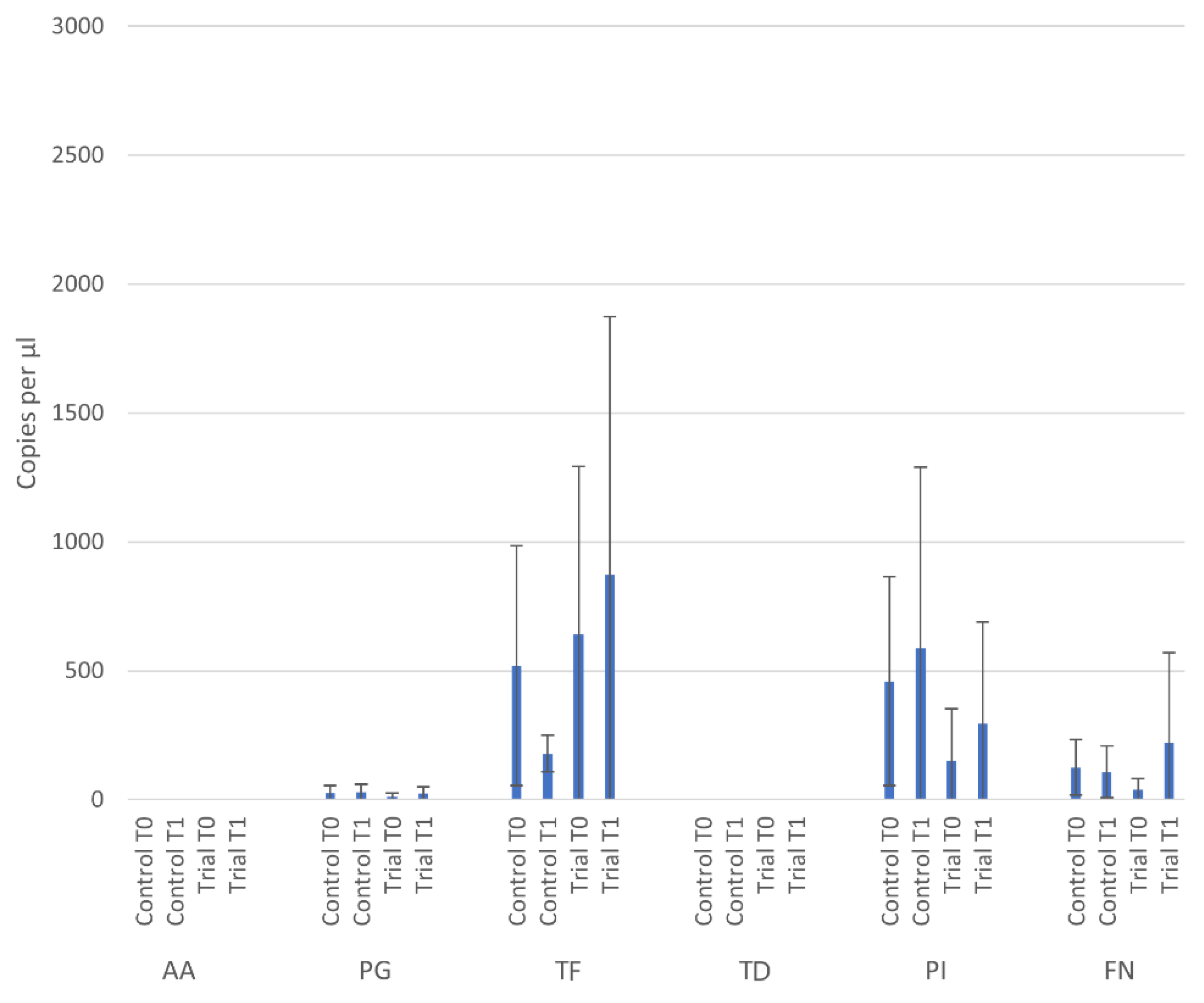
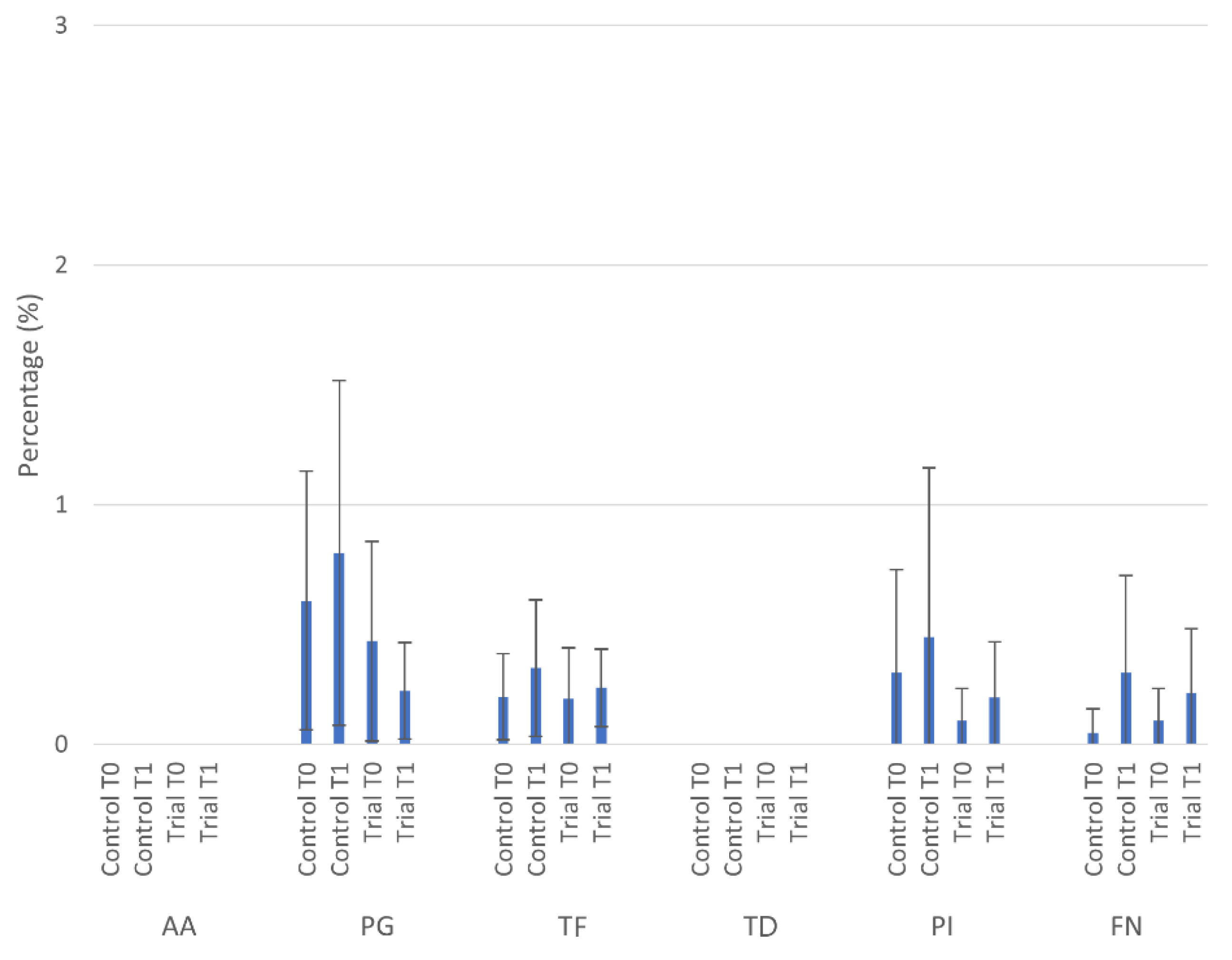
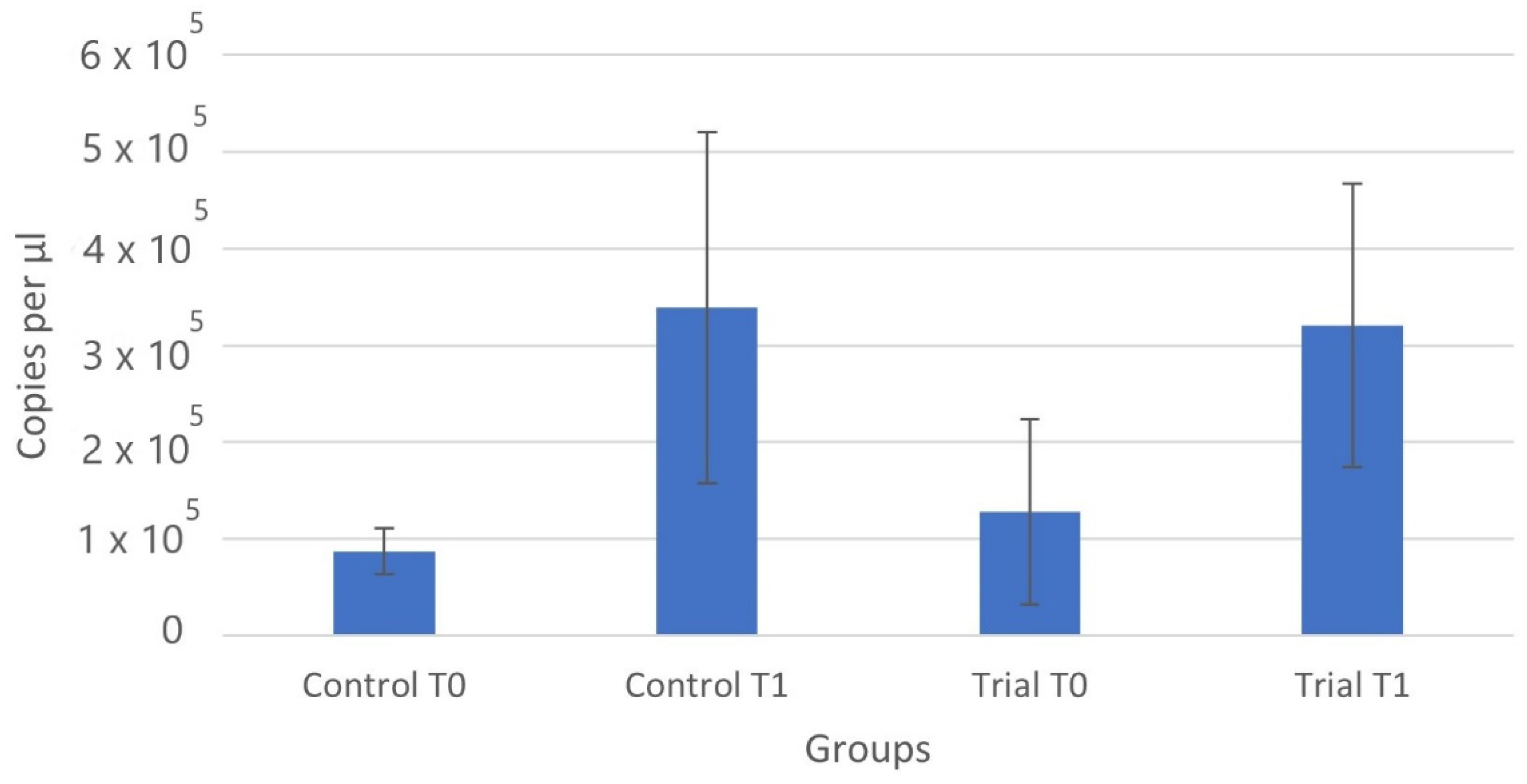
3.2. Microbiological Tests

As shown in Table 4 and Table 5 and Figure 2 and Figure 3, no significant inter- or intra-group differences were shown for the bacteria tested (p > 0.05), considering neither the copies/microlitre nor for the relative percentages.
Neither Aggregatibacter actinomycetemcomitans nor Treponema denticola were detected in the groups at any time. Considering the expression of the bacterial count as copies/microlitre, Porphyromonas gingivalis and Prevotella Intermedia increased in both groups from T0 to T1. Conversely, Tannerella forsythia and Fusobacterium nucleatum increased in the trial group but decreased in the control one. However, none of all these variations were shown to be statistically significant.
The table shows that the copies/microlitre of each bacterium tested did not significantly change, neither in the control group (no treatment) nor in the trial group (clear aligner treatment) from the beginning to the end of the study.
As regards the percentage expression of the bacteria detected (thus excluding Aggregatibacter actinomycetemcomitans and Treponema denticola), this value increased in both groups for Tannerella forsythia, Prevotella intermedia and Fusobacterium nucleatum. Conversely, the percentage of Porphyromonas gingivalis increased in the control group but decreased in the trial one. However, none of all these variations were shown to be statistically significant.
The table shows that the percentage of each bacterium tested did not significantly change, neither in the control group (no treatment) nor in the trial group (clear aligner treatment) from the beginning to the end of the study.
For both the control and the trial group, the total bacteria count was significantly increased compared to the values, respectively, assessed at T0 and at T1 (intragroup differences), as shown in Table 6 and Figure 4 (p < 0.05). No significant intergroup differences were assessed.
The table shows that the copies/microlitre of the total bacterial count significantly increased, both in the control group (no treatment) and in the trial group (clear aligner treatment) from the beginning to the end of the study. No significant differences were assessed compared to the respective time points in the two groups.

4. Discussion

Fixed orthodontic therapy has been related to a worsening of periodontal status and microbiological equilibrium several times; therefore, clinicians should be aware of the risk represented by the orthodontic treatment on these parameters [17]. Several studies in the literature have emphasised the impact of fixed orthodontic appliances on the arising of gingival inflammation, despite the fact that this event is generally regarded as a reversible condition with resolution after a few months from their removal, especially in patients with a good oral hygiene [5,18]. However, specific fixed orthodontic appliances, such as bands, are more likely to cause an impairment of both soft and hard tooth-supporting tissues [19]. In general, the appearance in the mouth of new retentive surfaces represented by the orthodontic appliances causes an increased accumulation of dental plaque with an inflammatory response [5]. At a later stage, quantitative and qualitative alterations of subgingival biofilm occur with a selection of periodontopathic microorganisms and an increase in inflammatory mediators [4,20]. Specific periodontal therapies have been proposed to prevent periodontitis and reduce the risk of gingivitis and discomfort in patients with orthodontic appliances. Cosola et al. [21] found that the non-surgical periodontal treatment combined with professional and domiciliary ozonised water could be effective in improving the clinical periodontal parameters in orthodontic patients because of the anti-inflammatory and anti-plaque effect, compared to the traditional non-surgical periodontal treatments combined with the home-care use of chlorhexidine therapy.
Clear aligners have been proposed in dentistry thanks to the higher aesthetic value and acceptability by patients, if compared to fixed appliances. Despite their wide diffusion, there is still a lack of solid scientific evidence as regards their impact on the periodontal status and microbial community, considering that controversial results have been reported. For instance, a study conducted by Miethke and Brauner [22] assessed the differences of periodontal indexes on patients treated with clear aligners and fixed lingual attachments, with a higher worsening of periodontal conditions in patients of the latter group, which is linked to the difficulties in cleaning lingual tooth surfaces when fitted with a fixed appliance. However, PPD was the only index with very similar values in both treatment groups. Conversely, several studies have stated a significantly higher PPD increase in patients with fixed appliances if compared to those under CAT [11,12]. Whereas a limited, but not irrelevant, increase in PPD was found in this last group of patients, the study conducted by Levrini et al. [10], considering a 3-month evaluation, reported no significant variation from the beginning to the end of the follow up. In accordance with this result, the increase in PPD from T0 to T1 in the trial group (from 1.89 to 1.99) was not even significant in our study.
The second periodontal index considered in our research protocol was Bleeding on Probing (BOP). A study conducted by Karkhanechi et al. [11] evaluated BOP in two groups of orthodontic patients (treated with fixed appliances or clear aligners) along three periodic follow ups: in both groups, BOP was similar after 6 weeks from the beginning of the treatment, but, subsequently, the former group started to show increased values of bleeding. At the 6-month follow up, the differences between the two groups increased, with a significant difference even higher at the 1-year follow up. Moreover, the latter group reported a progressive improvement for BOP. Similarly, Abbate et al. [12] reported a significant improvement of BOP after a year of treatment with clear aligners (probably due to the attention to oral hygiene and to the instructions received), differently from the one with fixed appliances. Azaripour et al. [23] also assessed significantly higher BOP values in patients treated with fixed therapy, despite no significant improvement of these values after the treatment with clear aligners. This last result is in accordance with our data: despite the fact that the BOP values in the trial group decreased from 4.55 to 4.08, the reduction was not statistically significant.
The last periodontal index considered in our study is related to plaque accumulation, considering that this event is generally linked to orthodontic therapy. Most authors have reported a significant difference between Plaque Index (PI) in orthodontic patients treated with fixed or removable appliances. Miethke and Brauner [22] found that the plaque values were almost double in the patients treated with lingual fixed attachments compared to those using Invisalign®; in addition to that, during the subsequent evaluations, a reduction in PI was reported for the latter. Comparable results emerged from further studies [11,12], despite the fact that some authors did not confirm a significant PI reduction in clear aligners-treated patients at the end of the follow up [24]. In our study, a certain reduction for this index was assessed in the trial group (0.39 at baseline and 0.30 after two months), but this variation did not turn out to be significant. Anyway, this difference might be attributed to easier oral hygiene procedures for the patients with removable aligners and to reduced plaque retention sites compared to fixed appliances [10].
According to the results obtained in our study concerning periodontal clinical indexes, treating orthodontic patients with CAT does not predispose a worsening of the periodontal condition, at least until the initial phase of the therapy. Furthermore, the periodontal status of these patients is similar to that of the control patients not under treatment. Therefore, the first null hypothesis of this study could not be rejected.
The second aim of this report was to verify the impact of clear aligners on the total bacterial load of the oral cavity and, in particular, on specific periodontal pathogens. Even in this case, the null hypothesis was accepted, since for all the bacteria tested, no significant increase in copies/microlitre or percentage was reported, neither between the trial and the control group nor within each one. This result is in accordance with the evidence reported by Levrini et al. [10], stating no significant alterations in the total bacterial load due to clear aligners and, consequently, a better impact of these appliances on periodontal health with respect to the traditional therapy. In addition, a literature review by Rossini et al. [25], confirmed that minimal quantitative and qualitative variations of the microbiota derive from the treatment with aligners. A subsequent study [26] analysed bacterial DNA from the plaque using 16S rRNA gene sequencing, both before the treatment, as well as one and three months after. The authors found a slightly decreasing microbial diversity with a significant change of microbial structure during the first three months of CAT. However, the patients were free from periodontitis and showed relatively stable levels of periodontal microorganisms and core microorganisms.
Our study suggests that using clear aligners causes no significant alterations in the bacterial count and bacterial percentage of neither red and orange complexes nor of A. actinomycetemcomitans, thus, avoiding a risk of periodontitis, at least during the first two months of the therapy. In particular, A. actinomycetemcomitans, one of the microorganisms most related to periodontal disease, was not detected at all in any group at any time. However, we cannot conclude that CAT is not associated with the growth of this pathogen, considering that a study conducted by Guo et al. [26] assessed its increase, despite not being statistically significant. This might be due to the longer follow up considered by the authors (three months instead of two) as well as to the fact that microbiological samples were collected at the beginning of the study without submitting patients to a professional oral hygiene, differently from our protocol.
Similar to A. actinomycetemcomitans, the microorganism T. denticola was not detected in any patient of this study. Considering that this bacterium belongs to the red complex of pathogens related to periodontitis, its absence further suggests that CAT does not represent a risk for the development of the disease. However, it must be taken into account that this result might be influenced again by the relative short follow up. As regards the other pathogens (P. gingivalis, T. forsythia, P. intermedia and F. nucleatum), a limited number of colonies were detected in both the trial and the control patients, with differences between T0 and T1 that were not statistically significant, anyway. Our data confirm the evidence of previous studies that did not assess the significant variations of these four microorganisms [10,26].
A significant difference assessed by us that deserves to be discussed concerns the total bacterial count, which encompasses not only the pathogens more strictly related to the periodontal disease, but also those that normally constitute the oral microbiota of healthy subjects. This value significantly varied from T0 to T1 both in the control and the trial group (intragroup significant differences). The increase was equal to 291% for controls and to 151% for trial, but the intergroup difference was not significant, allowing us, again, to consider clear aligners sure for the periodontium of the patients treated. These variations of the total bacterial count were obviously expected, considering the protocol chosen for the study: in fact, the first microbiological samples collection was carried out two weeks from the professional oral hygiene, which justifies the reductions in the total number of bacteria (independently of their pathogenetic action). Conversely, for the samples collection at T1, the patients were not submitted to a further hygiene treatment, therefore, explaining the higher values assessed for the total bacterial count. However, we cannot exclude the possibility that the bacterial increase might be independent from the protocol used and that the intergroup difference between trials and controls might become statistically significant on a longer follow up. Surely, it should be taken into account that periodontal diseases have multifactorial aetiology, encompassing not only the bacterial reservoir, but also the immune factors related to the host [27].
A major limitation of the present report is associated to the relatively short follow up considered. In order to confirm the results obtained here, further studies should be conducted including patients belonging to different countries and on a longer period, covering the entire length of the orthodontic treatment. Another limitation of this study is that only one category of clear aligners was tested, but further kinds of these appliances should be considered to detect the eventual different effects on periodontal health among them. Moreover, the results obtained in this report cannot be directly applied to the entire population. In fact, the participants selected had to show a good periodontal status and a dental malocclusion; therefore, the outcomes might be different in people already suffering from periodontal disease or reporting severe malocclusions. Another limitation was that we did not perform a calibration for the periodontal assessment. Only two groups were examined, encompassing participants under CAT and participants not treated; consequently, further evaluations should also compare the effects of clear aligners with those caused by fixed appliances in order to confirm the better impact of the former compared to the latter on periodontal status and microbiological equilibrium. Finally, it would be interesting to evaluate how different electric toothbrushes could influence the clinical and microbiological parameters, in order to understand how patients under CAT could benefit from these heterogenous devices [28].

5. Conclusions

During the first two months of CAT, the use of these devices did not significantly affect the periodontal and microbiological parameters with respect to patients not under treatment. Accordingly, the use of clear aligners should be considered as a valuable therapeutic option that has no significant impact on oral and microbiological parameters if compared with untreated patients over the 2-month time interval considered in the present report. Further studies with larger samples, a longer follow-up period and other laboratory parameters (e.g., quantification of MMP-8) are needed to investigate the efficacy of CAT and confute the results of the present pilot study.

Author Contributions

Conceptualisation, M.F.S., A.S. and A.B.; methodology, M.F.S., A.S. and A.B.; software, A.S.; validation, P.G.; formal analysis, A.S.; investigation, C.L., B.O., E.S. and M.P.; resources, P.D.M.; data curation, A.S., A.B., C.L., B.O. and E.S.; writing—original draft preparation, A.S. and S.G.; writing—review and editing, A.S. and M.P.; visualisation, M.F.S.; supervision, P.G.; project administration, A.B. and A.S.; funding acquisition, A.B. All authors have read and agreed to the published version of the manuscript.

Funding

This research received no external funding.

Institutional Review Board Statement

This study was approved by the Unit Internal Review Board (number of approval: IRB 2020-0129).

Informed Consent Statement

Informed consent was obtained from all subjects involved in the study.

Data Availability Statement

All data generated or analysed during this study are included in this published article.

Conflicts of Interest

The authors declare no conflict of interest.

References

  1. Xiao, J.; Fiscella, K.A.; Gill, S.R. Oral microbiome: Possible harbinger for children’s health. Int. J. Oral Sci. 2020, 12, 12. [Google Scholar] [CrossRef]
  2. Lamont, R.J.; Koo, H.; Hajishengallis, G. The oral microbiota: Dynamic communities and host interactions. Nat. Rev. Genet. 2018, 16, 745–759. [Google Scholar] [CrossRef] [PubMed]
  3. Caton, J.G.; Armitage, G.; Berglundh, T.; Chapple, I.L.; Jepsen, S.; Kornman, K.S.; Mealey, B.L.; Papapanou, P.N.; Sanz, M.; Tonetti, M.S. A new classification scheme for periodontal and peri-implant diseases and conditions—Introduction and key changes from the 1999 classification. J. Clin. Periodontol. 2018, 45, S1–S8. [Google Scholar] [CrossRef]
  4. Shirozaki, M.U.; Da Silva, R.A.B.; Romano, F.L.; Da Silva, L.A.B.; De Rossi, A.; Lucisano, M.P.; Messora, M.R.; Feres, M.; Júnior, A.B.N. Clinical, microbiological, and immunological evaluation of patients in corrective orthodontic treatment. Prog. Orthod. 2020, 21, 6. [Google Scholar] [CrossRef] [PubMed]
  5. Sfondrini, M.F.; Debiaggi, M.; Zara, F.; Brerra, R.; Comelli, M.; Bianchi, M.; Pollone, S.R.; Scribante, A. Influence of lingual bracket position on microbial and periodontal parameters in vivo. J. Appl. Oral Sci. 2012, 20, 357–361. [Google Scholar] [CrossRef] [PubMed] [Green Version]
  6. Sun, F.; Ahmed, A.; Wang, L.; Dong, M.; Niu, W. Comparison of oral microbiota in orthodontic patients and healthy individuals. Microb. Pathog. 2018, 123, 473–477. [Google Scholar] [CrossRef]
  7. Awartani, F.; Atassi, F. Oral Hygiene Status among Orthodontic Patients. J. Contemp. Dent. Pract. 2010, 11, 25–32. [Google Scholar] [CrossRef] [Green Version]
  8. Buschang, P.H.; Chastain, D.; Keylor, C.L.; Crosby, D.; Julien, K.C. Incidence of white spot lesions among patients treated with clear aligners and traditional braces. Angle Orthod. 2018, 89, 359–364. [Google Scholar] [CrossRef] [Green Version]
  9. D’Apuzzo, F.; Perillo, L.; Carrico, C.K.; Castroflorio, T.; Grassia, V.; Lindauer, S.J.; Shroff, B. Clear aligner treatment: Different perspectives between orthodontists and general dentists. Prog. Orthod. 2019, 20, 10. [Google Scholar] [CrossRef] [Green Version]
  10. Levrini, L.; Mangano, A.; Montanari, P.; Margherini, S.; Caprioglio, A.; Abbate, G.M. Periodontal health status in patients treated with the Invisalign® system and fixed orthodontic appliances: A 3 months clinical and microbiological evaluation. Eur. J. Dent. 2015, 9, 404–410. [Google Scholar] [CrossRef]
  11. Karkhanechi, M.; Chow, D.; Sipkin, J.; Sherman, D.; Boylan, R.J.; Norman, R.G.; Craig, R.G.; Cisneros, G.J. Periodontal status of adult patients treated with fixed buccal appliances and removable aligners over one year of active orthodontic therapy. Angle Orthod. 2013, 83, 146–151. [Google Scholar] [CrossRef]
  12. Abbate, D.G.; Caria, M.; Montanari, P.; Mannu, C.; Orrù, G.; Caprioglio, A.; Levrini, L. Periodontal health in teenagers treated with removable aligners and fixed orthodontic appliances. J. Orofac. Orthop. 2015, 76, 240–250. [Google Scholar] [CrossRef]
  13. Alqerban, A. Levels of proinflammatory chemokines and advanced glycation end products in patients with type-2 diabetes mellitus undergoing fixed orthodontic treatment. Angle Orthod. 2021, 91, 105–110. [Google Scholar] [CrossRef] [PubMed]
  14. Corbett, A.I.; Leggitt, V.L.; Angelov, N.; Olson, G.; Caruso, J.M. Periodontal health of anterior teeth with two types of fixed retainers. Angle Orthod. 2014, 85, 699–705. [Google Scholar] [CrossRef] [Green Version]
  15. Ross, M.C.; Campbell, P.M.; Tadlock, L.P.; Taylor, R.W.; Buschang, P.H. Effect of automated messaging on oral hygiene in adolescent orthodontic patients: A randomized controlled trial. Angle Orthod. 2019, 89, 262–267. [Google Scholar] [CrossRef] [Green Version]
  16. Sifakakis, I.; Papaioannou, W.; Papadimitriou, A.; Kloukos, D.; Papageorgiou, S.N.; Eliades, T. Salivary levels of cariogenic bacterial species during orthodontic treatment with thermoplastic aligners or fixed appliances: A prospective cohort study. Prog. Orthod. 2018, 19, 25. [Google Scholar] [CrossRef] [Green Version]
  17. Gomes, S.C.; Varela, C.C.; Da Veiga, S.L.; Rosing, C.; Oppermann, R.V. Periodontal conditions in subjects following orthodontic therapy. A preliminary study. Eur. J. Orthod. 2007, 29, 477–481. [Google Scholar] [CrossRef] [PubMed] [Green Version]
  18. Calniceanu, H.; Stratul, S.-I.; Rusu, D.; Jianu, A.; Boariu, M.; Nica, L.; Ogodescu, A.; Sima, L.; Bolintineanu, S.; Anghel, A.; et al. Changes in clinical and microbiological parameters of the periodontium during initial stages of orthodontic movement in patients with treated severe periodontitis: A longitudinal site-level analysis. Exp. Ther. Med. 2020, 20, 199. [Google Scholar] [CrossRef] [PubMed]
  19. Diedrich, P.; Rudzki-Janson, I.; Wehrbein, H.; Fritz, U. Effects of orthodontic bands on marginal periodontal tissues. A histologic study on two human specimens. J. Orofac. Orthop. 2001, 62, 146–156. [Google Scholar] [CrossRef]
  20. Ghijselings, E.; Coucke, W.; Verdonck, A.; Teughels, W.; Quirynen, M.; Pauwels, M.; Carels, C.; Van Gastel, J. Long-term changes in microbiology and clinical periodontal variables after completion of fixed orthodontic appliances. Orthod. Craniofacial Res. 2013, 17, 49–59. [Google Scholar] [CrossRef]
  21. Cosola, S.; Giammarinaro, E.; Genovesi, A.M.; Pisante, R.; Poli, G.; Covani, U.; Marconcini, S. A short-term study of the effects of ozone irrigation in an orthodontic population with fixed appliances. Eur. J. Paediatr. Dent. 2019, 20, 15–18. [Google Scholar] [PubMed]
  22. Miethke, R.-R.; Brauner, K. A Comparison of the Periodontal Health of Patients during Treatment with the Invisalign® System and with Fixed Lingual Appliances. J. Orofac. Orthop. 2007, 68, 223–231. [Google Scholar] [CrossRef]
  23. Azaripour, A.; Weusmann, J.; Mahmoodi, B.K.; Peppas, D.; Gerhold-Ay, A.; Van Noorden, C.J.F.; Willershausen, B. Braces versus Invisalign®: Gingival parameters and patients’ satisfaction during treatment: A cross-sectional study. BMC Oral Health 2015, 15, 69. [Google Scholar] [CrossRef] [PubMed] [Green Version]
  24. Lu, H.; Tang, H.; Zhou, T.; Kang, N. Assessment of the periodontal health status in patients undergoing orthodontic treatment with fixed appliances and Invisalign system: A meta-analysis. Medicine 2018, 97, e0248. [Google Scholar] [CrossRef]
  25. Rossini, G.; Parrini, S.; Castroflorio, T.; Deregibus, A.; Debernardi, C.L. Periodontal health during clear aligners treatment: A systematic review. Eur. J. Orthod. 2015, 37, 539–543. [Google Scholar] [CrossRef]
  26. Guo, R.; Zheng, Y.; Liu, H.; Li, X.; Jia, L.; Li, W. Profiling of subgingival plaque biofilm microbiota in female adult patients with clear aligners: A three-month prospective study. PeerJ 2018, 6, e4207. [Google Scholar] [CrossRef] [Green Version]
  27. Butera, A.; Gallo, S.; Maiorani, C.; Molino, D.; Chiesa, A.; Preda, C.; Esposito, F.; Scribante, A. Probiotic Alternative to Chlorhexidine in Periodontal Therapy: Evaluation of Clinical and Microbiological Parameters. Microorganisms 2021, 9, 69. [Google Scholar] [CrossRef]
  28. Preda, C.; Butera, A.; Pelle, S.; Pautasso, E.; Chiesa, A.; Esposito, F.; Oldoini, G.; Scribante, A.; Genovesi, A.; Cosola, S. The Efficacy of Powered Oscillating Heads vs. Powered Sonic Action Heads Toothbrushes to Maintain Periodontal and Peri-Implant Health: A Narrative Review. Int. J. Environ. Res. Public Health 2021, 18, 1468. [Google Scholar] [CrossRef] [PubMed]
Figure 1. Flow chart of the study.
Applsci 11 06758 g001
Figure 2. Number of copies/microlitre for the bacteria tested in the study (this figure shows the data reported in Table 4).
Applsci 11 06758 g002
Figure 3. Percentage of the bacteria tested in the study (this figure shows the data reported in Table 5).
Applsci 11 06758 g003
Figure 4. Total bacterial count expressed as copies/microlitre (this figure shows the data reported in Table 6).
Applsci 11 06758 g004
Table 1. Procedures/scores relative to the periodontal parameters assessed.
Probing Pocket Depth
(mm)
Measurement from the Free Gingival Margin to the Bottom of the Periodontal Pocket by Means of a Millimetre Probe
Bleeding on Probing (%)0—no bleeding upon 20 s after probing
1—bleeding upon 20 s after probing
BOP is calculated as the ratio between the number of bleeding sites and the total sites evaluated (expressed as percentage)
Plaque Index
(Score 0–3)
0—No plaque
1—Thin plaque layer at the margin, only detectable by scraping with a probe
2—Moderate layer of plaque along gingival margin; interdental spaces free, but plaque is visible to the naked eye
3—Abundant plaque along to the gingival margin; interdental spaces filled with plaque
Table 2. Protocol of the study.
GroupProcedures
GROUP 1
(Trial Group)
Signature of the informed consent for the study
Professional oral hygiene and motivation to the domiciliary oral hygiene
After 14 days from the professional oral hygiene, assessment of periodontal clinical indexes and execution of microbiological tests (T0)
Delivery of the first clear alignment and change each 14 days
After 2 months from T0, re-assessment of periodontal clinical indexes and re-execution of microbiological tests (T1)
GROUP 2
(Control Group)
Signature of the informed consent for the study
Professional oral hygiene and motivation to the domiciliary oral hygiene
After 14 days from the professional oral hygiene, assessment of periodontal clinical indexes and execution of microbiological tests (T0)
After 2 months from T0, re-assessment of periodontal clinical indexes and re-execution of microbiological tests (T1)
Table 3. Descriptive statistics of periodontal clinical indexes.
VariableGroupMeanSDMinMedianMaxSignificance *
PPD
(mm)
Control T02.260.401.532.422.61p > 0.05
Control T12.120.351.782.062.78
Trial T01.890.461.171.813.00
Trial T11.990.510.642.172.47
BOP
(%)
Control T05.9810.530.001.0038.91p > 0.05
Control T17.5710.500.004.9236.10
Trial T04.555.050.002.5215.00
Trial T14.085.500.001.8015.00
PI
(0–4)
Control T00.500.460.000.381.58p > 0.05
Control T10.570.540.000.461.78
Trial T00.390.220.000.360.78
Trial T10.300.230.000.250.65
Legend: T0: baseline; T1: after 2 months; * p > 0.05 means that there is no significant difference.
Table 4. Descriptive statistics of each bacterial count expressed as copies/microlitre.
Bacteria TypeGroupMeanSDMinMedianMaxSignificance *
Aggregatibacter
actinomycetemcomitans
Control T00.000.000.000.000.00p > 0.05
Control T10.000.000.000.000.00
Trial T00.000.000.000.000.00
Trial T10.000.000.000.000.00
Porphyromonas
gingivalis
Control T026.6361.500.000.00223.00p > 0.05
Control T129.4765.880.000.00223.00
Trial T012.1531.760.000.00102.00
Trial T123.1561.750.000.00199.00
Tannerella forsythiaControl T0520.001034.570.000.002470.00p > 0.05
Control T1178.74157.840.00158.00351.00
Trial T0641.001487.690.000.004720.00
Trial T1874.602280.120.000.007480.00
Treponema denticolaControl T00.000.000.000.000.00p > 0.05
Control T10.000.000.000.000.00
Trial T00.000.000.000.000.00
Trial T10.000.000.000.000.00
Prevotella intermediaControl T0459.16901.710.000.002650.00p > 0.05
Control T1586.841562.150.000.006600.00
Trial T0150.00461.690.000.001500.00
Trial T1294.40901.370.000.002930.00
Fusobacterium
nucleatum
Control T0125.21238.970.000.00712.00p > 0.05
Control T1107.58223.230.000.00766.00
Trial T038.4597.810.000.00305.00
Trial T1220.85797.480.000.003590.00
Legend: T0: baseline; T1: after 2 months; * p > 0.05 means that there is no significant difference.
Table 5. Descriptive statistics of each bacterial count expressed as percentage.
Bacteria TypeAAMeanSDMinMedianMaxSignificance *
Aggregatibacter actinomycetemcomitansControl T00.000.000.000.000.00p > 0.05
Control T10.000.000.000.000.00
Trial T00.000.000.000.000.00
Trial T10.000.000.000.000.00
Porphyromonas
gingivalis
Control T00.601.230.000.003.00p > 0.05
Control T10.801.640.000.004.00
Trial T00.430.950.000.003.00
Trial T10.220.460.000.002.00
Tannerella forsythiaControl T00.200.410.000.001.00p > 0.05
Control T10.320.650.000.001.59
Trial T00.190.490.000.001.59
Trial T10.240.370.000.000.87
Treponema denticolaControl T00.000.000.000.000.00p > 0.05
Control T10.000.000.000.000.00
Trial T00.000.000.000.000.00
Trial T10.000.000.000.000.00
Prevotella intermediaControl T00.300.980.000.004.00p > 0.05
Control T10.451.610.000.007.00
Trial T00.100.310.000.001.00
Trial T10.200.530.000.001.73
Fusobacterium
nucleatum
Control T00.050.220.000.001.00p > 0.05
Control T10.300.920.000.004.00
Trial T00.100.310.000.001.00
Trial T10.220.610.000.002.00
Legend: T0: baseline; T1: after 2 months; * p > 0.05 means that there is no significant difference.
Table 6. Descriptive statistics of the total bacterial count expressed as copies/microlitre.
GroupMeanSDMinMedianMaxIntergroup
Differences *
Intragroup
Differences *
Control T086,80054,59719,000100,000166,000p < 0.05T0–T0: p > 0.05
T1–T1: p > 0.05
Control T1338,960414,28167,800100,0001,130,000
Trial T0128,010219,41214804270546,000p < 0.05
Trial T1320,960334,47378,400260,0001,260,000
Legend: *: p > 0.05 means that there is no significant difference; p < 0.05 means that there is a significant difference.
Publisher’s Note: MDPI stays neutral with regard to jurisdictional claims in published maps and institutional affiliations.

