Abstract
Purpose
Glioblastoma, the most aggressive type of brain cancer, is composed of heterogeneous populations of differentiated cells, cancer stem cells and immune cells. Cystatin F, an endogenous inhibitor of lysosomal cysteine peptidases, regulates the function of cytotoxic immune cells. The aim of this study was to determine which type of cells expresses cystatin F in glioblastoma and to determine the role of cystatin F during disease progression.
Methods
RT-qPCR and immunohistochemistry were used to determine cystatin F mRNA and protein levels in glioblastoma tissue samples. The internalization of cystatin F was analyzed by Western blotting. Enzyme kinetics, real time invasion and calcein release cytotoxicity assays were used to assess the role of internalized cystatin F.
Results
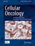
We found that cystatin F was not expressed in non-cancer brain tissues, but that its expression increased with glioma progression. In tumor tissues, extensive staining was observed in cancer stem-like cells and microglia/monocytes, which secrete cystatin F into their microenvironment. In trans activity of cystatin F was confirmed using an in vitro glioblastoma cell model. Internalized cystatin F affected cathepsin L activity in glioblastoma cells and decreased their invasiveness. In addition, we found that cystatin F decreased the susceptibility of glioblastoma cells to the cytotoxic activity of natural killer (NK) cells.
Conclusions
Our data implicate cystatin F as a mediator of immune suppression in glioblastoma. Increased cystatin F mRNA and protein levels in immune, glioblastoma and glioblastoma stem-like cells or trans internalized cystatin F may have an impact on decreased susceptibility of glioblastoma cells to NK cytotoxicity.


References
D.S. Nørøxe, H.S. Poulsen, U. Lassen, Hallmarks of glioblastoma: A systematic review. ESMO Open 1, e000144 (2017)
D.N. Louis, A. Perry, G. Reifenberger, A. von Deimling, D. Figarella-Branger, W.K. Cavenee, H. Ohgaki, O.D. Wiestler, P. Kleihues, D.W. Ellison, The 2016 World Health Organization classification of tumors of the central nervous system: A summary. Acta Neuropathol. 131, 803–820 (2016)
S. K. Ray, Glioblastoma: Molecular mechanisms of pathogenesis and current therapeutic strategies. Glioblastoma: Molecular Mechanisms of Pathogenesis and Current Therapeutic Strategies, 1st ed. (Springer, 2010)
H. Strobel, T. Baisch, R. Fitzel, K. Schilberg, M.D. Siegelin, G. Karpel-Massler, K.-M. Debatin, M.-A. Westhoff, Temozolomide and other alkylating agents in glioblastoma therapy. Biomedicines 7, 69 (2019)
S.H. Shen, K. Woroniecka, A.B. Barbour, P.E. Fecci, L. Sanchez-Perez, J.H. Sampson, CAR T cells and checkpoint inhibition for the treatment of glioblastoma. Expert. Opin. Biol. Ther. 20, 579–591 (2020)
B. Weenink, P.J. French, P.A.E.S. Smitt, R. Debets, M. Geurts, Immunotherapy in glioblastoma: Current shortcomings and future perspectives. Cancers (Basel) 12, 751 (2020)
J.H. Sampson, M.D. Gunn, P.E. Fecci, D.M. Ashley, Brain immunology and immunotherapy in brain tumours. Nat. Rev. Cancer 20, 12–25 (2020)
M. Lim, Y. Xia, C. Bettegowda, M. Weller, Current state of immunotherapy for glioblastoma. Nat. Rev. Clin. Oncol. 15, 422–442 (2018)
W. Tomaszewski, L. Sanchez-Perez, T.F. Gajewski, J.H. Sampson, Brain tumor microenvironment and host state: Implications for immunotherapy. Clin. Cancer Res. 25, 4202–4210 (2019)
M. Perišić Nanut, J. Sabotič, A. Jewett, J. Kos, Cysteine cathepsins as regulators of the cytotoxicity of NK and T cells. Front. Immunol. 5, 616 (2014)
B. Breznik, A. Mitrović, T.T. Lah, J. Kos, T.T. Lah, J. Kos, Cystatins in cancer progression: More than just cathepsin inhibitors. Biochimie 166, 233–250 (2019)
S. Halfon, J. Ford, J. Foster, L. Dowling, L. Lucian, M. Sterling, Y. Xu, M. Weiss, M. Ikeda, D. Liggett, A. Helms, C. Caux, S. Lebecque, C. Hannum, S. Menon, T. McClanahan, D. Gorman, G. Zurawski, Leukocystatin, a new class II cystatin expressed selectively by hematopoietic cells. J. Biol. Chem. 273, 16400–16408 (1998)
F. Cappello, E. Gatti, V. Camossetto, A. David, H. Lelouard, P. Pierre, Cystatin F is secreted, but artificial modification of its C-terminus can induce its endocytic targeting. Exp. Cell Res. 297, 607–618 (2004)
K. Maher, Š. Konjar, C. Watts, B. Turk, N. Kopitar-Jerala, S. Konjar, C. Watts, B. Turk, N. Kopitar-Jerala, Cystatin F regulates proteinase activity in IL-2-activated natural killer cells. Protein Pept. Lett. 21, 957–965 (2014)
T. Langerholc, V. Zavašnik-Bergant, B. Turk, V. Turk, M. Abrahamson, J. Kos, V. Zavaš Nik-Bergant, B. Turk, V. Turk, M. Abrahamson, J. Kos, Inhibitory properties of cystatin F and its localization in U937 promonocyte cells. FEBS J. 272, 1535–1545 (2005)
G. Hamilton, J.D. Colbert, A.W. Schuettelkopf, C. Watts, Cystatin F is a cathepsin C-directed protease inhibitor regulated by proteolysis. EMBO J. 27, 499–508 (2008)
Š. Konjar, V.R. Sutton, S. Hoves, U. Repnik, H. Yagita, T. Reinheckel, C. Peters, V. Turk, B. Turk, J.A. Trapani, N. Kopitar-Jerala, Human and mouse perforin are processed in part through cleavage by the lysosomal cysteine proteinase cathepsin L. Immunology 131, 257–267 (2010)
E. Dautović, M. Perišić Nanut, A. Softić, J. Kos, The transcription factor C/EBP α controls the role of cystatin F during the differentiation of monocytes to macrophages. Eur. J. Cell Biol. 97, 463–473 (2018)
J.D. Colbert, A. Plechanovová, C. Watts, Glycosylation directs targeting and activation of cystatin F from intracellular and extracellular sources. Traffic 10, 425–437 (2009)
Š. Magister, H.-C. Tseng, V.T. Bui, J. Kos, A. Jewett, Regulation of split anergy in natural killer cells by inhibition of cathepsins C and H and cystatin F. Oncotarget 6, 22310–22327 (2015)
M. Prunk, M. Perišić Nanut, J. Sabotič, U. Švajger, J. Kos, Increased cystatin F levels correlate with decreased cytotoxicity of cytotoxic T cells. Radiol. Oncol. 53, 57–68 (2019)
M. Perišić Nanut, J. Sabotič, U. Švajger, A. Jewett, J. Kos, Cystatin F affects natural killer cell cytotoxicity. Front. Immunol. 8, 1459 (2017)
T. Utsunomiya, Y. Hara, A. Kataoka, M. Morita, H. Arakawa, M. Mori, S. Nishimura, Cystatin-like metastasis-associated protein mRNA expression in human colorectal cancer is associated with both liver metastasis and patient survival. Clin. Cancer Res. 8, 2591 (2002)
V. Puxbaum, L. Mach, Proteinases and their inhibitors in liver cancer. World J. Hepatol. 1, 28–34 (2009)
C. Yang, T. Yu, Z. Liu, X. Ye, X. Liao, X. Wang, C. Han, G. Zhu, W. Qin, T. Peng, Cystatin F as a key family 2 cystatin subunit and prognostic biomarker for early-stage pancreatic ductal adenocarcinoma. Oncol. Rep. 42, 79 (2019)
R.L. Bowman, Q. Wang, A. Carro, R.G.W. Verhaak, M. Squatrito, GlioVis data portal for visualization and analysis of brain tumor expression datasets. Neuro-Oncology 19, 139–141 (2017)
M. Novak, M. Koprivnikar Krajnc, B. Hrastar, B. Breznik, B. Majc, M. Mlinar, A. Rotter, A. Porčnik, J. Mlakar, K. Stare, R.G. Pestell, T. Lah Turnšek, CCR5-mediated signaling is involved in invasion of glioblastoma cells in its microenvironment. Int. J. Mol. Sci. 21, 4199 (2020)
Š. Baebler, M. Svalina, M. Petek, K. Stare, A. Rotter, M. Pompe-Novak, K. Gruden, QuantGenius: Implementation of a decision support system for qPCR-based gene quantification. BMC Bioinforma 18, 276 (2017)
J. Kos, B. Stabuc, A. Schweiger, M. Krasovec, N. Cimerman, N. Kopitar-Jerala, I. Vrhovec, Cathepsins B, H, and L and their inhibitors stefin a and cystatin C in sera of melanoma patients. Clin. Cancer Res. 3, 1815 (1997)
J. Bryant, R. Day, T.L. Whiteside, R.B. Herberman, Calculation of lytic units for the expression of cell-mediated cytotoxicity. J. Immunol. Methods 146, 91–103 (1992)
M. Hüttenrauch, I. Ogorek, H. Klafki, M. Otto, C. Stadelmann, S. Weggen, J. Wiltfang, O. Wirths, Glycoprotein NMB : A novel Alzheimer ’ s disease associated marker expressed in a subset of activated microglia. Acta Neuropathol. Commun. 6, 1 (2018)
J.-V. Haure-Mirande, M. Wang, M. Audrain, T. Fanutza, S.H. Kim, S. Heja, B. Readhead, J.T. Dudley, R.D. Blitzer, E.E. Schadt, B. Zhang, S. Gandy, M.E. Ehrlich, Integrative approach to sporadic Alzheimer’s disease: Deficiency of TYROBP in cerebral Aβ amyloidosis mouse normalizes clinical phenotype and complement subnetwork molecular pathology without reducing Aβ burden. Mol. Psychiatry 24, 431–446 (2019)
Y. Wang, X. Zhang, Q. Song, Y. Hou, J. Liu, Y. Sun, P. Wang, Characterization of the chromatin accessibility in an Alzheimer’s disease (AD) mouse model. Alzheimers Res. Ther. 12, 29 (2020)
H. Keren-Shaul, A. Spinrad, A. Weiner, O. Matcovitch-Natan, R. Dvir-Szternfeld, T.K. Ulland, E. David, K. Baruch, D. Lara-Astaiso, B. Toth, S. Itzkovitz, M. Colonna, M. Schwartz, I. Amit, A unique microglia type associated with restricting development of Alzheimer’s disease. Cell 169, 1276–1290.e17 (2017)
J. Ma, K.F. Tanaka, G. Yamada, K. Ikenaka, Induced expression of cathepsins and cystatin C in a murine model of demyelination. Neurochem. Res. 32, 311–320 (2007)
W. Duan, H. Ran, Z. Zhou, Q. He, J. Zheng, Adenosine A2A receptor deficiency up-regulates cystatin F expression in white matter lesions induced by chronic cerebral hypoperfusion. PLoS One 7, e52566 (2012)
H. Ran, J. Yuan, J. Huang, J. Wang, K. Chen, Z. Zhou, Adenosine A2A receptors in bone marrow-derived cells attenuate cognitive impairment in mice after chronic hypoperfusion white matter injury. Transl. Stroke Res. 11, 1028–1040 (2020)
C.A. Baker, L. Manuelidis, Unique inflammatory RNA profiles of microglia in Creutzfeldt-Jakob disease. Proc. Natl. Acad. Sci. U. S. A. 100, 675–679 (2003)
M. Nuvolone, N. Schmid, G. Miele, S. Sorce, R. Moos, C. Schori, R.R. Beerli, M. Bauer, P. Saudan, K. Dietmeier, I. Lachmann, M. Linnebank, R. Martin, U. Kallweit, V. Kana, E.J. Rushing, H. Budka, A. Aguzzi, Cystatin F is a biomarker of prion pathogenesis in mice. PLoS One 12, e0171923 (2017)
J.I. Satoh, Gene expression profiles of M1 and M2 microglia characterized by comparative analysis of public datasets. Clin. Exp. Neuroimmunol. 9, 124–138 (2018)
J. Ma, K.F. Tanaka, T. Shimizu, C.C.A. Bernard, A. Kakita, H. Takahashi, S.E. Pfeiffer, K. Ikenaka, Microglial cystatin F expression is a sensitive indicator for ongoing demyelination with concurrent remyelination. J. Neurosci. Res. 89, 639–649 (2011)
J. Liang, N. Li, Y. Zhang, C. Hou, X. Yang, T. Shimizu, X. Wang, K. Ikenaka, K. Fan, J. Ma, Disinhibition of Cathepsin C caused by cystatin F deficiency aggravates the demyelination in a Cuprizone model. Front. Mol. Neurosci. 9, 152 (2016)
T. Shimizu, W. Wisessmith, J. Li, M. Abe, K. Sakimura, B. Chetsawang, Y. Sahara, K. Tohyama, K.F. Tanaka, K. Ikenaka, The balance between cathepsin C and cystatin F controls remyelination in the brain of Plp1 -overexpressing mouse, a chronic demyelinating disease model. Glia 65, 917–930 (2017)
J. Li, W.W. Durose, J. Ito, A. Kakita, Y. Iguchi, M. Katsuno, K. Kunisawa, T. Shimizu, K. Ikenaka, Exploring the factors underlying remyelination arrest by studying the post-transcriptional regulatory mechanisms of cystatin F gene. J. Neurochem. (2020)
D. Ofengeim, S. Mazzitelli, Y. Ito, J.P. DeWitt, L. Mifflin, C. Zou, S. Das, X. Adiconis, H. Chen, H. Zhu, M.A. Kelliher, J.Z. Levin, J. Yuan, RIPK1 mediates a disease-associated microglial response in Alzheimer’s disease. Proc. Natl. Acad. Sci. U. S. A. 114, E8788 (2017)
M. Nishikawa, A. Inoue, T. Ohnishi, S. Kohno, S. Ohue, S. Matsumoto, S. Suehiro, D. Yamashita, S. Ozaki, H. Watanabe, H. Yano, H. Takahashi, R. Kitazawa, J. Tanaka, T. Kunieda, Significance of glioma stem-like cells in the tumor periphery that express high levels of CD44 in tumor invasion, early progression, and poor prognosis in glioblastoma. Stem Cells Int. 2018, 1–15 (2018)
R.C. Gimple, S. Bhargava, D. Dixit, J.N. Rich, Glioblastoma stem cells: Lessons from the tumor hierarchy in a lethal cancer. Genes Dev. 33, 591–609 (2019)
S.Y. Ardebili, I. Zajc, B. Gole, B. Campos, C. Herold-Mende, S. Drmota, T.T. Lah, CD133/prominin1 is prognostic for GBM patient’s survival, but inversely correlated with cysteine cathepsins’ expression in glioblastoma derived spheroids. Radiol. Oncol. 45, 102 (2011)
J. Yin, K.L. Valin, M.L. Dixon, J.W. Leavenworth, The role of microglia and macrophages in CNS homeostasis, autoimmunity, and cancer. J. Immunol. Res. 2017, 1–12 (2017)
D.G. Walker, L.F. Lue, Immune phenotypes of microglia in human neurodegenerative disease: Challenges to detecting microglial polarization in human brains. Alzheimers Res. Ther. 7, 56 (2015)
K.L. Mooney, W. Choy, S. Sidhu, P. Pelargos, T.T. Bui, B. Voth, N. Barnette, I. Yang, The role of CD44 in glioblastoma multiforme. J. Clin. Neurosci. 34, 1–5 (2016)
T. Flannery, D. Gibson, M. Mirakhur, S. McQuaid, C. Greenan, A. Trimble, B. Walker, D. McCormick, P.G. Johnston, The clinical significance of cathepsin S expression in human astrocytomas. Am. J. Pathol. 163, 175–182 (2003)
T.T. Lah, M.B. Durán Alonso, C.J.F. Van Noorden, Antiprotease therapy in cancer: Hot or not? Expert Opin. Biol. Ther. 6, 257 (2006)
M. Sivaparvathi, M. Yamamoto, G.L. Nicolson, Z.L. Gokaslan, G.N. Fuller, L.A. Liotta, R. Sawaya, J.S. Rao, Expression and immunohistochemical localization of cathepsin L during the progression of human gliomas. Clin. Exp. Metastasis 14, 27–34 (1996)
N. Levičar, R.A. Dewey, E. Daley, T.E. Bates, D. Davies, J. Kos, G.J. Pilkington, T.T. Lah, Selective suppression of cathepsin L by antisense cDNA impairs human brain tumor cell invasion in vitro and promotes apoptosis. Cancer Gene Ther. 10, 141–151 (2003)
S. Kenig, R. Frangež, A. Pucer, T. Lah, Inhibition of cathepsin L lowers the apoptotic threshold of glioblastoma cells by up-regulating p53 and transcription of caspases 3 and 7. Apoptosis 16, 671–682 (2011)
T. Strojnik, R. Kavalar, M. Trinkaus, T.T. Lah, Cathepsin L in glioma progression: Comparison with cathepsin B. Cancer Detect. Prev. 29, 448–455 (2005)
N. Levičar, R.K. Nutall, T.T. Lah, Proteases in brain tumour progression. Acta Neurochir. 145(825), 1023 (2003)
S. Coniglio, I. Miller, M. Symons, J.E. Segall, Coculture assays to study macrophage and microglia stimulation of glioblastoma invasion. J. Vis. Exp. 2016 (2016)
X. Ye, S. Xu, Y. Xin, S. Yu, Y. Ping, L. Chen, H. Xiao, B. Wang, L. Yi, Q. Wang, X. Jiang, L. Yang, P. Zhang, C. Qian, Y. Cui, X. Zhang, X. Bian, Tumor-associated microglia/macrophages enhance the invasion of glioma stem-like cells via TGF-b1 signaling pathway. J. Immunol. 189, 444–453 (2012)
S.J. Coniglio, E. Eugenin, K. Dobrenis, E.R. Stanley, B.L. West, M.H. Symons, J.E. Segall, Microglial stimulation of glioblastoma invasion involves epidermal growth factor receptor (EGFR) and colony stimulating factor 1 receptor (CSF-1R) signaling. Mol. Med. 18, 519–527 (2012)
H.C. Tseng, A. Inagaki, V.T. Bui, N. Cacalano, N. Kasahara, Y.G. Man, A. Jewett, Differential targeting of stem cells and differentiated glioblastomas by NK cells. J. Cancer 6, 866–876 (2015)
A.K. Kozlowska, H.C. Tseng, K. Kaur, P. Topchyan, A. Inagaki, V.T. Bui, N. Kasahara, N. Cacalano, A. Jewett, Resistance to cytotoxicity and sustained release of interleukin-6 and interleukin-8 in the presence of decreased interferon-γ after differentiation of glioblastoma by human natural killer cells. Cancer Immunol. Immunother. 65, 1085–1097 (2016)
Acknowledgements
We would like to acknowledge Metka Novak, Bernarda Majc and Mateja Mlinar for technical support in processing tissue biopsies, RNA isolation and Fluidigm gene expression analysis and Eva Lasič for critical reading of the manuscript.
Availability of data and materials
The TCGA datasets used in this study can be accessed at: http://gliovis.bioinfo.cnio.es/. The datasets generated during the current study are available from the corresponding author upon reasonable request.
Code availability
Not applicable.
List of abbreviations
BSA bovine serum albumin.
C/EBPα CCAAT/enhancer-binding protein-α.
CTL cytotoxic T lymphocyte.
GBM glioblastoma.
GSC glioblastoma stem-like cell.
MpL Macrolepiota procera.
NK natural killer cell.
PBS phosphate buffered saline.
RT room temperature.
TCGA The Cancer Genome Atlas.
Funding
This research was funded by the Slovenian Research Agency, grant numbers P4–0127, J4–1776 to J.K., J3–2516 to M.P.N., P1–0245 to T.L.T., Z3–1870 to B.B. and the European Program of Cross-Border Cooperation for Slovenia-Italy Interreg TRANS-GLIOMA to T.L.T. The funders had no role in the design of the study, in the collection, analyses or interpretation of data, in the writing of the manuscript, or in the decision to publish the results.
Author information
Authors and Affiliations
Contributions
Conceptualization, M.P.N., J.K., E.S.; methodology, M.P.N., B.B., E.S., A.M.; validation, M.P.N.; resources J.M., A.P.; formal analysis, A.R., E.S.; investigation, E.S.; writing and original draft preparation, E.S.; writing - review and editing, M.P.N., B. B, A.M., J.M., A.P., T.L.T, J.K.; visualization, E.S.; supervision, M.P.N., B.B., J.K.; project administration M.P.N., J.K., B.B., T.L.T.; funding acquisition, M.P.N., J.K., B.B., T.L.T. All authors have read and agreed to the published version of the manuscript.
Corresponding author
Ethics declarations
Ethics approval and consent to participate
The study was approved by the National Medical Ethics Committee of the Republic of Slovenia (Approval no. 0120–179 190/2018/4).
Consent for publication
Not applicable.
Competing interests
The authors declare that they have no competing interests.
Additional information
Publisher’s note
Springer Nature remains neutral with regard to jurisdictional claims in published maps and institutional affiliations.
Supplementary Information
ESM 1
(DOCX 1861 kb)
Rights and permissions
About this article

Cite this article
Senjor, E., Perišić Nanut, M., Breznik, B. et al. Cystatin F acts as a mediator of immune suppression in glioblastoma. Cell Oncol. 44, 1051–1063 (2021). https://doi.org/10.1007/s13402-021-00618-9
Accepted:
Published:
Issue Date:
DOI: https://doi.org/10.1007/s13402-021-00618-9