Article Text

Statistics from Altmetric.com
Colonic stricture is a rare complication of hemolytic uremic syndrome (HUS). Risk factors include severe colitis, female gender and younger age. A clinical presentation of bowel obstruction in children, with previous known HUS, should prompt a contrast enema to rule out areas of stenosis. Surgical treatments consist of laparotomy, resection of the affected bowel and primary anastomosis.
A 4 year-old girl was referred to the pediatric surgery emergency service presenting with subacute abdominal occlusion for 1 week prior to admission.
The patient had a known history of HUS complicated by extensive colitis 3 months previously that required ventilation, transfusion support and renal replacement therapy. Verotoxin [also known as Shiga toxin (Stx)], which is associated with HUS enterocolitis from producing bacteria, mainly Escherichia coli O157:H7 and Shigella dysenteriae type 1, had been isolated, confirming the diagnosis of Shiga-like toxin producing E coli hemolytic-uremic syndrome (STEC-HUS or typical HUS).1 2 The patient was discharged after 48 days, with normal renal function (creatinine level of 0.4 mg/dL) but still with ongoing signs of mesenteritis on abdominal ultrasound, which were assumed residual.
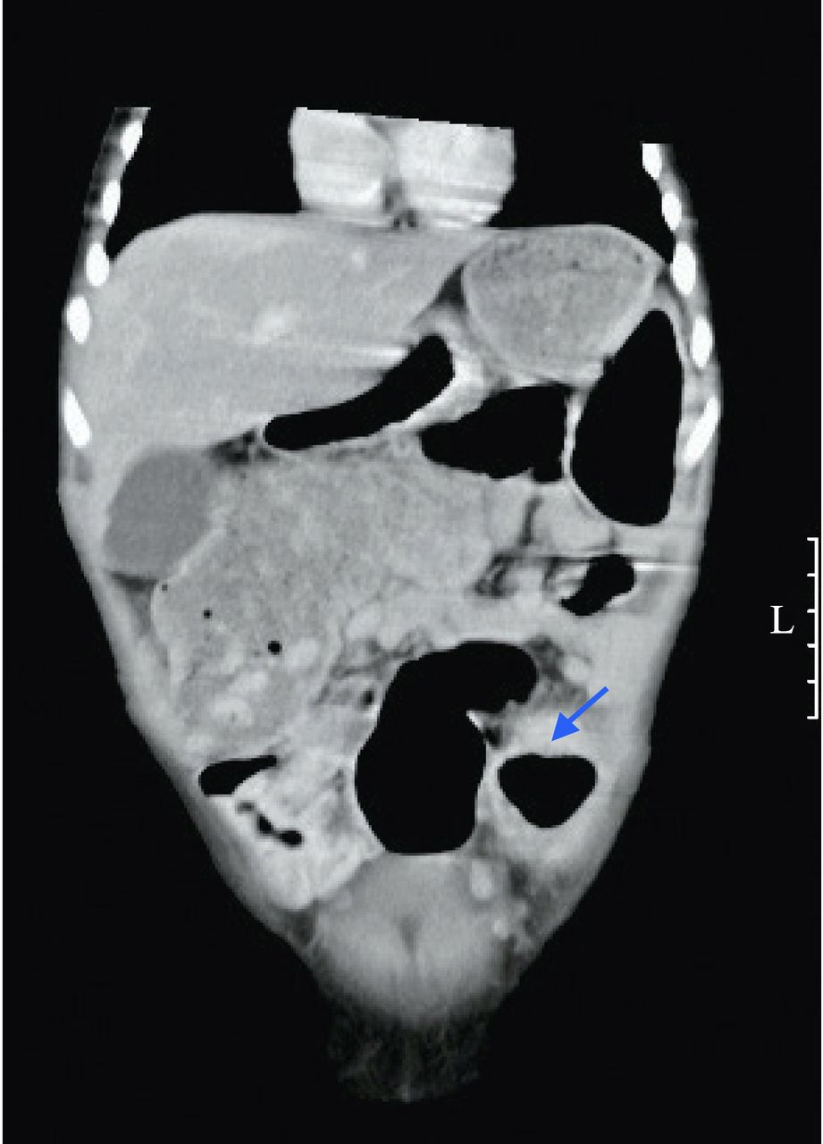
On admission, physical examination of the patient was unremarkable except for a marked abdominal distension. Hematological evaluation was normal. Abdominal CT scan revealed a 2 cm stenotic segment between the descending and sigmoid colons (figure 1). A contrast enema was performed, confirming colonic stenosis (figure 2).

CT scan, after intravenous contrast injection, showing dilated descending colon and stenotic sigmoid colon segment (blue arrow). L, left.
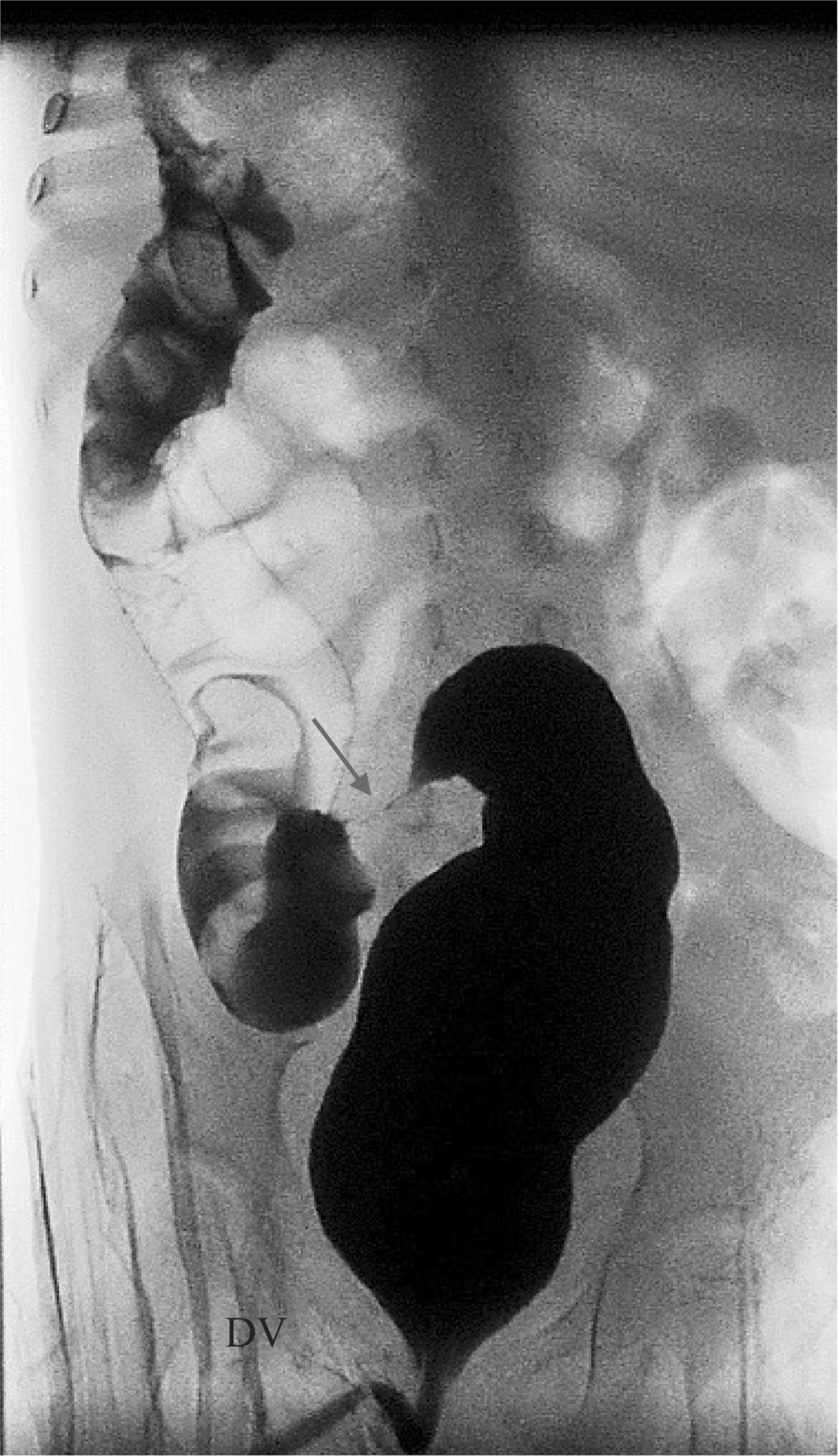
Inverted contrast enema displaying stenotic sigmoid segment (arrow).
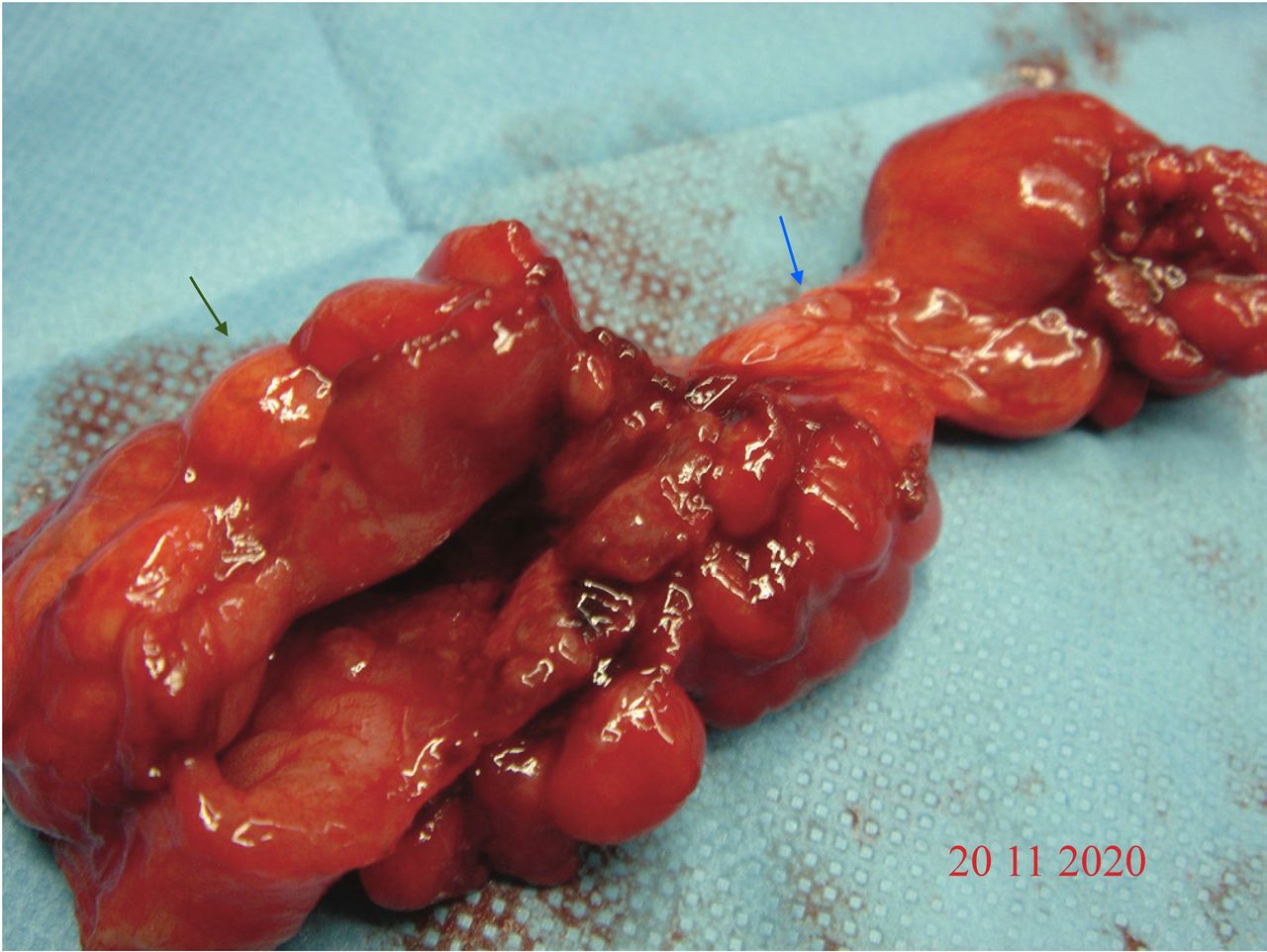
The patient was taken to the operating room and laparotomy was performed by a left paramedian incision. A hard mass was found on the descending colon, with adhesions to the anterior abdominal wall; after release, a 2 cm perforation was noted (figure 3). There were several adhesions between the left bladder wall, the left ovary and the sigmoid colon. A segmental resection of nearly 10 cm of the descending colon, including the perforated and stenotic segments, was performed, followed by manual end-to-end colon anastomosis. Recovery after surgery was uneventful. Histopathology confirmed the presence of a 1.4 cm stenotic segment (lumen diameter between 0.3 and 0.7 cm), with severe inflammation, chronic ischemia and necrosis.

Surgical resected segment showing stenotic and perforated segments (blue and green arrow, respectively).
HUS is a thrombotic microangiopathy characterized by thrombocytopenia, hemolytic anaemia and acute kidney injury. Abdominal complaints are common in the prodromic phase, mimicking intussusception, appendicitis or acute inflammatory bowel disease.3
Extrarenal manifestations of acute HUS are protean and seem to relate to microvascular lesions affecting arterioles and capillaries of target organs, ultimately leading to ischemic damage.4 Although neurological and gastrointestinal (GI) manifestations are considered to be the most common extrarenal manifestations, with GI complications ranging from 13% to 38% of cases, surgical complications are rare and occur most frequently in patients with severe hemorrhagic colitis (HC).4 5 Leukocyte count of >2000/μL and hematocrit of >30% were found to be more common in patients with HC.6 In this case, when the patient was first admitted to the intensive care unit (ICU) for HUS, hemoglobin level was 93 g/L with 21.7% hematocrit and leukocyte count 17840/μL. Although these values were lower than the reported limit, intravenous fluids had been previously administered, which could have acted as a misleading factor. Interestingly, leukocyte levels remained high at first admission for the first 15 days. This patient also tested positive for SARS-CoV-2 40 days before being admitted for stenosis. In retrospect, these might have contributed to the development of the stricture, given that prolonged inflammatory conditions seemed to have played an important role in the development of HUS complications.7
Post-HUS colonic strictures are rare and two occurrence peaks have been identified: the first, 1–2 months after HUS and the second peak at more than 1 year.8 In a recent report, two cases of colonic stricture after STEC-HUS were described, together with a review of the literature on post-HUS GI strictures: there were a total of 22 cases, including those presented by the authors.9 In the same report, the authors proposed that segmental colonic ischemia without necrosis could be a mechanism for subsequent fibrosis, resulting in stricture formation.
In the present case, several proposed risk factors for stricture development were present; female gender, young age and extensive colitis.9 Of note, colitis was a prominent feature in every ultrasound performed in the acute setting, together with high leukocyte counts, which was consistent with a prolonged inflammatory period. Although we do not know whether the perforation occurred in the acute phase or after stenosis development, the histological findings were consistent with a chronic process.
Treatment of STEC-HUS is mainly supportive.2 In the present case, eculizumab treatment was considered at first but was dismissed after Stx identification because it had not been shown to improve outcomes in STEC-HUS.1 Broad-spectrum antibiotics were administered because leukocytosis and colitis were prominent features. Although anti-inflammatory therapy with intravenous immunoglobulin and methylprednisolone pulses has been reported with good results, additional studies are needed to implement this approach.10
This case highlights several important key messages for pediatric surgeons. Although GI involvement is common in the prodromic phase, extensive colitis during acute HUS can result in bowel ischemia and perforation. These complications must be actively sought through serial abdominal examinations and, if necessary, imaging studies.
In patients with severe GI involvement in the acute setting, especially if other risk factors are present (younger age, female gender, high leukocyte count and high hematocrit), the possibility of a late stenosis must be kept in mind. Acute or subacute abdominal occlusion symptoms should prompt the need for further investigation. Contrast studies showing narrowing of a colon segment have diagnostic value; surgical intervention with segmental resection and anastomosis is the treatment option.8
Data availability statement
All data relevant to the study are included in the article or uploaded as supplemental information.
Ethics statements
Ethics approval
Approved by hospital's ethics committee.
Acknowledgments
The authors thank Dr João Pimentel, Dr João Dias, Dr Lúcia Nascimento and nurse Ana Nunes for their cooperation in the review of this article
Footnotes
Contributors SML contributed to conceptualization, data curation, investigation, writing – original draft. LC, PSC and AR contributed to formal analysis, resources, supervision, validation, writing – review & editing. All authors contributed to the article and approved the submitted version.
Funding The authors have not declared a specific grant for this research from any funding agency in the public, commercial or not-for-profit sectors.
Competing interests None declared.
Provenance and peer review Not commissioned; externally peer reviewed.