
Late Menarche, Not Reproductive Period, Is Associated with Poor Cognitive Function in Postmenopausal Women in Taiwan

 ,
,  , and
, and
Abstract
:1. Introduction
2. Materials and Methods
2.1. Ethics Statement
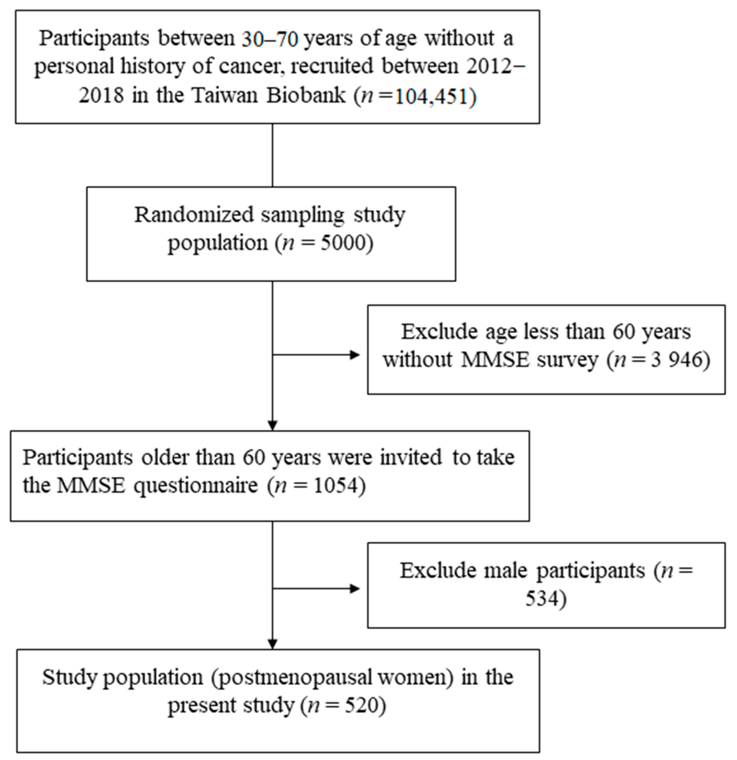
2.2. TWB
2.3. Collection of Demographic, Medical and Laboratory Data
2.4. Assessment of Age at Menarche and Menopause
2.5. Evaluation of Cognitive Function
2.6. Statistical Analysis
3. Results
3.1. Association between MMSE Total Score and SubScores According to Age at Menarche
3.2. Determinants of Total MMSE Score
3.3. Correlations between Age at Menarche and Reproductive Period and Each MMSE Subdomain
4. Discussion
Author Contributions
Funding
Institutional Review Board Statement
Informed Consent Statement
Data Availability Statement
Acknowledgments
Conflicts of Interest
References
- Dalal, P.K.; Agarwal, M. Postmenopausal syndrome. Indian J. Psychiatry 2015, 57 (Suppl. S2), S222. [Google Scholar] [CrossRef]
- The North American Menopause Society. The 2017 hormone therapy position statement of The North American Menopause Society. Menopause 2017, 24, 728–753. [Google Scholar] [CrossRef]
- Fait, T. Menopause hormone therapy: Latest developments and clinical practice. Drugs Context 2019, 8, 1–9. [Google Scholar] [CrossRef]
- De Franciscis, P.; Colacurci, N.; Riemma, G.; Conte, A.; Pittana, E.; Guida, M.; Schiattarella, A. A Nutraceutical Approach to Menopausal Complaints. Medicina 2019, 55, 544. [Google Scholar] [CrossRef] [PubMed] [Green Version]
- De Franciscis, P.; Conte, A.; Schiattarella, A.; Riemma, G.; Cobellis, L.; Colacurci, N. Non-hormonal Treatments For Menopausal Symptoms and Sleep Disturbances: A Comparison Between Purified Pollen Extracts and Soy Isoflavones. Curr. Pharm. Des. 2020, 26, 4509–4514. [Google Scholar] [CrossRef]
- Green, P.S.; Simpkins, J.W. Neuroprotective effects of estrogens: Potential mechanisms of action. Int. J. Dev. Neurosci. 2000, 18, 347–358. [Google Scholar] [CrossRef]
- Yao, J.; Brinton, R.D. Estrogen Regulation of Mitochondrial Bioenergetics; Elsevier: Amsterdam, The Netherlands, 2012; pp. 327–371. [Google Scholar]
- Sherwin, B.B. Estrogen and cognitive functioning in women. Endocr. Rev. 2003, 24, 133–151. [Google Scholar] [CrossRef] [Green Version]
- Henderson, V.W. Menopause and disorders of the central nervous system. Minerva Ginecol. 2005, 57, 579–592. [Google Scholar] [PubMed]
- Maki, P.M. Estrogen effects on the hippocampus and frontal lobes. Int. J. Fertil. Womens Med. 2005, 50, 67–71. [Google Scholar] [PubMed]
- Joffe, H.; Hall, J.E.; Gruber, S.; Sarmiento, I.A.; Cohen, L.S.; Yurgelun-Todd, D.; Martin, K.A. Estrogen therapy selectively enhances prefrontal cognitive processes: A randomized, double-blind, placebo-controlled study with functional magnetic resonance imaging in perimenopausal and recently postmenopausal women. Menopause 2006, 13, 411–422. [Google Scholar] [CrossRef]
- Krug, R.; Born, J.; Rasch, B. A 3-day estrogen treatment improves prefrontal cortex-dependent cognitive function in postmenopausal women. Psychoneuroendocrinology 2006, 31, 965–975. [Google Scholar] [CrossRef]
- Wroolie, T.E.; Kenna, H.A.; Williams, K.E.; Powers, B.N.; Holcomb, M.; Khaylis, A.; Rasgon, N.L. Differences in Verbal Memory Performance in Postmenopausal Women Receiving Hormone Therapy: 17β-Estradiol Versus Conjugated Equine Estrogens. Am. J. Geriatr. Psychiatry 2011, 19, 792–802. [Google Scholar] [CrossRef] [Green Version]
- Sherwin, B.B.; Henry, J.F. Brain aging modulates the neuroprotective effects of estrogen on selective aspects of cognition in women: A critical review. Front. Neuroendocrinol. 2008, 29, 88–113. [Google Scholar] [CrossRef]
- Karim, R.; Dang, H.; Henderson, V.W.; Hodis, H.N.; St. John, J.; Brinton, R.D.; Mack, W.J. Effect of Reproductive History and Exogenous Hormone Use on Cognitive Function in Mid- and Late Life. J. Am. Geriatr. Soc. 2016, 64, 2448–2456. [Google Scholar] [CrossRef] [PubMed] [Green Version]
- Liu, L.; Wang, J.; Zhao, L.; Nilsen, J.; McClure, K.; Wong, K.; Brinton, R.D. Progesterone increases rat neural progenitor cell cycle gene expression and proliferation via extracellularly regulated kinase and progesterone receptor membrane components 1 and 2. Endocrinology 2009, 150, 3186–3196. [Google Scholar] [CrossRef]
- Brinton, R.D. The healthy cell bias of estrogen action: Mitochondrial bioenergetics and neurological implications. Trends Neurosci. 2008, 31, 529–537. [Google Scholar] [CrossRef] [PubMed]
- Brinton, R.D. Estrogen-induced plasticity from cells to circuits: Predictions for cognitive function. Trends Pharmacol. Sci. 2009, 30, 212–222. [Google Scholar] [CrossRef] [PubMed] [Green Version]
- McLay, R.N.; Maki, P.M.; Lyketsos, C.G. Nulliparity and Late Menopause Are Associated With Decreased Cognitive Decline. J. Neuropsychiatry Clin. Neurosci. 2003, 15, 161–167. [Google Scholar] [CrossRef]
- Henderson, V.W. Alzheimer’s disease: Review of hormone therapy trials and implications for treatment and prevention after menopause. J. Steroid Biochem. Mol. Biol. 2014, 142, 99–106. [Google Scholar] [CrossRef] [Green Version]
- Paganini-Hill, A. Estrogen Replacement Therapy and Risk of Alzheimer Disease. Arch. Intern. Med. 1996, 156, 2213. [Google Scholar] [CrossRef] [PubMed]
- Tang, M.-X.; Jacobs, D.; Stern, Y.; Marder, K.; Schofield, P.; Gurland, B.; Andrews, H.; Mayeux, R. Effect of oestrogen during menopause on risk and age at onset of Alzheimer’s disease. Lancet 1996, 348, 429–432. [Google Scholar] [CrossRef]
- Baldereschi, M.; Di Carlo, A.; Lepore, V.; Bracco, L.; Maggi, S.; Grigoletto, F.; Scarlato, G.; Amaducci, L. Estrogen-replacement therapy and Alzheimer’s disease in the Italian Longitudinal Study on Aging. Neurology 1998, 50, 996. [Google Scholar] [CrossRef]
- Roberts, R.O.; Cha, R.H.; Knopman, D.S.; Petersen, R.C.; Rocca, W.A. Postmenopausal Estrogen Therapy and Alzheimer Disease: Overall Negative Findings. Alzheimer Dis. Assoc. Disord. 2019, 53, 12594–12601. [Google Scholar] [CrossRef] [PubMed]
- Shimizu, Y.; Sawada, N.; Iwasaki, M.; Shikimoto, R.; Nozaki, S.; Mimura, M.; Tsugane, S. Reproductive history and risk of cognitive impairment in Japanese women. Maturitas 2019, 128, 22–28. [Google Scholar] [CrossRef]
- Low, L.F.; Anstey, K.J.; Jorm, A.F.; Rodgers, B.; Christensen, H. Reproductive period and cognitive function in a representative sample of naturally postmenopausal women aged 60–64 years. Climacteric 2005, 8, 380–389. [Google Scholar] [CrossRef] [PubMed]
- Georgakis, M.K.; Kalogirou, E.I.; Diamantaras, A.-A.; Daskalopoulou, S.S.; Munro, C.A.; Lyketsos, C.G.; Skalkidou, A.; Petridou, E.T. Age at menopause and duration of reproductive period in association with dementia and cognitive function: A systematic review and meta-analysis. Psychoneuroendocrinology 2016, 73, 224–243. [Google Scholar] [CrossRef]
- Heys, M.; Jiang, C.; Cheng, K.K.; Zhang, W.; Yeung, S.L.A.; Lam, T.H.; Leung, G.M.; Schooling, C.M. Life long endogenous estrogen exposure and later adulthood cognitive function in a population of naturally postmenopausal women from Southern China: The Guangzhou Biobank Cohort Study. Psychoneuroendocrinology 2011, 36, 864–873. [Google Scholar] [CrossRef]
- Paganini-Hill, A.; Corrada, M.M.; Kawas, C.H. Prior endogenous and exogenous estrogen and incident dementia in the 10th decade of life: The 90+ Study. Climacteric 2020, 23, 311–315. [Google Scholar] [CrossRef]
- Matyi, J.M.; Rattinger, G.B.; Schwartz, S.; Buhusi, M.; Tschanz, J.T. Lifetime estrogen exposure and cognition in late life: The Cache County Study. Menopause 2019, 26, 1366–1374. [Google Scholar] [CrossRef] [PubMed]
- Raghava, N.; Das, B.C.; Ray, S.K. Neuroprotective effects of estrogen in CNS injuries: Insights from animal models. Neurosci. Neuroeconomics 2017, 6, 15. [Google Scholar] [CrossRef] [Green Version]
- Lin, J.; Kroenke, C.H.; Epel, E.; Kenna, H.A.; Wolkowitz, O.M.; Blackburn, E.; Rasgon, N.L. Greater endogenous estrogen exposure is associated with longer telomeres in postmenopausal women at risk for cognitive decline. Brain Res. 2011, 1379, 224–231. [Google Scholar] [CrossRef] [PubMed] [Green Version]
- Lin, J.; Epel, E.S.; Blackburn, E.H. Telomeres, telomerase, stress, and aging. In Handbook of Neuroscience for the Behavioral Sciences; John Wiley & Sons Inc.: Hoboken, NJ, USA, 2009. [Google Scholar]
- Rierdan, J.; Koff, E. Age at menarche and cognitive functioning. Bull. Psychon. Soc. 1984, 22, 174–176. [Google Scholar] [CrossRef]
- Gilsanz, P.; Lee, C.; Corrada, M.M.; Kawas, C.H.; Quesenberry, C.P.; Whitmer, R.A. Reproductive period and risk of dementia in a diverse cohort of health care members. Neurology 2019, 92, e2005–e2014. [Google Scholar] [CrossRef]
- Kiliçaslan, E.E.; Erol, A.; Zengin, B.; Aydin, P.Ç.; Mete, L. Şizofreni Başlangıç Yaşı ile Menarş Yaşı Arasındaki İlişki. Nöro Psikiyatr. Arşivi 2014, 211–215. [Google Scholar] [CrossRef]
- Jung, S.J.; Shin, A.; Kang, D. Menarche age, menopause age and other reproductive factors in association with post-menopausal onset depression: Results from Health Examinees Study(HEXA). J. Affect. Disord. 2015, 187, 127–135. [Google Scholar] [CrossRef] [Green Version]
- Rao, M.L.; Kölsch, H. Effects of estrogen on brain development and neuroprotection—implications for negative symptoms in schizophrenia11Part of this review was presented at the Seventh International Congress of Biological Psychiatry in Berlin, Germany, 1–7 July 2001. Psychoneuroendocrinology 2003, 28, 83–96. [Google Scholar] [CrossRef]
- Folstein, M.F.; Folstein, S.E.; McHugh, P.R. “Mini-mental state”. A practical method for grading the cognitive state of patients for the clinician. J. Psychiatr. Res. 1975, 12, 189–198. [Google Scholar] [CrossRef]
- Chen, C.H.; Yang, J.H.; Chiang, C.W.K.; Hsiung, C.N.; Wu, P.E.; Chang, L.C.; Chu, H.W.; Chang, J.; Song, I.W.; Yang, S.L.; et al. Population structure of Han Chinese in the modern Taiwanese population based on 10,000 participants in the Taiwan Biobank project. Hum. Mol. Genet. 2016, 25, 5321–5331. [Google Scholar] [CrossRef] [Green Version]
- Fan, C.T.; Hung, T.H.; Yeh, C.K. Taiwan Regulation of Biobanks. J. Law Med. Ethics J. Am. Soc. Law Med. Ethics 2015, 43, 816–826. [Google Scholar]
- Directorate General of Budget, Accounting and Statistics, Executive Yuan, Republic of China (Ed.) SOCIAL INDICATORS 2010; Directorate General of Budget, Accounting and Statistics of Executive Yuan: Taipei City, Taiwan, 2011. [Google Scholar]
- Levey, A.S.; Bosch, J.P.; Lewis, J.B.; Greene, T.; Rogers, N.; Roth, D. A more accurate method to estimate glomerular filtration rate from serum creatinine: A new prediction equation. Modification of Diet in Renal Disease Study Group. Ann. Intern. Med. 1999, 130, 461–470. [Google Scholar] [CrossRef]
- Piekarski, D.J.; Boivin, J.R.; Wilbrecht, L. Ovarian Hormones Organize the Maturation of Inhibitory Neurotransmission in the Frontal Cortex at Puberty Onset in Female Mice. Curr. Biol. 2017, 27, 1735–1745.e3. [Google Scholar] [CrossRef] [Green Version]
- Almey, A.; Milner, T.A.; Brake, W.G. Estrogen receptors in the central nervous system and their implication for dopamine-dependent cognition in females. Horm. Behav. 2015, 74, 125–138. [Google Scholar] [CrossRef] [Green Version]
- Alexander, A.; Irving, A.J.; Harvey, J. Emerging roles for the novel estrogen-sensing receptor GPER1 in the CNS. Neuropharmacology 2017, 113, 652–660. [Google Scholar] [CrossRef] [Green Version]
- Yager, L.M.; Garcia, A.F.; Wunsch, A.M.; Ferguson, S.M. The ins and outs of the striatum: Role in drug addiction. Neuroscience 2015, 301, 529–541. [Google Scholar] [CrossRef] [PubMed] [Green Version]
- Uytun, M.C. Development period of prefrontal cortex. In Prefrontal Cortex; IntechOpen: Rijeka, Croatia, 2018. [Google Scholar] [CrossRef]
- Koppitz, E.M. Diagnosing brain damage in young children with the Bender Gestalt test. J. Consult. Psychol. 1962, 26, 541. [Google Scholar] [CrossRef] [PubMed]
- Tseng, W.-J.; Hung, L.-W.; Lin, J. Time Orientation and Visual Construction Subdomains of the MMSE as Independent Risk Factors for Hip Fractures. Orthopedics 2013, 36, e869–e876. [Google Scholar] [CrossRef] [Green Version]
- Peper, J.S.; Brouwer, R.M.; Schnack, H.G.; van Baal, G.C.; van Leeuwen, M.; van den Berg, S.M.; Delemarre-Van de Waal, H.A.; Boomsma, D.I.; Kahn, R.S.; Pol, H.E.H. Sex steroids and brain structure in pubertal boys and girls. Psychoneuroendocrinology 2009, 34, 332–342. [Google Scholar] [CrossRef] [Green Version]
- Lafer-Sousa, R.; Conway, B.R. Parallel, multi-stage processing of colors, faces and shapes in macaque inferior temporal cortex. Nat. Neurosci. 2013, 16, 1870–1878. [Google Scholar] [CrossRef] [Green Version]
- Szymczak, J.; Milewicz, A.; Thijssen, J.H.H.; Blankenstein, M.A.; Daroszewski, J. Concentration of sex steroids in adipose tissue after menopause. Steroids 1998, 63, 319–321. [Google Scholar] [CrossRef]
- Nelson, L.R.; Bulun, S.E. Estrogen production and action. J. Am. Acad. Dermatol. 2001, 45 (Suppl. S3), S116–S124. [Google Scholar] [CrossRef] [PubMed]
- Prickett, C.; Brennan, L.; Stolwyk, R. Examining the relationship between obesity and cognitive function: A systematic literature review. Obes. Res. Clin. Pract. 2015, 9, 93–113. [Google Scholar] [CrossRef]
- Newman, A.B.; Fitzpatrick, A.L.; Lopez, O.; Jackson, S.; Lyketsos, C.; Jagust, W.; Ives, D.; DeKosky, S.T.; Kuller, L.H. Dementia and Alzheimer’s disease incidence in relationship to cardiovascular disease in the Cardiovascular Health Study cohort. J. Am. Geriatr. Soc. 2005, 53, 1101–1107. [Google Scholar] [CrossRef] [PubMed]
- Fox, M.; Berzuini, C.; Knapp, L.A. Cumulative estrogen exposure, number of menstrual cycles, and Alzheimer’s risk in a cohort of British women. Psychoneuroendocrinology 2013, 38, 2973–2982. [Google Scholar] [CrossRef]
- Tombaugh, T.N.; McIntyre, N.J. The Mini-Mental State Examination: A Comprehensive Review. J. Am. Geriatr. Soc. 1992, 40, 922–935. [Google Scholar] [CrossRef]


| Characteristics | All (n = 520) | MMSE ≥ 24 (n = 445) | MMSE < 24 (n = 75) | p |
|---|---|---|---|---|
| Age (year) | 63.7 ± 2.9 | 63.6 ± 2.9 | 64.3 ± 3.0 | 0.046 |
| Smoking history (%) | 3.7 | 3.8 | 2.7 | 1.000 |
| Alcohol history (%) | 0.4 | 0.4 | 0 | 1.000 |
| DM (%) | 16.3 | 16.4 | 16.0 | 0.930 |
| Hypertension (%) | 20.8 | 20.2 | 24.0 | 0.456 |
| Coronary artery disease (%) | 2.5 | 2.5 | 2.7 | 1.000 |
| Cerebrovascular disease (%) | 0.8 | 0.7 | 1.3 | 0.465 |
| Education higher than senior high schools (%) | 56.0 | 62.7 | 16.0 | <0.001 |
| Living alone (%) | 14.6 | 15.5 | 9.3 | 0.162 |
| Having job (%) | 18.8 | 17.4 | 27.5 | 0.046 |
| Regular exercise habits (%) | 68.1 | 67.4 | 72.0 | 0.431 |
| Midnight snack habits (%) | 16.7 | 16.9 | 16.0 | 0.855 |
| BMI (kg/m2) | 24.0 ± 3.3 | 23.9 ± 3.2 | 24.8 ± 3.6 | 0.016 |
| SBP (mmHg) | 124.2 ± 17.7 | 123.8 ± 17.6 | 126.7 ± 18.3 | 0.190 |
| DBP (mmHg) | 70.0 ± 10.3 | 69.9 ± 10.5 | 70.2 ± 9.2 | 0.846 |
| Laboratory parameters | ||||
| Fasting glucose (mg/dL) | 99.7 ± 22.0 | 99.3 ± 20.8 | 102.0 ± 27.8 | 0.327 |
| Triglyceride (mg/dL) | 100 (76–133) | 99 (75–133) | 102 (79–134) | 0.967 |
| Total cholesterol (mg/dL) | 210.0 ± 36.4 | 211.0 ± 63.7 | 204.2 ± 34.5 | 0.133 |
| HDL-cholesterol (mg/dL) | 58.3 ± 13.4 | 58.2 ± 13.4 | 58.8 ± 13.4 | 0.743 |
| LDL-cholesterol (mg/dL) | 130.1 ± 33.4 | 130.9 ± 33.8 | 124.9 ± 30.6 | 0.148 |
| Hemoglobin (g/dL) | 13.3 ± 1.0 | 13.3 ± 1.0 | 13.4 ± 1.1 | 0.365 |
| eGFR (mL/min/1.73 m2) | 107.6 ± 24.9 | 107.8 ± 24.9 | 106.1 ± 25.1 | 0.589 |
| Uric acid (mg/dL) | 5.2 ± 1.2 | 5.1 ± 1.2 | 5.2 ± 1.2 | 0.523 |
| Characteristics | All (n = 520) | MMSE ≥ 24 (n = 445) | MMSE < 24 (n = 75) | p |
|---|---|---|---|---|
| MMSE | ||||
| G1 (Orientation) | 9.4 ± 0.9 | 9.6 ± 0.7 | 8.5 ± 1.3 | <0.001 |
| G2 (Registration) | 2.9 ± 0.3 | 3.0 ± 0.3 | 2.8 ± 0.5 | 0.076 |
| G3 (Attention & Calculation) | 3.6 ± 1.8 | 3.9 ± 1.6 | 1.3 ± 1.1 | <0.001 |
| G4 (Recall) | 2.2 ± 0.9 | 2.4 ± 0.8 | 1.3 ± 1.1 | <0.001 |
| G5 (Language, construction & obey) | 8.4 ± 0.9 | 8.6 ± 0.7 | 7.2 ± 1.3 | <0.001 |
| MMSE total | 26.5 ± 2.9 | 27.4 ± 2.0 | 21.2 ± 1.9 | <0.001 |
| Menstruation related conditions | ||||
| Age of menarche (year) | 14.3 ± 1.6 | 14.2 ± 1.5 | 15.1 ± 1.9 | <0.001 |
| Reproductive period (years) | 36.0 ± 5.2 | 36.2 ± 5.2 | 35.0 ± 5.4 | 0.070 |
| Irregular menstrual cycle (%) | 14.3 | 14.8 | 10.8 | 0.360 |
| Birth history (%) | 99.4 | 99.5 | 98.6 | 0.373 |
| Birth times | 2.7 ± 1.0 | 2.7 ± 1.0 | 3.1 ± 1.1 | 0.005 |
| Breast feeding history (%) | 75.3 | 83.1 | 74.0 | 0.100 |
| Breastfeeding period (months) | 16.2 ± 19.6 | 15.0 ± 19.5 | 23.5 ± 18.5 | 0.001 |
| Hormone therapy history (%) | 34.9 | 35.4 | 32.0 | 0.563 |
| Contraceptive use history (%) | 3.1 | 3.4 | 1.3 | 0.489 |
| Contraceptive use period (years) | 4.5 ± 1.2 | 4.3 ± 6.5 | 6.0 ± 0.0 | 0.809 |
| Menarche Age (Year) | ≦12 (n = 54) | 13 (n = 104) | 14 (n = 162) | 15 (n = 82) | 16 (n = 68) | ≧17 (n = 50) | p | |
|---|---|---|---|---|---|---|---|---|
| MMSE | ||||||||
| G1 (Orientation) | 9.5 ± 0.8 | 9.4 ± 0.8 | 9.6 ± 0.7 | 9.5 ± 0.9 | 9.3 ± 1.1 | 9.1 ± 1.1 | 0.011 | |
| G2 (Registration) | 3.0 ± 0.0 | 3.0 ± 0.3 | 2.9 ± 0.3 | 2.9 ± 0.4 | 2.9 ± 0.4 | 2.8 ± 0.5 | 0.110 | |
| G3 (Attention &Calculation) | 3.6 ± 1.7 | 3.7 ± 1.8 | 3.8 ± 1.7 | 3.5 ± 1.7 | 2.9 ± 1.9 | 3.4 ± 2.0 | 0.030 | |
| G4 (Recall) | 2.3 ± 0.7 | 2.3 ± 0.9 | 2.3 ± 0.9 | 2.2 ± 0.9 | 2.3 ± 1.0 | 1.9 ± 1.0 | 0.190 | |
| G5 (Language, construction & obey) | 8.5 ± 0.8 | 8.6 ± 0.7 | 8.3 ± 1.0 | 8.4 ± 1.0 | 8.2 ± 1.1 | 8.1 ± 1.0 | 0.010 | |
| MMSE total | 26.9 ± 2.5 | 26.9 ± 2.7 | 26.9 ± 2.6 | 26.6 ± 3.0 | 25.6 ± 3.2 | 25.3 ± 3.8 | 0.001 | |
| Characteristics | Univariable | Multivariable | ||
|---|---|---|---|---|
| Coefficient β (95% CI) | p | Coefficient β (95% CI) | p | |
| Age (per one year) | −0.151 (−0.238, −0.064) | 0.001 | −0.040 (−0.127, 0.047) | 0.371 |
| Smoking history (ever vs. never) | 0.105 (−1.246, 1.455) | 0.879 | - | - |
| Alcohol history (ever vs. never) | −0.533 (−4.626, 3.561) | 0.798 | - | - |
| DM | −0.255 (−0.940, 0.430) | 0.465 | - | - |
| Hypertension | −0.483 (−1.106, 0.140) | 0.129 | - | - |
| Coronary artery disease | −0.229 (−1.852, 1.394) | 0.782 | - | - |
| Cerebrovascular disease | −3.306 (−6.193, −0.420) | 0.025 | −2.013 (−4.617, 0.591) | 0.129 |
| Education higher than senior high schools | 2.252 (1.780, 2.724) | <0.001 | 1.591 (1.052, 2.130) | <0.001 |
| Living alone | 0.750 (0.035, 1.464) | 0.040 | 0.585 (−0.122, 1.292) | 0.105 |
| Having job | −0.335 (−0.983, 0.314) | 0.311 | - | - |
| Regular exercise habits | −0.326 (−0.869, 0.216) | 0.238 | - | - |
| Midnight snack habits | 0.053 (−0.626, 0.732) | 0.879 | - | - |
| BMI (per 1 kg/m2) | −0.166 (−0.242, −0.089) | <0.001 | −0.103 (−0.183, −0.023) | 0.011 |
| SBP (per 1 mmHg) | −0.007 (−0.021, 0.008) | 0.350 | - | - |
| DBP (per 1 mmHg) | 0.002 (−0.023, 0.026) | 0.889 | - | - |
| Laboratory parameters | ||||
| Fasting glucose (per 1 mg/dL) | −0.011 (−0.022, 0.001) | 0.066 | - | - |
| Triglyceride (log per 1mg/dL) | −0.718 (−2.025, 0.589) | 0.281 | - | - |
| Total cholesterol (per 1 mg/dL) | 0.007 (0, 0.014) | 0.037 | −0.005 (−0.020, 0.009) | 0.448 |
| HDL-cholesterol (per 1 mg/dL) | 0.003 (−0.016, 0.022) | 0.759 | - | - |
| LDL-cholesterol (per 1 mg/dL) | 0.008 (0.001, 0.016) | 0.030 | 0.013 (−0.002, 0.028) | 0.094 |
| Hemoglobin (per 1 g/dL) | −0.085 (−0.328, 0.159) | 0.496 | - | - |
| eGFR (per 1 mL/min/1.73 m2) | 0.002 (−0.008, 0.012) | 0.741 | - | - |
| Uric acid (per 1 mg/dL) | −0.159 (−0.366, 0.048) | 0.131 | - | - |
| Menstruation related conditions | ||||
| Age of menarche (per one year) | −0.347 (−0.501, −0.192) | <0.001 | −0.189 (−0.348, −0.030) | 0.020 |
| Reproductive period (per one year) | 0.045 (−0.004, 0.094) | 0.069 | 0.008 (−0.040, 0.056) | 0.733 |
| Menstrual cycle (irregular vs. regular) | 0.193 (−0.524, 0.910) | 0.598 | - | - |
| Birth history | 0.170 (−3.161, 3.500) | 0.920 | - | - |
| Birth times (per one time) | −0.667 (−0.922, −0.412) | <0.001 | −0.121 (−0.404, 0.162) | 0.402 |
| Breastfeeding period (per one month) | −0.035 (−0.048, −0.023) | <0.001 | −0.007 (−0.022, 0.099) | 0.398 |
| Hormone therapy history | 0.037 (−0.496, 0.570) | 0.893 | - | - |
| Contraceptive use history | 0.041 (−1.428, 1.510) | 0.957 | - | - |
| Contraceptive use period (per one year) | −0.131 (−0.377, 0.115) | 0.269 | - | - |
| Characteristics | Multivariable | |
|---|---|---|
| Coefficient β (95% CI) | p | |
| G1 (Orientation) | ||
| Age of menarche (per one year) | −0.028 (−0.078, 0.023) | 0.281 |
| Reproductive period (per one year) | 0.006 (−0.009, 0.022) | 0.403 |
| G2 (Registration) | ||
| Age of menarche (per one year) | −0.022 (−0.042, −0.002) | 0.035 |
| Reproductive period (per one year) | 0 (−0.006, 0.007) | 0.904 |
| G3 (Attention &Calculation) | ||
| Age of menarche (per one year) | −0.073 (−0.178, 0.032) | 0.174 |
| Reproductive period (per one year) | 0.001 (−0.030, 0.033) | 0.937 |
| G4 (Recall) | ||
| Age of menarche (per one year) | −0.013 (−0.066, 0.040) | 0.637 |
| Reproductive period (per one year) | −0.003 (−0.019, 0.013) | 0.693 |
| G5 (Language, construction and obey) | ||
| Age of menarche (per one year) | −0.054 (−0.107, 0) | 0.047 |
| Reproductive period (per one year) | 0.003 (−0.013, 0.020) | 0.674 |
Publisher’s Note: MDPI stays neutral with regard to jurisdictional claims in published maps and institutional affiliations. |
© 2021 by the authors. Licensee MDPI, Basel, Switzerland. This article is an open access article distributed under the terms and conditions of the Creative Commons Attribution (CC BY) license (http://creativecommons.org/licenses/by/4.0/).
Share and Cite
Chou, H.-T.; Wu, P.-Y.; Huang, J.-C.; Chen, S.-C.; Ho, W.-Y. Late Menarche, Not Reproductive Period, Is Associated with Poor Cognitive Function in Postmenopausal Women in Taiwan. Int. J. Environ. Res. Public Health 2021, 18, 2345. https://doi.org/10.3390/ijerph18052345
Chou H-T, Wu P-Y, Huang J-C, Chen S-C, Ho W-Y. Late Menarche, Not Reproductive Period, Is Associated with Poor Cognitive Function in Postmenopausal Women in Taiwan. International Journal of Environmental Research and Public Health. 2021; 18(5):2345. https://doi.org/10.3390/ijerph18052345
Chicago/Turabian StyleChou, Hung-Tse, Pei-Yu Wu, Jiun-Chi Huang, Szu-Chia Chen, and Wan-Yi Ho. 2021. "Late Menarche, Not Reproductive Period, Is Associated with Poor Cognitive Function in Postmenopausal Women in Taiwan" International Journal of Environmental Research and Public Health 18, no. 5: 2345. https://doi.org/10.3390/ijerph18052345