Article

In Planta Analysis of the Radial Movement of Minerals from Inside to Outside in the Trunks of Standing Japanese Cedar (Cryptomeria japonica D. Don) Trees at the Cellular Level

Forestry and Forest Products Research Institute, 1 Matsunosato, Tsukuba, Ibaraki 305-8687, Japan
*
Author to whom correspondence should be addressed.
Forests 2021, 12(2), 251; https://doi.org/10.3390/f12020251
Submission received: 13 January 2021 / Revised: 17 February 2021 / Accepted: 18 February 2021 / Published: 22 February 2021
(This article belongs to the Section Forest Ecophysiology and Biology)

Abstract

:
Although the radial movement of minerals in tree trunks is a widely accepted phenomenon, experimental evidence of their movement in standing trees and underlying mechanisms is very limited. Previously, we clarified that cesium (Cs) artificially injected into the outer part of the sapwood of standing Japanese cedar (Cryptomeria japonica D. Don) trunks moved to the inner part of the sapwood, including the intermediate wood, via active transport by xylem parenchyma cells and diffusion through cell walls and then moved into the heartwood by diffusion. To understand the mechanism underlying the radial movement of minerals in the standing tree trunk, it is necessary to clarify their movement in the opposite direction. Therefore, the present study aimed to determine the radial movement of minerals from inside to outside in the trunks of standing trees at the cellular level. For this, a long hole across the center part of the trunk, which reached the heartwood, intermediate wood, and sapwood, was made in standing Japanese cedar trunks, and a solution of stable isotope Cs was continuously injected into the hole for several days as a tracer. The injected part of the trunk was collected after being freeze-fixed with liquid nitrogen, and the frozen sample was subjected to analysis of Cs distribution at the cellular level using cryo-scanning electron microscopy/energy-dispersive X-ray spectroscopy. The Cs injected into the inner sapwood or intermediate wood rapidly moved toward the outer sapwood via xylem ray parenchyma cells together with diffusion through the cell walls. In contrast, the Cs injected into the heartwood barely moved to the sapwood, although it reached a part of the inner intermediate wood. These results suggest that minerals in xylem ray parenchyma cells in the sapwood are bidirectionally supplied to each other; however, the minerals accumulated in the heartwood may not be supplied to living cells.

1. Introduction

Minerals are absolutely necessary for tree growth and are generally absorbed by the roots along with water from the soil and transported throughout the tree [1]. This transport is thought to occur via the combination of the apoplasmic system through the sap solution in the longitudinal direction and the symplasmic system using living cells in the radial direction [2,3,4,5]. The longitudinal movement of the sap solution has long been studied [6,7,8,9,10]; however, there are few studies on long-distance radial movements in tree trunks, although short-distance radical movements have been experimentally shown using tomato [11], Eucalyptus saligna stem [12], and tree branches [13,14]. Okada et al. [15,16] showed that rubidium injected into the sapwood of Japanese cedar trunk was detected in the heartwood. They concluded that the movement was caused by the function of ray parenchyma cells; however, no experiments have been conducted to provide direct evidence of radial movement via parenchyma cells. Because there is a network of parenchyma cells in the xylem [5,17], it is natural to imagine that the minerals move in a radial direction in the trunk using this network. Nevertheless, this has not been experimentally proven because it is difficult to directly analyze the phenomena occurring inside a large trunk. Therefore, we proposed a novel experimental method to directly analyze minerals inside standing trees to elucidate the radial movement of minerals in the trunk [18,19]. In previous studies, we found that, when the stable isotope Cs was injected into the outer part of the sapwood in the trunks of standing Japanese cedar as a tracer, it moved to the inner part of the sapwood, including the intermediate wood, via rapid active transport by xylem parenchyma cells and diffusion through the cell walls of parenchyma cells and tracheids, and then moved to the heartwood via diffusion [19]. On the other hand, to elucidate the entire mechanism underlying mineral movement in standing tree trunks with heartwood, it is necessary to clarify mineral movement from inside to outside in trunks, a question that remains unelucidated in previous studies.
Regarding mineral movement from the inner to the outer parts of the trunk, minerals should move via diffusion and active transport by xylem parenchyma cells if this movement occurs via a mechanism similar to that from the outer to inner trunk [18,19]. The xylem parenchyma cells are known to store nutrients in winter to form new cells in spring [20,21]. For this purpose, accumulated nutrients must be supplied to the cambium and/or differentiating cells. To provide such a supply, it is necessary to transfer nutrients from the storage compartments (inner part) to the outer part. The transport of chemicals from parenchyma cells to the phloem in plants has been reported in review papers [22,23]. In tree species, however, studies on chemical movement from the sapwood to the cambium are very limited [13,14]. Trees must have the function of mineral movement from the inner sapwood to the outer parts via parenchyma cells. Therefore, proof of this movement is essential for a complete understanding of mineral transport in standing trees.
The movement of minerals from the heartwood to the sapwood has also not been clarified. The formation of heartwood is a phenomenon peculiar to trees. Further, the formation of large heartwood with high decay resistance is important for tree stability [24,25,26]. One of the important features of heartwood is its accumulation of minerals. The elements in tree trunks have been investigated in many trees [27,28,29,30,31]. Okada et al. [27,28] classified the radial pattern of the mineral distribution of trees into three types. In the first, the concentration in the heartwood is higher than that in the sapwood; in the second, the concentration in the sapwood is higher than that in the heartwood; in the third, there is a peak in the concentration in the boundary between the heartwood and sapwood. Among the 8 softwood and 21 hardwood species studied, the pattern was non-uniform among elements or species. For example, potassium concentration in the heartwood of Japanese cedar was higher than that in the sapwood, whereas an opposite pattern was observed for manganese concentration in the same species as well as for the same elements in other species, such as Japanese larch. This kind of non-uniformity was also found in the lists of other articles [29,30,31]. What causes these differences among species and elements? If mineral movement from the heartwood to the sapwood occurred only via diffusion, the minerals would move to the sapwood whenever mineral concentration in the heartwood was higher than that in the sapwood. In this case, the tree trunk might have a mechanism to keep accumulated minerals in the heartwood. On the other hand, if the minerals in the heartwood were actively transported to the sapwood, trees might strategically use the heartwood for mineral storage to reuse minerals in the sapwood. This would be a new discovery and is worth exploring.
The aim of this study was to elucidate the radial movement of minerals from inside to outside in trunks of standing trees. For cellular-level analysis, we used a previously developed method with modifications in the injection protocol, which allowed direct analysis of mineral movement in freeze-fixed trunks to reflect the standing tree state as much as possible [18,19]. Then we examined radial mineral movement from the inner sapwood to the outer sapwood and that from the heartwood to the sapwood to understand the mechanism underlying mineral movement in the trunks of standing trees with developed heartwood.

2. Materials and Methods

2.1. Plant Materials

Japanese cedar trees, which had developed heartwood planted at the Chiyoda nursery of the Forestry and Forest Products Research Institute (Kasumigaura, Ibaraki, Japan), were used in this study (Table 1).
The intermediate wood was defined as the part inside the sapwood that developed a white color under frozen conditions [18,19]. The intermediate wood and the heartwood were also distinguished using cellular-level water distribution by cryo-scanning electron microscopy (cryo-SEM) observation [32].

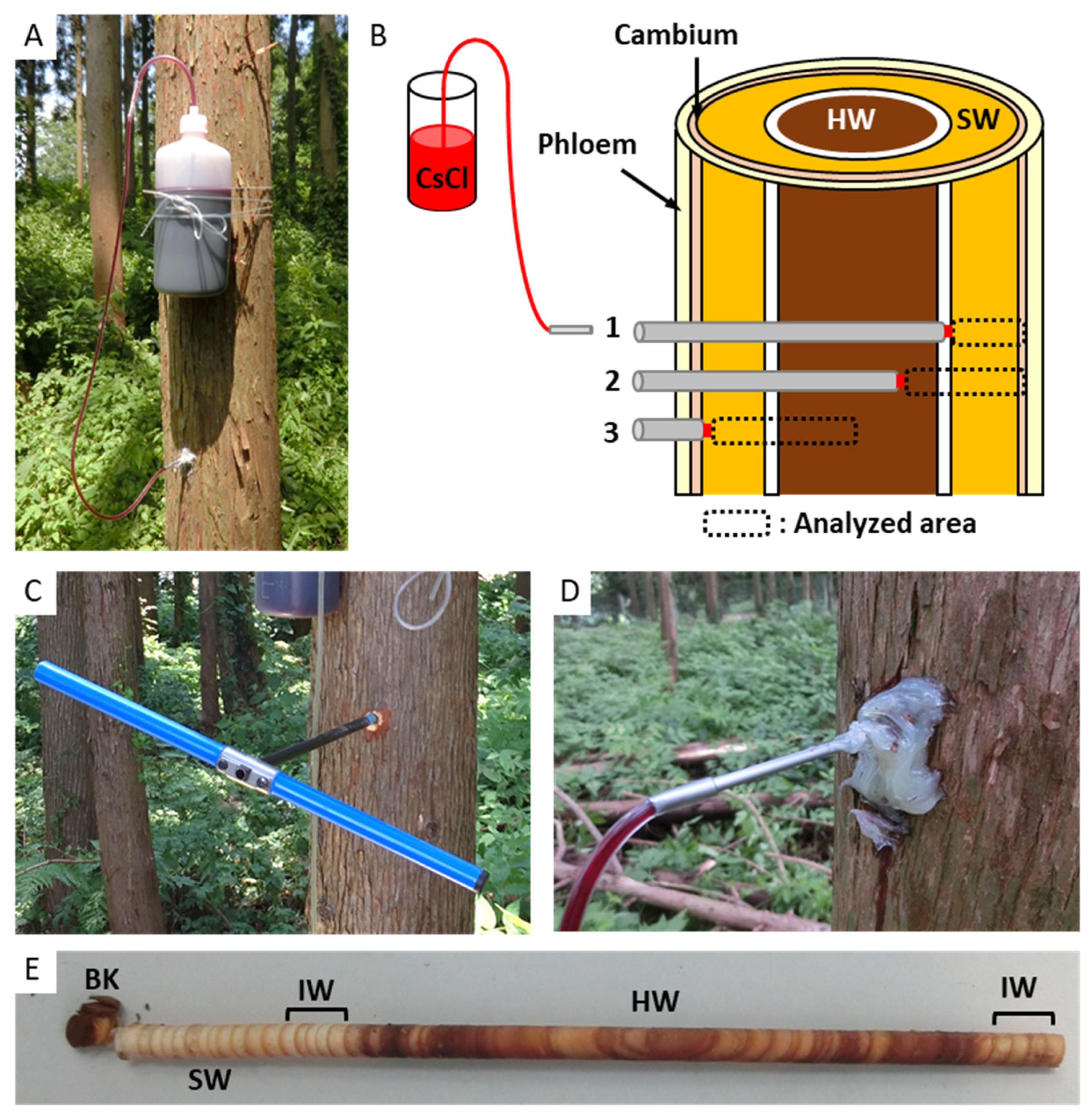
2.2. Cs Injection in Standing Tree Trunk and Sample Collection

Cs injection and sample collection were performed according to Kuroda et al. [18,19] with modifications. A cesium chloride solution (final concentration, 1.0 M; CsCl, FUJIFILM Wako Pure Chemical, Osaka, Japan) was prepared in aqueous acid fuchsin (final concentration, 0.1% w/v; Nacalai Tesque Inc., Kyoto, Japan). Acid fuchsin was used to readily identify the solution movement area in the trunk.
To trace the Cs from the inner trunk to the outer part, a long hole was made in each tree approximately 1.2 m above the ground using an increment borer (3-threaded borer with 5.15-mm diameter, Haglöf Sweden AB; Långsele, Västernorrland, Sweden) to reach the xylem of the opposite side across the center part (near the pith) of the trunk (Figure 1). The depth of the hole was identified after cutting the tree trunk (Figure 2). A 1000 mL polyethylene bottle filled with Cs solution was set on the trunk of each sample tree approximately 20 cm above the drilled hole. The solution was injected into the hole through a plastic tube (Tygon LMT-55; Saint-Gobain, Tokyo, Japan) with a stainless steel tube (20 cm long, 2.84 mm ⌀) attached to the end. The hole was filled with a silicone sealant to prevent leakage of the solution. The solution was continuously injected for a set period (Table 1), with the termination of injection <20 min before the trunk was frozen.
Following injection, the trunk part was frozen with liquid nitrogen (LN2) for approximately 30 min, and the tree was felled [18,19]. Serial disks (1-cm-thick cross-sections) were cut from the frozen part of the trunk, immediately immersed in LN2, and stored at −80 °C in a deep freezer.

2.3. Cryo-SEM/Energy-Dispersive X-ray Spectroscopy (EDX)

Sample preparation and cryo-SEM/EDX analysis were performed according to Kuroda et al. [18,19]. Small blocks, approximately 3 mm × 3 mm × 10 mm, were made from the frozen disks obtained from 1–3 cm above the Cs injection hole, and the fresh transverse surfaces of these disks were smoothly cut using a cryostat (Cryostar NX70; Thermofisher Scientific, Tokyo, Japan) at approximately −30 °C. The observations and analyses were performed using the cryo-SEM/EDX system with the Analysis Station software ver. 3.8 (JSM6510A, JED-2300; JEOL, Tokyo, Japan). Secondary electron (SE) images were obtained at acceleration voltages of 3 and 15 kV with gold coating in the cryo-SEM/EDX system. For point analysis, several points of the lumen of tracheid (TL), which was filled with water, cell wall between tracheids (TW), lumen of ray parenchyma cell (PL), and cell wall between ray parenchyma cells (PW) were selected on each SE image, and characteristic X-rays were collected for 300 s at an acceleration voltage of 15 kV. The Cs-Lα peak (4.286 keV) at each point was defined in terms of the presence of Cs.

3. Results

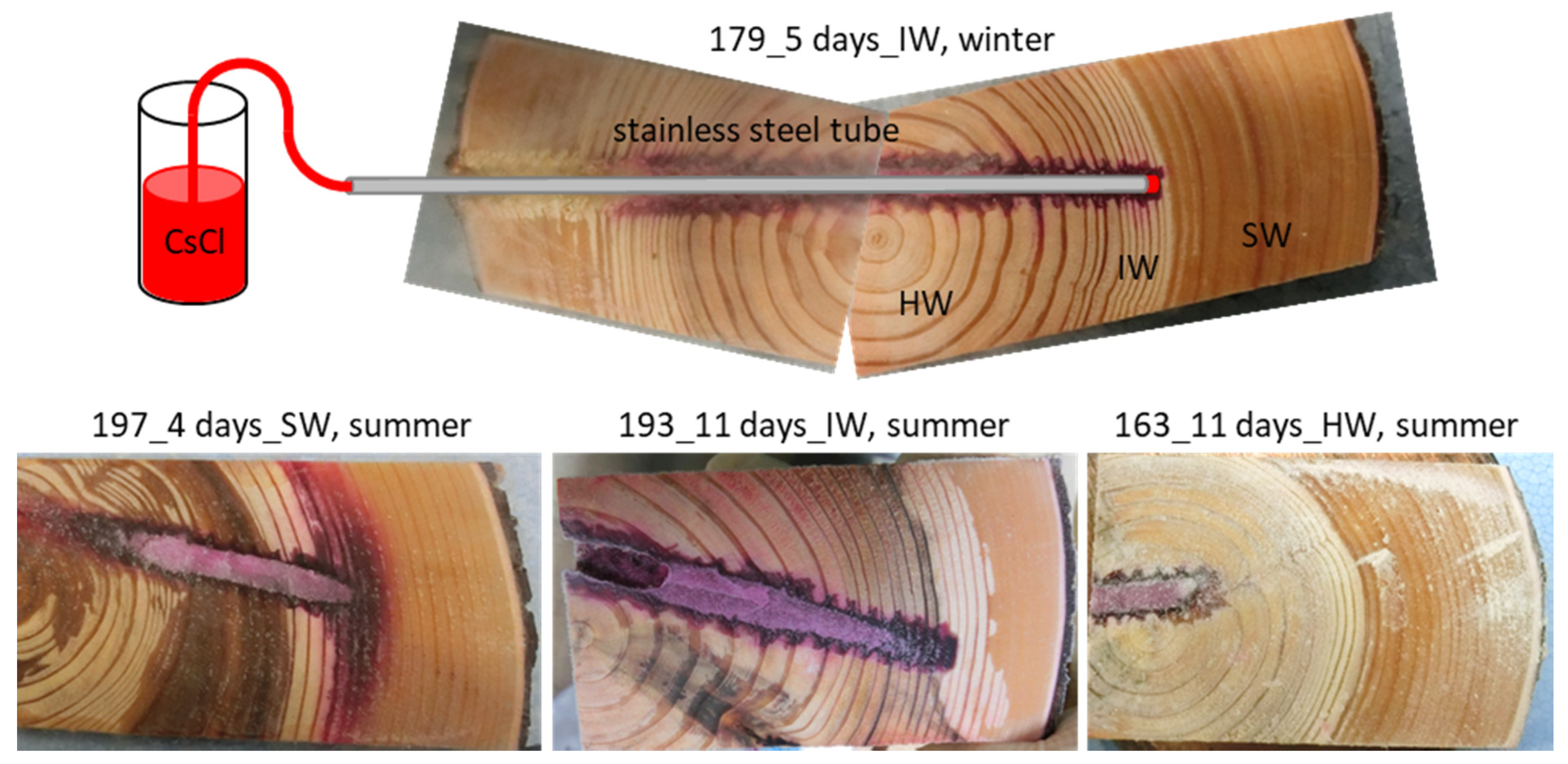
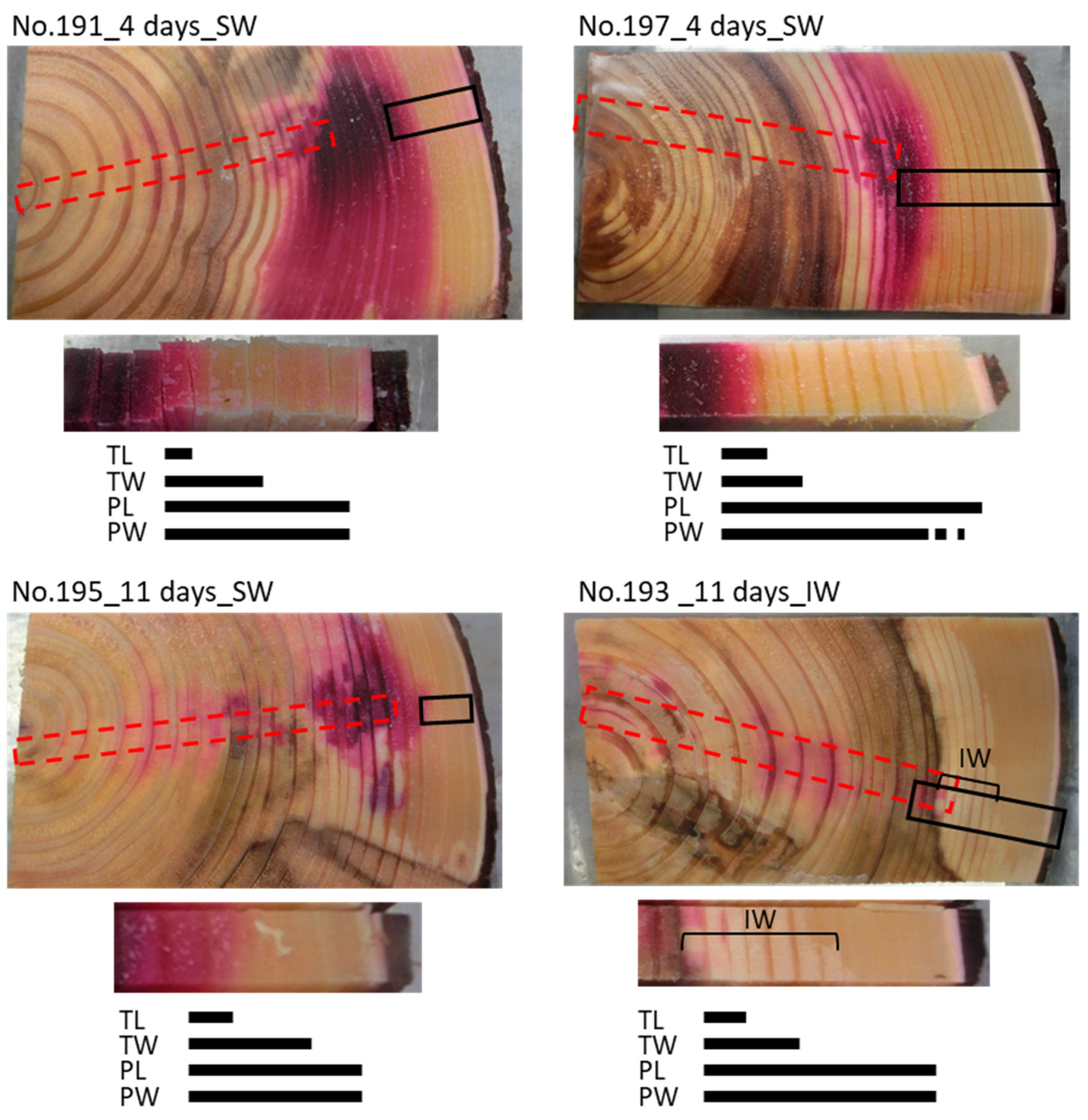
3.1. Cs Distribution in the Xylem Following Cs Injection into the Sapwood and Intermediate Wood in Summer

The Cs solution injected into the hole reached the inner sapwood or the outer part of intermediate wood at the opposite side from the injection. Outward Cs movement in the sapwood was investigated using cryo-SEM/EDX (Figure 3). In the 4-day (No. 191 and 197) and 11-day (No. 195) injection samples, the red-colored area which was stained by acid fuchsin was distributed over the intermediate wood and inner sapwood (Figure 2 and Figure 3). Cs distribution was analyzed along a radial position from this area to the phloem. In the TL, the Cs peaks were detected in the red-colored area. In the TW, the Cs peaks were detected approximately a few annual rings from the border of red-colored area. In the PL and PW, the Cs peaks were detected farther from that in the TW, which reached the phloem. In the 11-day injection sample, in which the hole reached the middle part of the intermediate wood (No. 193), the red-colored area was distributed in the middle part of the intermediate wood (Figure 2 and Figure 3). In the TL, the Cs peaks were detected only in the red-colored area and not detected outward from this area in the intermediate wood. In the TW, the Cs peaks were detected in all areas of the intermediate wood but not in the sapwood. In the PL and PW, the Cs peaks were detected in all areas from the intermediate wood to the sapwood.

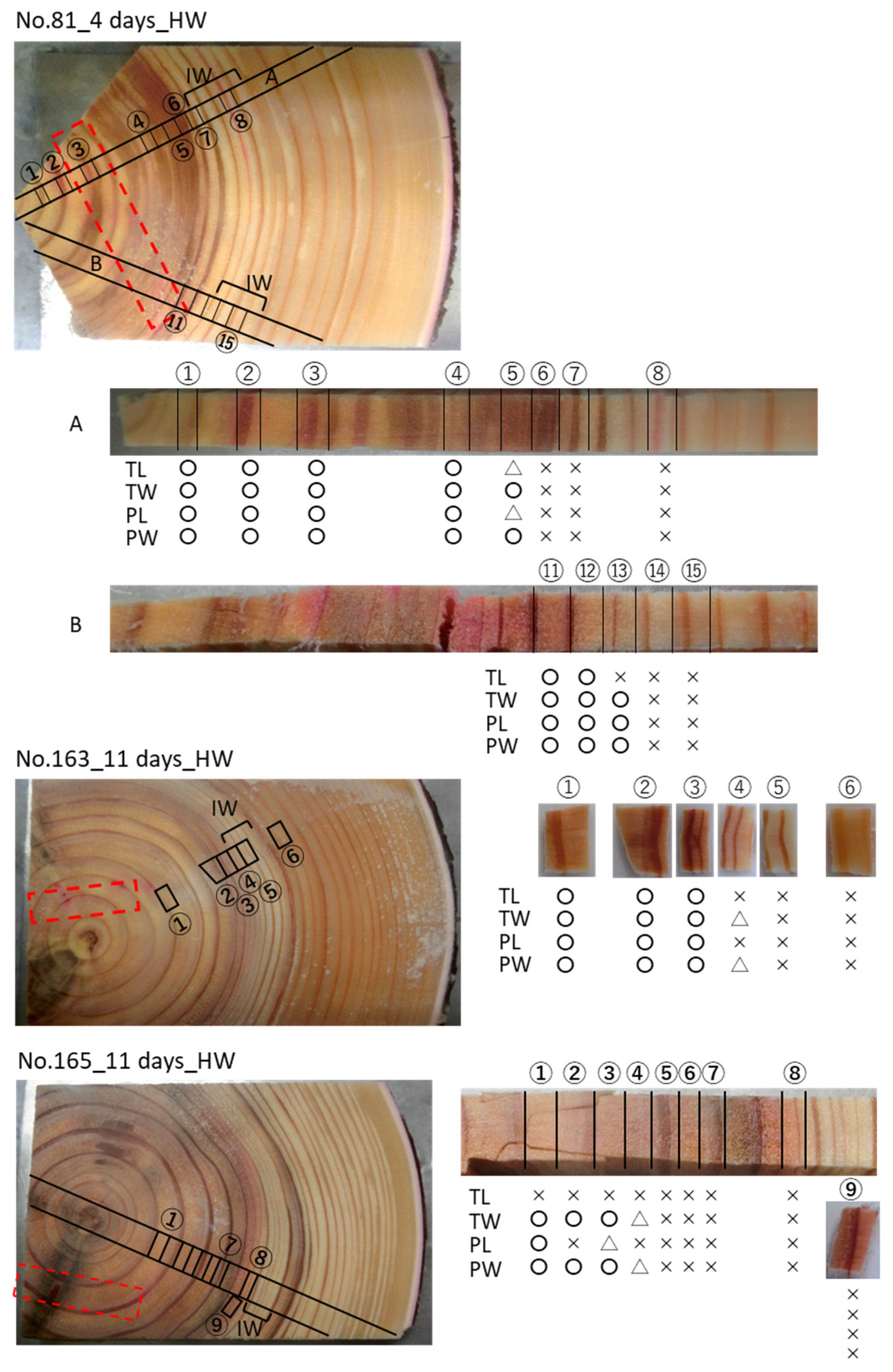
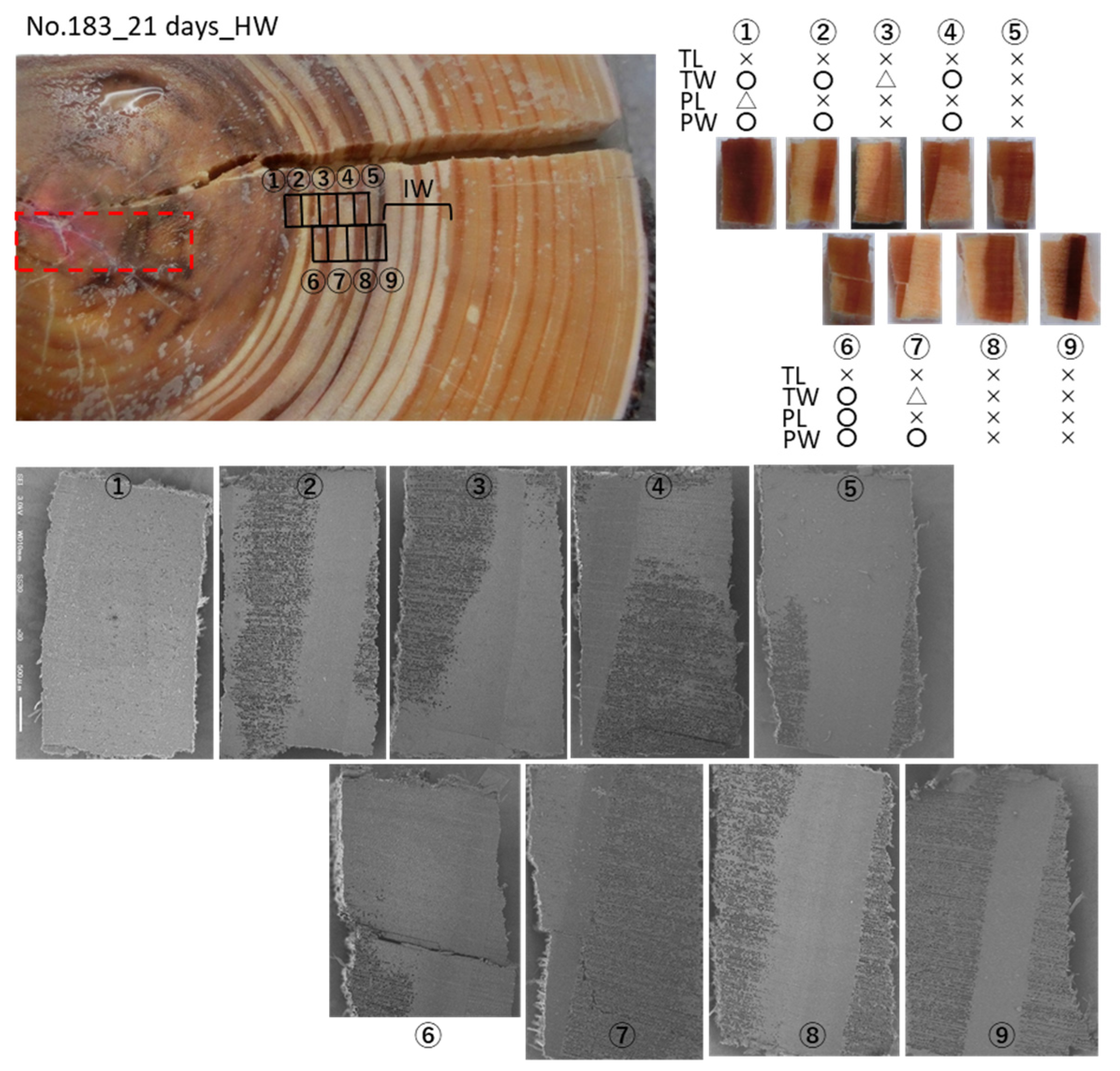
3.2. Cs Distribution in the Xylem Following Cs Injection into the Heartwood in Summer

To clarify Cs movement from the heartwood to the sapwood, the Cs solution was injected into the heartwood and its Cs distribution was investigated using cryo-SEM/EDX (Figure 4). In the 4-day injection sample (No. 81), two radial directions were analyzed: line A was from the middle part of the stainless steel injection tube and line B was from the outlet of injection to the outside. In line A, Cs peaks were detected in all structures in the heartwood, except for the outer part (blocks 6, 7). In line B, Cs peaks were detected in all structures in the heartwood (blocks 11, 12) as well as in the TW, PL, and PW in the boundary between the heartwood and intermediate wood (block 13). Cs peaks were not detected in any structures in the outer part of the intermediate wood or sapwood (blocks 14, 15).
In the 11-day injection sample (No. 163), Cs peaks were detected in the TL, TW, PL, and PW in the heartwood. Weaker Cs peaks were detected in the TW and PW of the innermost annual ring of the intermediate wood (the inner annual ring of block 4), although Cs peaks were not detected in any structures in the outer part. In the other 11-day injection sample (No. 165), Cs peaks were detected in the TW and PW in the heartwood, except in the outer part, which was farther from the injection outlet compared with PL. No Cs peak was detected in any structure in the outer part of the heartwood.
The 21-day injection sample (No. 183) had a wide whitish color area between the intermediate wood and heartwood, in which the outer part with white color was determined as intermediate wood and the inner part with a brownish white color was identified as heartwood because of its color and water distribution. In the middle part of the heartwood, Cs peaks were detected in the TW, PL, and PW, but not in the TL (blocks 1, 6). With increasing distance from the outlet in the heartwood, the Cs peaks in TW and PW were detected farther than those in the PL (blocks 1–4, 6–7).
The water distribution of the analyzed sample parts is shown in Figure 4. Water distribution helps identify the area of the intermediate wood because the earlywood tracheid loses water from its lumen in the intermediate wood and some of them are refilled in the heartwood [32]. In addition, we hypothesized that water distribution might become the key property of sap (solution) movement between the heartwood and sapwood [19].

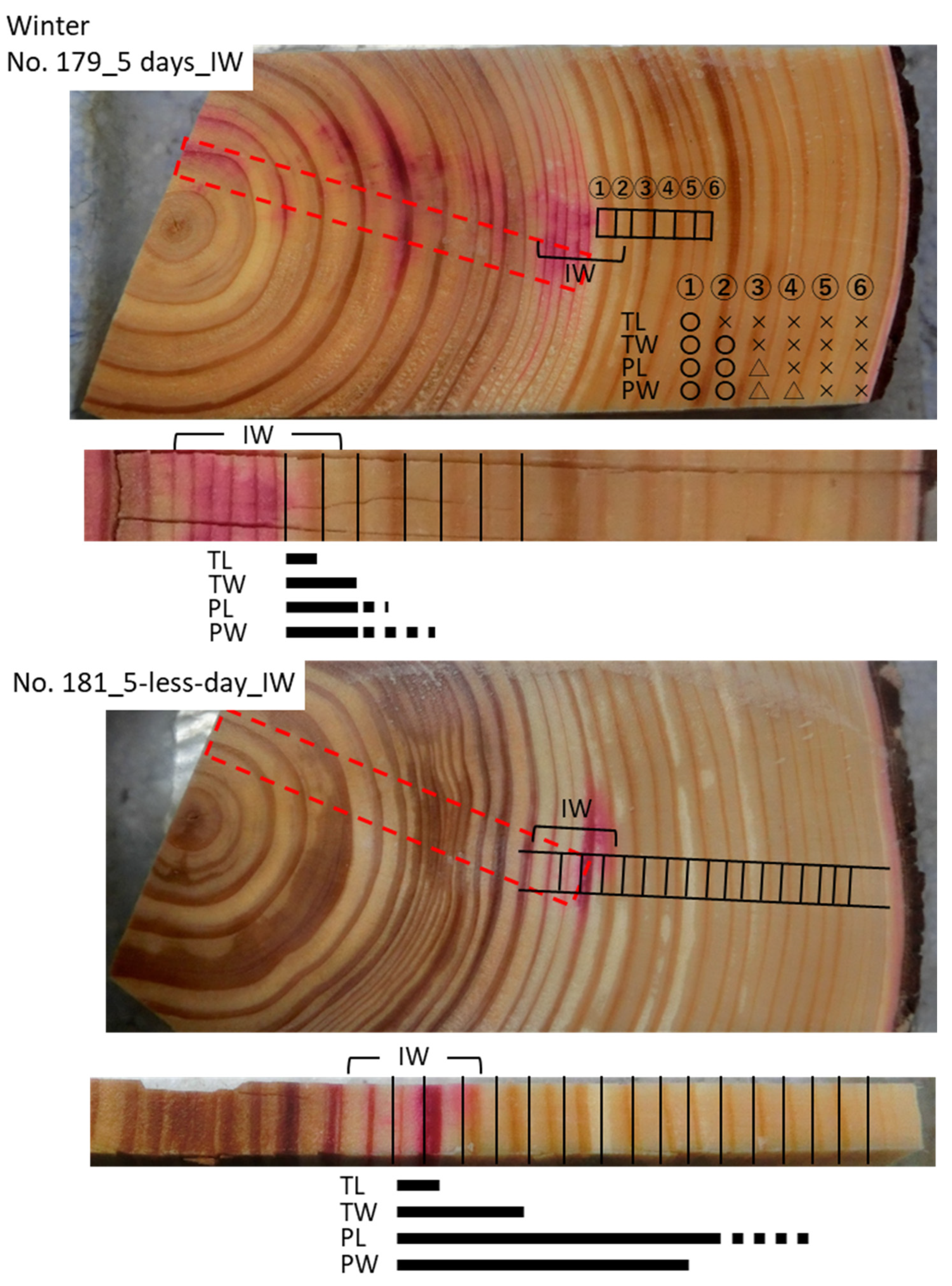
3.3. Cs Distribution in the Xylem Following Cs Injection into the Intermediate Wood in Winter

To clarify the seasonal difference in Cs movement in the sapwood, the Cs solution was injected into the intermediate wood of Japanese cedar trunks in winter and its Cs distribution was investigated using cryo-SEM/EDX (Figure 5). Although we tried to inject the Cs solution for 5 days using two trees in winter, one bottle of the Cs solution became empty before 5 days (No. 181). Because Cs was detected in the sapwood, this sample was used to trace Cs so as to analyze mineral movement in winter. Both samples had injection holes that reached past the middle part of the intermediate wood. The red-colored area was distributed over the outer part of the intermediate wood, whereas it was not distributed in the sapwood (Figure 2 and Figure 5). In the 5-day injection sample (No. 179), clear Cs peaks were detected in the TL, TW, PL, and PW in the red-colored area. The Cs peaks were analyzed along a radial position from the red-colored area to the outer xylem. However, the Cs peak in the TL was not detected outside the red-colored area. The Cs peaks were detected in the TW, PL and PW in the inner sapwood, and the Cs detection area reached farther in the PL and PW than that in the TW. Additionally, Cs reached farther in the PW than that in the PL. In the 5-less-day injection sample (No. 181), clear Cs peaks were detected in all structures in the middle part of the intermediate wood where fuchsin red was observed. In the outer intermediate wood, even in the red-colored area, Cs peaks were not detected in the TL but were detected in the TW, PL, and PW. The Cs peaks in the TW were detected only in the innermost part of the sapwood. The Cs peaks in the PL and PW were detected farther into the sapwood than in the TW, whereas that in the PL reached farther than that in the PW.

4. Discussion

Although the radial movement of minerals in tree trunks is a well-known function among the many functions of ray parenchyma cells [2,4,5,17,20,22], the underlying mechanisms have not been completely elucidated. Our previous studies [19] revealed that mineral movement from the outer sapwood toward the heartwood in standing Japanese cedar trunk occurs through a combination of active transport by parenchyma cells and diffusion in the cell walls from the sapwood to the intermediate wood, followed by movement to the heartwood by diffusion. Then, a new question arose as to how the movement of minerals occurs in the opposite direction. Therefore, we designed the present study to follow the movement of a tracer, stable isotope Cs, from the inner trunk of standing Japanese cedar trees outward (Figure 1). We injected Cs solution using a long hole across the central part of the trunk, and samples were taken from the opposite side of the injection. The difficulty of this injection method was in controlling the position of the outlet of the tube. Thus, the injection hole and fuchsin red-colored area were checked after the trees were cut to identify the solution movement area (Figure 2). Next, the Cs distribution of frozen samples was analyzed using cryo-SEM/EDX. For clear interpretation of the results, we evaluated both movement from inner to outer sapwood and movement from the heartwood to the sapwood (Figure 6).

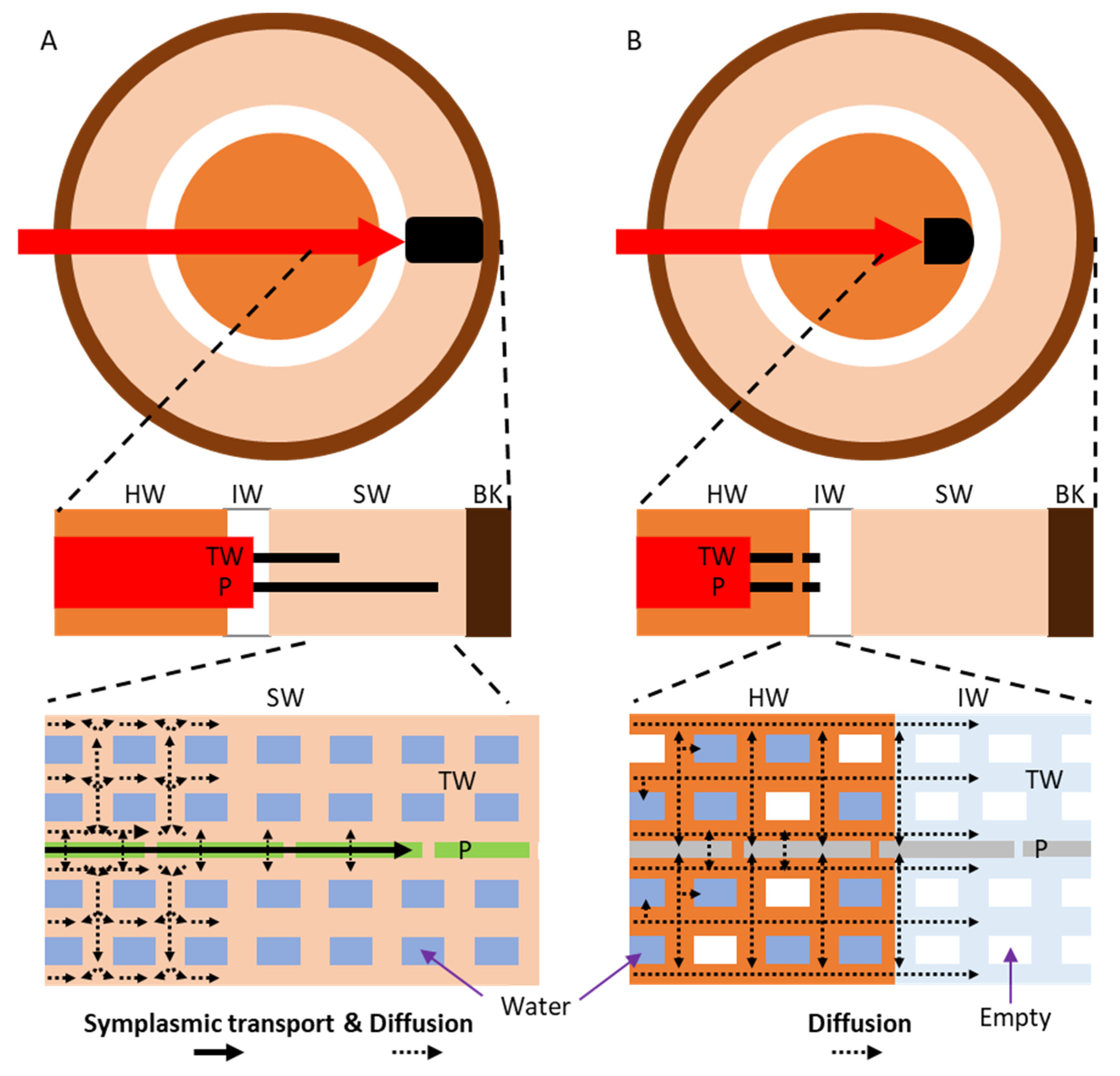
4.1. Mineral Transport in Both Directions in the Sapwood by Ray Parenchyma Cells

When the Cs solution was injected into the innermost sapwood, Cs was detected in the outer sapwood along with the radial position from the fuchsin red color area in the inner sapwood. When we compared the Cs distribution of the TW to that of ray parenchyma cells (together with the PL and PW), Cs had moved farther in the ray parenchyma cells than in the TW. If Cs moved in the ray parenchyma cells via diffusion, the Cs detection area should be the same as that of the tracheid cell walls because the diffusion of Cs in the TW and ray parenchyma cells occurs in a similar manner [19]. In addition, when the Cs solution was injected into the middle part of the intermediate wood, where about half of ray parenchyma cells are alive [33,34], Cs was detected farther in the ray parenchyma cells than in the TW in the sapwood. This indicates that ray parenchyma cells symplasmically transport Cs from the inner to the outer sapwood and that this process is faster than diffusion in the cell walls. Together with our previous result, ray parenchyma cells in the trunks of standing Japanese cedar trees have the function of mineral transport in both directions: from outer toward inner sapwood and from inner toward outer sapwood.
The activity of parenchyma cells is known to change with season [20,21,35]; for example, parenchyma cells store nutrients in winter and provide them as sources for newly forming cells in the cambial zone and developing areas in spring. The present study showed that ray parenchyma cells can transport minerals in both directions in either summer or winter, although the number of the studied trees was limited; this suggests that bidirectional transport can occur in any season during the life of a tree, including during the tree’s response to insect attack [36] or to sudden environmental changes [37]. Therefore, what exactly determines the direction of transport? Our studies showed that Cs migrated from the injected side, i.e., from the higher concentration side to the lower concentration side. So, detecting high-concentration sites might be a key for starting Cs transport. On the other hand, some switches are required to initiate active transport from lower concentration sites to higher concentration sites. Further studies are warranted to obtain information on these switches to determine the entire bidirectional transport mechanism of ray parenchyma cells.

4.2. Minerals Rarely Move from the Heartwood to the Sapwood

The color and size of the heartwood are one of the features that identify tree species, and the properties of the heartwood affect the usage and value of the trunk. Many tree species have higher mineral concentration in the heartwood than in the sapwood [27,28,29,30,31]. In the case of Japanese cedar, high concentration of potassium is accumulated in the heartwood [27,38,39]. For minerals to accumulate at a high concentration in the heartwood after being transported from the sapwood, minerals in the heartwood should not move to the sapwood, or the amount of mineral movement from the sapwood to the heartwood should be greater than that from the heartwood to the sapwood. When Cs was injected into the heartwood in this study, it moved to the vicinity of the heartwood–intermediate wood boundary but did not spread to the outer part; that is, Cs did not move to the intermediate wood and sapwood. This result indicates that there is some sort of a barrier that prevents the free diffusion of Cs between the heartwood and sapwood, which may determine the direction of the movement.
The anatomical features of the intermediate wood may be involved in this phenomenon. Regarding the movement from the sapwood to the intermediate wood, the red color of acidic fuchsin did not enter to the intermediate wood, even though Cs entered when it was injected into the sapwood for a long period of time [19]. The results indicated that movement solely via diffusion rarely occurs from the sapwood to the intermediate wood; this indicated that diffusion was prevented at the boundary between the sapwood and intermediate wood. In addition, Cs, as well as fuchsin red, did not move from the heartwood to the intermediate wood when the Cs solution was injected into the heartwood. Interestingly, Cs moved to the sapwood when the Cs solution was injected into the middle part of the intermediate wood, indicating that Cs was taken up by living ray parenchyma cells when it reached the periphery of those cells and then moved to the sapwood via the function of ray parenchyma cells. Because the number of living ray parenchyma cells is small and some heartwood substances start accumulating at the inner part of the intermediate wood [34], it is difficult for Cs to reach living ray parenchyma cells. Therefore, Cs diffusion was prevented before Cs reached the area where there were many living ray parenchyma cells, that is, the blocking of this direction of diffusion occurred at the boundary between the heartwood and intermediate wood. In the intermediate wood of Japanese cedar trunks, either tracheids or parenchyma cells dehydrate, particularly in the earlywood (Figure 5) [18,32]; therefore, diffusion of the solute from the heartwood to the intermediate wood is difficult [40]. This may be one of the reasons why it is difficult for Cs to move toward living parenchyma cells. Thus, anatomical features of and the water environment at the intermediate wood may act as a barrier to mineral movement between the heartwood and sapwood.
On the other hand, gradual diffusion can occur from the heartwood to the intermediate wood and the sapwood because the cell wall and latewood tracheids contain water, even in the intermediate wood [32]. Therefore, it is impossible to completely stop high-concentration minerals in the heartwood from diffusing to the periphery of living parenchyma cells in the intermediate wood. Nevertheless, minerals are kept at a high concentration in the heartwood, suggesting the existence of an unknown mechanism that limits mineral movement. Although we hypothesized that the decrease in water content of the intermediate wood has an important role in limiting diffusion, not all tree species exhibit a decrease in water content when the sapwood is transformed into heartwood, such as Acacia spp. [41] and Cunninghamia lanceolata [42]. To determine the unknown mechanism underlying mineral accumulation in the heartwood, it is necessary to investigate mineral movement in tree species that do not show a decrease in water content at the boundary between the sapwood to heartwood.

5. Conclusions

We clarified mineral movement from inside to outside in the trunks of Japanese cedar trees by directly analyzing mineral distribution in freeze-fixed trunks to reflect the state of standing trees as much as possible. Together with our previous results, we experimentally showed that, in the sapwood, bidirectional movement occurs, owing to the function of ray parenchyma cells, regardless of the season. To determine the mechanism by which xylem ray parenchyma cells actively accumulate minerals and discharge them when needed, it is necessary to find the key determinant of the direction of this transport. We also clarified that minerals move from the intermediate wood to the heartwood but not in the opposite direction. Although the presence of the intermediate wood is important for inhibiting mineral movement from the heartwood to the sapwood, our results suggest that there is an unknown mechanism that prevents diffusion from the heartwood to the intermediate wood. This unknown mechanism may control the properties of the heartwood. Controlling heartwood formation is an important strategy in the longevity of trees. We hypothesized that accumulated minerals in the heartwood are stored for use when they are needed by the living cells in trees, but this might not be the case. Is there any physiological significance for tree species in the accumulation of high mineral concentration in the heartwood? Further elucidation of the mechanism underlying mineral movement, including the elucidation of this unknown mechanism, will lead to an understanding of the nature of the formation of large tree trunks.

Author Contributions

Conceptualization and design, K.K.; methodology and investigation, K.K., K.Y. and Y.I.; writing—original draft preparation, K.K.; writing—review and editing, K.K., K.Y. and Y.I.; funding acquisition, K.K. All authors have read and agreed to the published version of the manuscript.

Funding

This work was supported by JSPS KAKENHI (Grant Numbers JP25292110, JP16H04936, JP18H02258), and by the Support program of FFPRI for researchers having family obligations.

Institutional Review Board Statement

Not applicable.

Informed Consent Statement

Not applicable.

Data Availability Statement

Not applicable.

Acknowledgments

We appreciate the editors and anonymous reviewers for their helpful comments on the manuscript.

Conflicts of Interest

The authors declare no conflict of interest.

References

  1. Pallardy, S. Physiology of Woody Plants, 3rd ed.; Academic Press: London, UK, 2008. [Google Scholar]
  2. Sauter, J.J.; Kloth, S. Plasmodesmatal frequency and radial translocation rates in ray cells of poplar (Populus × canadensis Moench ‘robusta’). Planta 1986, 168, 7–380. [Google Scholar] [CrossRef]
  3. Holbrook, N.M.; Zwieniecki, M.A. Integration of long distance transport systems in plants: Perspectives and prospects for future research. In Vascular Transport in Plants; Holbrook, N.M., Zwieniecki, M.A., Eds.; Elsevier Academic Press: San Diego, CA, USA, 2005; pp. 537–545. [Google Scholar]
  4. Sokołowska, K. Symplasmic transport in wood: The importance of living xylem cells Symplasmic Transport in Vascular Plants. In Symplasmic Transport in Vascular Plants; Sokołowska, K., Sowiński, P., Eds.; Springer Science-Business Media: New York, NY, USA, 2013; pp. 101–132. [Google Scholar]
  5. Spicer, R. Symplasmic networks in secondary vascular tissues: Parenchyma distribution and activity supporting long-distance transport. J. Exp. Bot. 2014, 65, 1829–1848. [Google Scholar] [CrossRef] [Green Version]
  6. Baker, H.; James, W.O. The behaviour of dyes in the transpiration stream of sycamores (Acer pseudoplatanus L.). New Phytol. 1933, 32, 245–260. [Google Scholar] [CrossRef]
  7. Stout, P.R.; Hoagland, D.R. Upward and lateral movement of salt in certain plants as indicated by radioactive isotopes of potassium, sodium, and phosphorus absorbed by roots. Am. J. Bot. 1939, 26, 320–324. [Google Scholar] [CrossRef]
  8. Tyree, M.T.; Zimmermann, M.H. Xylem Structure and the Ascent of Sap; Springer: Berlin, Germany, 2002. [Google Scholar]
  9. Umebayashi, T.; Utsumi, Y.; Koga, S.; Inoue, S.; Shiiba, Y.; Arakawa, K.; Matsumura, J.; Oda, K. Optimal conditions for visualizing water-conducting pathways in a living tree by the dye injection method. Tree Physiol. 2007, 27, 993–999. [Google Scholar] [CrossRef] [PubMed] [Green Version]
  10. Umebayashi, T.; Utsumi, Y.; Koga, S.; Inoue, S.; Fujikawa, S.; Arakawa, K.; Matsumura, J.; Oda, K. Conducting pathways in north temperate deciduous broadleaved trees. IAWA J. 2008, 29, 247–263. [Google Scholar] [CrossRef]
  11. van der Schoot, C.; van Bel, A.J.E. Mapping membrane potential differences and dye-coupling in internodal tissues of tomato (Solanum lycopersicum L.). Planta 1990, 182, 9–21. [Google Scholar] [CrossRef]
  12. Pfautsch, S.; Renard, J.; Tjoelker, M.G.; Salih, A. Phloem as capacitor: Radial transfer of water into xylem of tree stems occurs via symplastic transport in ray parenchyma. Plant Physiol. 2015, 167, 963–971. [Google Scholar] [CrossRef] [PubMed] [Green Version]
  13. Sokołowska, K.; Zagórska-Marek, B. Seasonal changes in the degree of symplasmic continuity between the cells of cambial region of Acer pseudoplatanus and Ulmus minor. Acta Soc. Bot. Pol. 2007, 76, 277–286. [Google Scholar] [CrossRef]
  14. Sokołowska, K.; Zagórska-Marek, B. Symplasmic, long-distance transport in xylem and cambial regions in branches of Acer pseudoplatanus (Aceraceae) and Populus tremula × P. tremuloides (Salicaceae). Am. J. Bot. 2012, 99, 1745–1755. [Google Scholar]
  15. Okada, N.; Hirakawa, Y.; Katayama, Y. Application of activable tracers to investigate radial movement of minerals in the stem of Japanese cedar (Cryptomeria japonica). J. Wood Sci. 2011, 57, 421–428. [Google Scholar] [CrossRef]
  16. Okada, N.; Hirakawa, Y.; Katayama, Y. Radial movement of sapwood-injected rubidium into heartwood of Japanese cedar (Cryptomeria japonica) in the growing period. J. Wood Sci. 2012, 58, 1–8. [Google Scholar] [CrossRef]
  17. Chaffey, N.; Barlow, P. The cytoskeleton facilitates a three-dimensional symplasmic continuum in the long-lived ray and axial parenchyma cells of angiosperm trees. Planta 2001, 213, 811–823. [Google Scholar] [CrossRef]
  18. Kuroda, K.; Yamane, K.; Itoh, Y. Cellular level in planta analysis of radial movement of artificially injected caesium in Cryptomeria japonica xylem. Trees 2018, 32, 1505–1517. [Google Scholar] [CrossRef]
  19. Kuroda, K.; Yamane, K.; Itoh, Y. Radial movement of minerals in the trunks of standing Japanese cedar (Cryptomeria japonica D. Don) trees in summer by tracer analysis. Forests 2020, 11, 562. [Google Scholar] [CrossRef]
  20. Sauter, J.J.; van Cleve, B. Storage, mobilization and interrelations of starch, sugars, protein and fat in the ray storage tissue of poplar trees. Trees 1994, 8, 297–304. [Google Scholar] [CrossRef]
  21. Miyazaki, Y.; Hiura, T.; Kato, E.; Funada, R. Allocation of Resources to Reproduction in Styrax obassia in a Masting Year. Ann. Bot. 2002, 89, 767–772. [Google Scholar] [CrossRef] [Green Version]
  22. van Bel, A.J.E. Xylem-phloem exchange via the rays: The undervalued route of transport. J. Exp. Bot. 1990, 41, 631–644. [Google Scholar] [CrossRef]
  23. Aubry, E.; Dinant, S.; Vilaine, F.; Bellini, C.; Le Hir, R. Lateral transport of organic and inorganic solutes. Plants 2019, 8, 20. [Google Scholar] [CrossRef] [Green Version]
  24. Stewart, C.M. Excretion and heartwood formation in living trees. Science 1966, 153, 1068–1074. [Google Scholar] [CrossRef] [PubMed]
  25. Hillis, W.E. Heartwood and Tree Exudates; Springer: Berlin/Heidelberg, Germany, 1987. [Google Scholar]
  26. Taylor, A.M.; Gartner, B.L.; Morrell, J.J. Heartwood formation and natural durability-a review. Wood Fiber Sci. 2002, 34, 587–611. [Google Scholar]
  27. Okada, N.; Katayama, Y.; Nobuchi, T.; Ishimaru, Y.; Aoki, A. Trace elements in the stems of trees V. Comparisons of radial distributions among softwood stems. Mokuzai Gakkaishi 1993, 10, 1111–1118. [Google Scholar]
  28. Okada, N.; Katayama, Y.; Nobuchi, T.; Ishimaru, Y.; Aoki, A. Trace elements in the stems of trees VI. Comparisons of radial distributions among hardwood stems. Mokuzai Gakkaishi 1993, 10, 1119–1127. [Google Scholar]
  29. Penninckx, V.; Glineur, S.; Gruber, W.; Herbauts, J.; Meerts, P. Radial variations in wood mineral element concentrations: A comparison of beech and pedunculate oak from the Belgian Ardennes. Ann. For. Sci. 2001, 58, 253–260. [Google Scholar] [CrossRef]
  30. Meerts, P. Mineral nutrient concentrations in sapwood and heartwood: A literature review. Ann. For. Sci. 2002, 59, 713–722. [Google Scholar] [CrossRef] [Green Version]
  31. Tsuchiya, Y.; Shimogaki, H.; Abe, H.; Kagawa, A. Inorganic elements in typical trees for woody biomass fuel. J. Wood Sci. 2010, 56, 53–63. [Google Scholar] [CrossRef]
  32. Kuroda, K.; Yamashita, K.; Fujiwara, T. Cellular level observation of water loss and the refilling of tracheids in the xylem of Cryptomeria japonica during heartwood formation. Trees 2009, 23, 1163–1172. [Google Scholar] [CrossRef]
  33. Nobuchi, T.; Harada, H. Physiological features of the ‘white zone’ of sugi (Cryptomeria japonica D. Don) -cytological structure and moisture content. Mokuzai Gakkaishi 1983, 29, 824–832. [Google Scholar]
  34. Arakawa, I.; Funada, R.; Nakaba, S. Changes in the morphology and functions of vacuoles during the death of ray parenchyma cells in Cryptomeria japonica. J. Wood Sci. 2018, 64, 177–185. [Google Scholar] [CrossRef] [Green Version]
  35. Kean-Jim, L.; Paasela, T.; Harju, A.; Venäläinen, M.; Paulin, L.; Auvinen, P.; Kärkkäinen, K.; Teeriet, T. Developmental changes in Scots pine transcriptome during heartwood formation. Plant Physiol. 2016, 172, 1403–1417. [Google Scholar]
  36. Nakaba, S.; Morimoto, H.; Arakawa, I.; Yamagishi, Y.; Nakada, R.; Funada, R. Responses of ray parenchyma cells to wounding differ between earlywood and latewood in the sapwood of Cryptomeria japonica. Trees 2017, 31, 27–39. [Google Scholar] [CrossRef]
  37. Begum, S.; Kudo, K.; Rahman, M.H.; Nakaba, S.; Yamagishi, Y.; Nabeshima, E.; Nugroho, W.D.; Oribe, Y.; Kitin, P.; Jin, H.-O.; et al. Climate change and the regulation of wood formation in trees by temperature. Trees 2018, 32, 3–15. [Google Scholar] [CrossRef]
  38. Okada, N.; Katayama, Y.; Nobuchi, T.; Ishimaru, Y.; Yamashita, H.; Aoki, A. Trace elements in the stems of trees I. Radial distributions in Sugi (Cryptomeria japonica D. Don). Mokuzai Gakkaishi 1987, 12, 913–920. [Google Scholar]
  39. Kubo, T.; Ataka, S. Blackening of Sugi (Cryptomeria japonica D. Don) heartwood in relation to metal content and moisture content. J. Wood Sci. 1998, 44, 137–141. [Google Scholar] [CrossRef]
  40. Nakada, R.; Okada, N.; Nakai, T.; Kuroda, K.; Nagai, S. Water potential gradient between sapwood and heartwood as a driving force in water accumulation in wetwood in conifers. Wood Sci. Technol. 2019, 53, 407–424. [Google Scholar] [CrossRef]
  41. Yamamoto, K. Moisture distribution in stems of Acacia mangium, A. auriculiformis and hybrid Acacia trees. Jpn. Agric. Res. Q. 2003, 37, 207–212. [Google Scholar] [CrossRef] [Green Version]
  42. Nakada, R. Within-stem water distribution in living trees of some conifers. IAWA J. 2006, 27, 313–327. [Google Scholar] [CrossRef]
Figure 1. Schematic presentation of sample preparation. (A) Cs solution was injected into a standing Japanese cedar (Cryptomeria japonica) tree trunk. (B) A hole was designed to reach the opposite sapwood or intermediate wood (1) or the opposite heartwood (2) through the center part of trunk, and these were compared to the normal injection (3) used in our previous reports [18,19]. (C) Holes were drilled using an increment borer. (D) Each hole was filled with silicone sealant after setting the tube. (E) The length of the hole was checked by measuring the core before the injection. BK, bark; SW, sapwood; IW, intermediate wood; HW, heartwood.
Forests 12 00251 g001
Figure 2. Typical images representing the injection holes in each trunk. A stainless steel tube was inserted into the hole, and Cs solution was injected from the outlet of the tube. The fuchsin red color spread widely when the hole reached the sapwood, which was not the case when the hole reached the intermediate wood or the heartwood. Abbreviations are the same as described in Figure 1.
Forests 12 00251 g002
Figure 3. Schematic presentation of radially distributed cesium (Cs) in the xylem of Japanese cedar (Cryptomeria japonica) trees after Cs was injected into the sapwood or the intermediate wood in summer, as observed using cryo-scanning electron microscopy/energy-dispersive X-ray spectroscopy (cryo-SEM/EDX). Bars below the sample image represent the area where Cs was detected. Dotted red rectangles represent the area of the injection hole (about 2 cm below this plane); black rectangles represent the analyzed area for cryo-SEM/EDX; TL, lumen of tracheid; TW, cell wall of tracheid; PL, lumen of ray parenchyma cell; PW, cell wall of ray parenchyma cell. Other abbreviations are the same as described in Figure 1.
Forests 12 00251 g003
Figure 4. Schematic presentation of radially distributed cesium (Cs) in the xylem of Japanese cedar (Cryptomeria japonica) trees after Cs was injected into the heartwood in summer, as observed using cryo-scanning electron microscopy/energy-dispersive X-ray spectroscopy (cryo-SEM/EDX). Water distribution in some samples was analyzed by observing secondary electron images, in which the cells filled with water and cell walls appeared light gray and the cell lumen without water appeared dark gray. Circle, Cs was detected; triangle, Cs was detected in part; Cross, Cs was not detected. Other abbreviations are the same as described in Figure 1 and Figure 3.
Forests 12 00251 g004aForests 12 00251 g004b
Figure 5. Schematic presentation of radially distributed cesium (Cs) in the xylem of Japanese cedar (Cryptomeria japonica) trees after Cs was injected into the intermediate wood in winter, as observed using cryo-scanning electron microscopy/energy-dispersive X-ray spectroscopy (cryo-SEM/EDX). Abbreviations are the same as described in Figure 1, Figure 3 and Figure 4.
Forests 12 00251 g005
Figure 6. Schematic drawing of the radial movement of cesium (Cs) in the trunk of Japanese cedar (Cryptomeria japonica) trees. (A) Movement of Cs after injection into the intermediate wood or the sapwood. (B) Movement of Cs after injection into the heartwood. In the top row, red arrows represent the direction of Cs injection, and black areas represent the areas where Cs moved. In the middle row, red boxes represent the injection holes, and black areas and black bars represent the areas where Cs moved. In the bottom row, the solid arrows and dotted arrows represent symplasmic transport and diffusion, respectively. P, ray parenchyma cell. Other abbreviations are the same as described in Figure 1, Figure 3, and Figure 4.
Forests 12 00251 g006
Table 1. Basic information about the samples.
Injection
Tree Number* Injection Hole DepthPeriod of InjectionStarting Date (D/M/Y)Harvesting Date (D/M/Y)AgeTree Height (m)Girth at 1.2 m (cm)
Summer
191SW4 days03/08/202007/08/20203817.255
197SW4 days03/08/202007/08/20203918.258
195SW11 days03/08/202014/08/20213716.052
193IW11 days03/08/202014/08/20203814.857
81HW4 days10/07/201714/07/20173416.352
163HW11 days18/06/201829/06/20183516.547
165HW11 days18/06/201829/06/20183514.248
185HW16 days10/07/201926/07/20193817.356
183HW21 days26/06/201917/07/20193517.445
Winter
179IW5 days08/02/201913/02/20193718.257.5
181IW5-less days08/02/201913/02/20193816.459
* The injection hole depth was checked after the trees were cut. SW, sapwood; IW, intermediate wood; HW, heartwood.
Publisher’s Note: MDPI stays neutral with regard to jurisdictional claims in published maps and institutional affiliations.

