
Abstract
The review highlights the need of non-antibacterial, non-antifungal and non-anticancer characters of metal or metal oxide nanoparticles. The usage of nanoparticles as a part of therapeutic measures results in certain unfavourable effects. The nanoparticles can disturb healthy gut microorganisms that may bring about some health damages regarding pathogenic diseases, obesity, and inflammation likewise. Even the nonspecific interactions of nanoparticles with healthy cells and tissues can cause altered expressions of various pro-inflammatory factors and stress related genes. This review indicates and prospect about the demand of nanoparticles with non-antibacterial, non-antifungal and non-anticancer properties. Such nanoparticles will be effective in various remedial and diagnostic purposes.
Export citation and abstract BibTeX RIS

Original content from this work may be used under the terms of the Creative Commons Attribution 4.0 licence. Any further distribution of this work must maintain attribution to the author(s) and the title of the work, journal citation and DOI.
1. Introduction
Recently, the utilization of nanoparticles has increased to a high extent, since these nanoparticles possess high surface to volume ratio and peculiar characteristics regarding physical, chemical, structural, mechanical, electrical and optical properties. The metal nanoparticles hold wide-ranging applications in many fields, for example, catalysis, bioimaging, diagnosis, therapy and environmental remediation. Besides, nanoparticles have noteworthy usages in the fields of apparel, cosmetics, paints, construction, food, energy, and agriculture. The demand of nanoparticles has increased exceedingly because of their antimicrobial, antifungal, and anticancer activities. These activities can be observed by several types of nanoparticles (NPs) such as silver (AgNPs), gold (AuNPs), silica oxide (SiO2NPs), zinc oxide (ZnONPs), carbon nanotubes (CNTs), titanium oxide (TiO2 NPs), and magnesium oxide (MgONPs) [1–7]. Metal and metal oxide nanoparticles can act against many microorganisms. These nanoparticles can show antimicrobial activities against several microorganisms such as B. subtilis, E. coli, Staphylococcus aureus, A. niger, F. oxysporum, A. fumigatus, Candida albicans [8]. The applications of nanoparticles are not only restricted towards their bactericidal and fungicidal activities but also demand drug delivery, bioimaging and gene delivery to target tissues or cancer cells and other diagnostic purposes. Drug delivery is the release of a specific drug at a desired site with a certain speed. Therefore, this is one of the intriguing fields of research which draws a great interest of the researchers. The potential usage of nanoparticles for the delivery of genetic materials, including single stranded DNA or RNA, plasmid DNA as a part of gene therapy could be within reach. Some nanoparticles also exert influential applications such as protein delivery and vaccine delivery [9, 10].
The field of nanotechnology is growing to a greater extent as these nanoparticles have multifaceted applications. While considering all these applications, the nanoparticles should have some stringent properties such as biologically inert, low inherent toxicity, lower toxicological effects on microbiota/microbiome of living things, specific in action and abiding in vivo toxic effects, and that should be taken into scrutiny. The review mainly outlines the need of non-antibacterial, non-antifungal and non-anticancer properties of nanoparticles which consecutively are more helpful for various diagnostic approaches, including other vital applications. Such nanoparticles will exert limited or no side effects on the normal functioning of the living cells and microbiota of the living body.
2. Impact of nanoparticles on the microflora of biological system
Since the field of nanotechnology has expanded extensively and has application in almost everyday products of lifestyle which subsequently augments a chronic exposure to nanoparticles. These nanoparticles have serious health issues. Metallic nanoparticles have become prerequisites for a range of therapeutic applications. Therefore, the safety and possible toxic effects are the major factors of nanoparticles, which need to be perceived. The living body is composed of several cells and tissues besides the microflora which naturally exist in the body. The microbiota mainly plays the role in nutrition, improvement of immune system, metabolic equilibrium and anti-pathogenic action for humans and animals. The rich colonisation of essential microorganism occurs in the digestive tract, specifically the large intestine. The gut microflora which lives in the gastrointestinal tract of human body has an indispensable role in regulating innumerable physiological functions. The malfunctioning of these functions may cause illness of the human body. These gut microflora play a remarkable role even in the energy harvest and storage. Some recent reports intimated the importance of gut microbial cells as major contributors in evolution as well. These microorganisms also provide different vitamins and amino acids for the body. These microorganisms are also involved in various physiological functions such as fermentation, transformations of different substrates as shown in figure 1 [11–16].
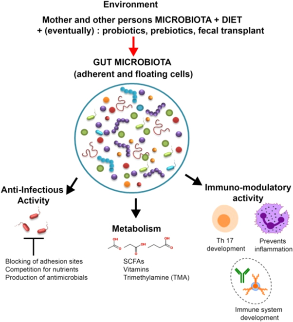
Figure 1. Role of gut microflora in the digestion and synthesis of varied metabolites; vital role of microbes in enhancement of defence system of host through production of anti-pathogenic components which can block the adhesion sites of pathogens Reprinted with permission from [12]. Copyright (2019).
Download figure:
Standard image High-resolution imageThe microbiota get affected by various factors such as antibiotics, diet and probiotics. When the nanoparticles are administered in the living system with the intension of offering a therapy, there are fair chances of interactions with the other healthy body cells and microorganisms. The nanoparticles can pass through the alimentary tract and digestive tract, where the particles might cause structural and functional changes, before reaching the desired site of action. In vivo applications of metallic nanoparticles may show harmful effects because of nonspecific associations with body cells and organisms. Wide array of nanoparticles has recently been used as a part of diagnosis and remedy of various pathogenic states, including cancer. There are few reports which illustrate that the nanoparticles, for example; carbon nanotubes (CNTs), titanium dioxide (TiO2), cerium dioxide (CeO2), zinc oxide (ZnO), silica and silver may impinge on the microbiota followed by other clinical disorders for instance colitis, obesity and immunological dysfunctions [17–22]. The fate and bioavailability of ingested nanoparticles primarily depend upon the various characteristic e.g. size, shape, physical and chemical properties of nanoparticles during transition through the gastrointestinal tract (GIT). The dimensions and active groups of CNTs have significant activity in the growth's obstruction of GIT organisms. CNTs exemplified the inhibitory activity against the microbes; Lactobacillus acidofilus, Bifidobacterium adolescentis, Escherichia coli, Enterococcus faecalis and Staphylococcus aureus [23]. The mechanistic approach behind the antibacterial activity of CNTs is the cell wall disruption, which results in the leakage of cellular contents of bacterial cells. The diminished number of gut microbial cells may have harmful effects, which alternatively culminate into various disease conditions; obesity, type II diabetes, asthma and inflammatory bowel disease [24]. Obesity which can be caused due to the alterations in the microbial contents of coliforms in intestine. Researchers have established the linear relation into the dosage of AgNPs and the body weights of weaned pigs [25]. Consequently, the decrease in the coliform count was also observed. The exposure of mice with AgNPs also resulted in the unevenness of gut microflora, which is often related to the pathogenic conditions such as obesity, diabetes, inflammatory bowel disease and even asthma. The in vivo study, mice were exposed to silver nanoparticles or silica nanoparticles, where silica for 28 days exposure exhibited the decrease in the bacterial richness [26].
Occasionally the administration of nanoparticles can affect the phenotype of microbes. The impact of the TiO2 nanoparticles on the composition of the microorganisms is still an enigma. In vitro studies reveal that the phenotypic alterations in the gut microflora occurred due to the exposure of TiO2 along with ZnO and CeO2 nanoparticles on the model colon for five days in absence of light. The changes mainly observed regarding formation of short-chain fatty acid (SCFA), cell hydrophobicity, sugar percentage of polymers, cell size and electrophoretic movability [27]. The enzymatic expression of microbial cells was found to be affected by the usage of zinc oxide nanoparticles. The nanoparticles affect the specific growth rate and alter the physiological functions of soil microorganisms. The widespread synthesis and the applications of metallic nanoparticles and eventual disposal in the environment result in harmful effects on predominant microorganisms, thus posing a serious hazard to soil inhabitants [28]. Extensively used ZnONPs also show some inhibitory activities towards the Lactobacillus present in ileum of hens. The decline in the microbial richness in the ileum has correspondence with different metabolic activities. A detailed study is required to understand the key mechanism in which nanoparticles affect the microbiota [29].
Introduction of nanotechnology in the various fields have raised multiple questions. This is because of the presence of high reactivity of nanoparticles which enables the nanoparticles to travel across biological barriers. The relation between the human body and nanoparticles has a dual effect with probable unfavourable side effects and with positive, beneficial applications. The interaction between nanoparticles and gut microorganisms is not an anomaly for this. Various factors are responsible for triggering toxicity by nanoparticles on the living system such as size, shape, reactivity, aggregations, charge and surface chemistry. The nanoparticles can be used as a cargo which releases specific drugs awfully selectively at the desired site as a part of therapy without affecting the normal microbiome of human and animal body [30–32].
3. Toxicity of nanoparticles
As the size of nanoparticles reducing, the surface to volume ratio eventually increases. This property of nanoparticles having a high surface to volume ratio makes the nanoparticles more reactive and toxic as well. Small particles have more chance to penetrate the plant and animal cells. Therefore, these nanoparticles at this stage behave unusually, as these nanoparticles have more cellular uptake. The nanoparticles can generate reactive oxygen species (ROS) that result in oxidative damage, ultimately causing cellular damage. Along with these factors, stable nanoparticles also cause toxicity. The pH of the biological system controls the solubility of organic and inorganic nanoparticles. As the concentration of metal and metal oxide nanoparticles within the cell increases, it leads to the stress within the cells. These aggregated nanoparticles do it distinctly that may cause toxic effects on the cells [33–41]. The nanoparticles also affect the cell cycle. Researchers validated ZnONPs caused the cell death of A431 cells by ROS. The nanoparticles can induce cell cycle arrest in S and G2/M phase of cell division, in which the higher uptake of nanoparticles was observed in G2/M phase as compared with other phases of cell division. ZnONPs are cytotoxic at high concentrations and generate oxidative stress because of the increase in intracellular ROS and causes cell cycle arrest [42]. The reactive oxygen species plays a significant role in various physiological and cellular activities inclusive of oxidative stress, apoptosis, proteins and DNA damage [43]. The production of free radicals observed in the cells because of the NPs persuade the oxidative stress. The ZnONPs have wide usages in various fields such as paints, medicines, apparels, electronics, cosmetics, food industry because of their characteristic properties such as UV absorption, antimicrobial activity, catalytic and semi-conductive nature. In recent years researchers have strengthened concerns about the health risks because of ZnONPs. These are in vitro studies also which show the toxic effects on the mammalian cells. ZnONPs caused severe toxicity to in vitro culture of mouse leukemic monocyte macrophage cells in dose and time-dependent way. In the ZnO-induced cytotoxicity, the intracellular dissociation of zinc oxide nanoparticles into zinc ions would disrupt the homeostasis of cells and increase the ROS level that ultimately affects the membrane integrity of the cell [44].
Nanoparticles can cross the pulmonary epithelial barriers. Further, the nanoparticles can interact with vascular endothelium. The nanoparticles can meet various immune cells along with other proteins present in the body fluids. When the nanoparticles interact with the cells, they may induce harmful effects by enhanced manifestations of pro-inflammatory cytokines, DNA strand breakages. Immune cells recognise nanoparticles as foreign bodies as soon as they enter the host system leading to toxicity in the host system. Inflammatory response is observed because of the uptake of nanoparticles by different immune cells. Mostly macrophages are involved in the uptake of nanoparticles. Phagocytosis and macropinocytosis, receptor-mediated endocytosis, and passive penetration are the plausible mechanisms by which nanoparticles are internalised into these cells. Table 1 describes some examples of nanoparticles with the effect of nanoparticles on expressions of Interleukins (ILs) and Toll-like Receptors (TLRs).
Table 1. Effect of nanoparticles on pro-inflammatory factors and Toll like receptors.
| Type of nanoparticles | Model cell lines or animal models as target | Interleukins (ILs), Toll-like Receptors | Reference |
|---|---|---|---|
| Gold | Apo E-/- mice | Enhanced mRNA, monocyte chemoattractant protein-I (Mcp-I), and interleukin -6) | [45] |
| Gold | Bovine retinal pigment epithelial cells | Inhibit vascular endothelial growth factor, IL-1 caused in proliferation and migration | [46] |
| Silver | Peripheral blood mononuclear cell (PBMC), human mesenchymal stem cells (hMSCs), J774 A1 macrophages | Decreased Interleukin activities of 5, 1, 6, 8, 11 | [47, 48] |
| Titanium oxide | CD-1 mice | Macrophage inflammatory protein-1 and 2,(eotaxin, monocytechemotactic protein-1, interferon-, vascular cell adhesion molecule-1, IL-13, Interferon (IFN)-inducible protein-10, migration inhibitory factor | [49] |
| Zinc oxide | Human amniotic epithelial cells (HAECs) | Increased levels of Tumor Necrosis Factor-α, MIP-2, IL-6, 8 and MCP-1 mRNA | [50] |
| Zinc oxide | Peripheral blood mononuclear cell (PBMC) | Increased levels of IFN-, TNF-, and IL-12 | [51] |
| Amorphous silica | Human umbilical vein endothelial cells (HUVECs) | Increased levels of IL6, 8 | [52] |
When the nanoparticles are exposed to cells or organs, some genes get expressed related to inflammation. The nanoparticles induced the genes responsible for the stress. ZnO (13 nm) nanoparticles tested on the RAW 264.7 and BEAS-2B cell lines which responded with the increased levels of ROS, oxidant injury, excitation of inflammation, and cell death [53] when RAW 264.7 macrophages ZnO (20 nm) nanoparticles manifested the higher levels of p47phox NADPH oxidase-mediated superoxide radical generation. DNA damage was also caused because of oxidative damages. Zinc oxide nanoparticles can cause the production of ROS, which was found because of activation of p47phox NADPH oxidase enzyme in macrophages. This is expendable to caspase-9/3-mediated apoptosis. The zinc oxide nanoparticles can cause p47phox NADPH oxidase-dependent superoxide generation that results in cell death by apoptosis [54]. Besides this HeLa cells when exposed to gold nanorods of dimension of 1.4 nm, the expressions of genes causing stress and inflammation increased [55]. Some other effects of gold nanoparticles of length 20 nm were observed on different physiological functions such as detoxification, fat metabolism, cell cycle, defence mechanism and biological clock on the male Wistar rats [56]. Increased level of messenger RNA of p53 and apoptotic proteins were observed [57]. Therefore, there are many kinds of abnormal activities resulting because of the interactions between nanoparticles and cells.
A thorough knowledge about toxicity of the functionalized nanoparticles plays an important role to ease the trend of nanoparticles usage in diverse applications. The independent toxicity testing of nanoparticles should be carried out to lead safer designing of nanoparticles. These in vitro studies will help in the designing of safer nanomaterials with no side effects to human cells. The studies about the effect of nanoparticles in vitro and in vivo show that these nanoparticles can activate pro-inflammatory cytokines, chemokines. These alterations may affect the homeostasis of immune system and may lead to severe pathological conditions of autoimmune, allergic and even neoplastic diseases. Thus, it is necessary to understand the immunotoxicity of metal and metal oxide nanoparticles, which will then help in designing nanoparticles with low or with no hazardous side effects on the living system.
The engineered nanoparticles can interact with genetic materials of cells that instigate in toxicity called genotoxicity. The direct and indirect are two types of genotoxicity in which the former involves the direct interactions of nanoparticles with the genetic material, while the latter explains the generations of reactive oxygen species or toxic ions that may cause the toxicity to the DNA. The production of ROS causes secondary genotoxicity, resulting in DNA damage. These ROS which results from activated immune cells show the oxidative damages to the DNA. The nanoparticles which could pass through the membrane and directly interact with genetic material. These nanoparticles may enter through the diffusion process through nuclear pores or through mitosis of cells. During the cell cycle, the nanoparticles associate with DNA molecules and affect the DNA replication and transcription processes. The nanoparticles can induce the breakages in DNA strands mechanically, or may cause chemical changes in the DNA structures. The nanoparticles such as TiO2, ZnO2 can induce oxidative damages to genetic material by producing micronuclei. Nanoparticles caused apoptosis in human lung epithelial cells along with rat lung alveolar macrophages. Research carried out on the study of genotoxicity and cytotoxicity of various nanoparticles suggests they are always not linked to each other as the exposure levels or the nature of nanoparticles is different. The exposure to nanoparticles can cause toxicity and environmental impact through direct and indirect ways. The effect of nanoparticles on the genotoxicity need to be explored to prevent the side effects of nanoparticles on the human health and animals [58, 59].
When human health is concerned, the nanoparticles exert toxic effects at the cellular levels and biomolecular levels. The biomolecules such as genetic material and proteins get affected [60–62]. The nanoparticles interact with the plasma membrane or cell membrane. It is the first barrier for the nanoparticles internalization. There are various mechanisms by which nanoparticles get internalised into the cell depending upon the type of nanoparticles, nature, shape and size of nanoparticles as shown in figure 2. The nanoparticles such as AuNPs, CNTs can penetrate the cell membranes directly into the cell with no mechanism. The nanoparticles with size ranging in micrometres are internalised by the cell by the mechanism called pinocytosis [63–67]. The size of endocytotic vesicles can differentiate phagocytosis and pinocytosis. The particles with the size of 250 nm are internalised into the cell by the method of phagocytosis, while the particles with few nanometres to 100 nm are internalised by pinocytosis. Pinocytosis is further categorised into clathrin dependent and independent endocytosis, caveolae dependent and independent endocytosis, and macropinocytosis. The nanoparticles which are already inside the cell get enclosed within the endosomes. The nanoparticles escape from endosomes and freely get dispersed within the cell cytoplasm. These nanoparticles then interact with the nuclear envelop. Depending upon the size and nature of nanoparticles, particles interact with nuclear pore complex [68–72].

Figure 2. Step-by-step entry of nanoparticles within the nucleus from extracellular space. (1) endocytosis (2) escaping from the endosomes and (3) crossing the nuclear envelope through the nuclear pore complex. This may differ in type, shape and size of nanoparticles. Reprinted with permission from [73]. Copyright (2019).
Download figure:
Standard image High-resolution imageReports confirm that TiO2 nanoparticles can cause swelling, redness, and damage to lungs. The fibrosis along with pulmonary cancer was observed in rodents. Because of high surface area and extreme reactivity of TiO2 nanoparticles as compared to bulk TiO2 cause several oxidative damages to the cells. The mitochondrial enzymatic activity was also hampered because of TiO2 nanoparticles and later resulted in an amendment in the mitochondrial membrane potential. The TiO2 nanoparticles generated free radicals interact with DNA causing genotoxicity. The antioxidant compounds and enzymes such as glutathione, catalase and superoxide dismutase are essential for nullifying reactive oxygen species formed, but depletion in the enzymatic activities ultimately results in oxidative damages. Genotoxicity was also reported for widely used AgNPs. AgNPs can bring about ROS that results in DNA damage, apoptosis and necrosis. The apoptosis and necrosis are different modes of cell death. The apoptosis is pre-defined cell death, while necrosis is accidental cell death. In apoptosis, the cell undergoes suicide without affecting the normal body functions. Hence, apoptosis is called programmed cell death. Necrosis involves the premature death of cells by injury by autolysis. Various factors such as trauma, infections are responsible for the necrosis. The third mode of cell death is autophagy in which the cell removes the unwanted, non-functional components through natural, regulated mode of action. Figure 3 ascribes the three modes of cell death [74].

Figure 3. The attributes of three modes of cell death; apoptosis, autophagy and necrosis. The apoptosis causes cells to shrink, blebbing in cell membranes, condensation of chromatin, nuclear fragmentation. Autophagy involves the formation of autophagosomes and autophagolysosomes (formed by the fusion of autophagosomes with lysosomes) for cytoplasm, organelle, and protein degradation. Necrosis causes the swelling of the cells and organelles even early membrane damage. Reprinted with permission from [74]. Copyright (2017).
Download figure:
Standard image High-resolution imageThe nanoparticles can be tested in vitro and in vivo for the genomic toxicity. Ames test is based on the principle of bacterial reverse mutation phenomenon, while the comet test is the single cell gel electrophoresis. These are common genotoxicity tests performed. Chromosomal mutations, DNA strand breakages, chromatid breaks can be detected by Chromosomal aberration (CHA) and Micronuclei (MN). These tests are essential to explore the toxicity of nanoparticles before use in therapeutics. The nanoparticles ideally should not exert any adverse effects on the living cells and tissues. Therefore, nanoparticles can be fabricated in such a way that would not cause any deleterious effects on the body [75–79].
Therefore, the growing field of nanotechnology has lifted a lot of concern about the safety issues regarding human health and environmental approach. To avoid the hazardous effects of nanoparticles on the living organism it is indispensable to focus on the synthesis approach which will design the nanoparticles with better activities and fewer deleterious effects. Nanoparticles need to be designed in such a way that they will exert less toxicity towards the healthy cells of body and should not affect the helpful microorganisms which are often called as commensals. This kind of perspective will definitely be helpful in the fabrication of nanomaterials which are inevitably helpful in various therapeutic applications, even this could cut the non-specific interactions of nanoparticles. Nanoparticles are being exploited for drug delivery in two different means such as inert, stable carrier of drug and other one as pharmaceutically active agent.
4. Non-antibacterial, non-antifungal, non-anticancer and biocompatibility properties of nanoparticles
As far as this perspective is concerned, researchers are trying to meet the nanoparticles for better drug delivery and other therapeutic applications with diminished side effects. Ankamwar et al. [80] reported first ever report of synthesis of non-antibacterial and non-anti-cancerous AgNPs using Albizia lebbeck flowers extract. Albizia lebbeck flowers extract was mainly utilised as the source of reducing and stabilising agents. The nanoparticles along with flower extract tested in vitro for their antibacterial activity against S. aureus, E. coli, P. aeruginosa and B. subtilis. The organisms exposed to AgNPs at concentrations of lower (100–400 μg per ml as shown in figure 4) and higher (500–2500 μg per ml as shown in figure 5) and nanoparticles did not show any inhibitory activities towards the organisms. An antibacterial activity of AgNPs was not observed against any of the four organisms. An antibacterial activity of AgNPs was tested by using three different growth media.
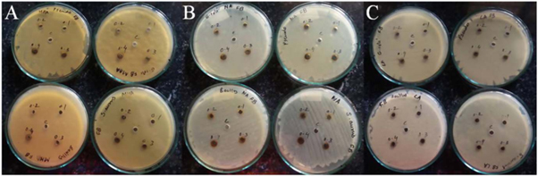
Figure 4. Bacterial inhibition of silver nanoparticles synthesized using Albizia lebbeck flowers extract tested against E. coli, S. aureus, P. aeruginosa, B. subtilis at low concentration (100–400 μg per ml) in Mueller Hinton agar (A), NA (B) and Luria Bertani agar (C).Reprinted with permission from [80]. Copyright (2019).
Download figure:
Standard image High-resolution image
Figure 5. Bacterial inhibition of silver nanoparticles synthesized using Albizia. lebbeck flowers extract tested against E. coli, S. aureus, P. aeruginosa, B. subtilis at high concentration (500–2500 μg per ml) in Mueller Hinton agar (A), NA (B) and Luria Bertani agar (C). Reprinted with permission from [80]. Copyright (2019).
Download figure:
Standard image High-resolution imageThe study of biosynthesized AgNPs against cancer cell lines A549 revealed the biocompatible nature of AgNPs as the nanoparticles did not affect viability of cell lines as shown in figure 6. The cell viability of A549 cancer cells were not affected even on treatment with different concentrations of AgNPs. The difference in the morphologies of treated and untreated cells was also not observed. At higher concentration of AgNPs precipitation was observed.

Figure 6. Cytotoxicity studies of silver nanoparticles of concentrations 2 μg per ml (A)–(D), 10 μg per ml (E)–(H), 50 μg per ml (I)–(L) against A549 cancer cells. Reprinted with permission from [80]. Copyright (2019).
Download figure:
Standard image High-resolution imageThe AgNPs did not show significant toxicity to the cells, even at higher concentrations. This signifies that these biologically synthesized AgNPs are biocompatible which could be best carrier for drug delivery, gene delivery and diagnosis as these nanoparticles exert no notable toxic effects on the biological system regarding the healthy microorganisms cells and tissues of the body. The flower extract has several phytochemicals such as fatty acids, esters, polyamines, the firm binding of these phytochemicals restricts the conversion of AgNPs to silver ions, thus impeding the antibacterial and antitumor activity. This excessive coating of phytochemicals onto the AgNPs affects the release of silver ions and consequently diminish availability of silver ions, thus, they do not support toxicity.
The biocompatibility studies of iron oxide (Fe3O4) nanoparticles performed by utilizing normal cell lines, glia cell lines and breast cancer cell lines [81]. The toxicity studies of Fe3O4 nanoparticles on human glia cells (D54MG, G9T, SF126, U87, U251, U373), human breast cancer (MB157, SKBR3, T47D) and normal (H184B5F5/M10, WI-38, SVGp12) cell lines reveal that the nanoparticles manifest no toxic effects towards these cell lines in the concentration range of 0.1–10 μg ml per ml as shown in figure 7.
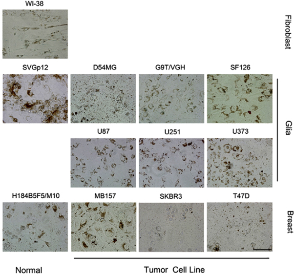
Figure 7. Bright field microscopy images of cell lines after incubation of cell lines with iron oxide nanoparticles of maximum concentration at maximum of 100 μg per ml cells for 72 h. Reprinted with permission from [81]. Copyright (2010).
Download figure:
Standard image High-resolution imageFe3O4 nanoparticles coated with bipolar surfactant tetramethylammonium 11-aminoundecanoate are found to be biocompatible in nature and could derive potential applications in drug delivery, magnetic resonance imaging (MRI) and magnetic hyperthermia. The graphs of treatment of cell lines with Fe3O4 nanoparticles revealed the nanoparticles did not affect the cell lines up to concentration of 0.1–10 μg per ml while the nanoparticles with concentration at 100 μg per ml exhibited the detectable toxicity as shown in figure 8.

Figure 8. Graphical representation of effect of Fe3O4 nanoparticles on the different cell lines exposed with different concentrations of iron oxide nanoparticles for 72 h (doses in the range of 0.1 to 100 μg per ml). Reprinted with permission from [81]. Copyright (2010).
Download figure:
Standard image High-resolution imageAlong with cytotoxicity studies, bactericidal activities of nanoparticles were also studied. The AuNPs synthesized using different concentrations of aqueous extract of white flowers from the Albizia lebbeck did not show any inhibitory activities against B. subtilis, S. aureus, and P. aeruginosa, E. coli as shown in figure 9 [82]. The organisms were exposed to different concentrations of AuNPs along with flowers extract as a control. The polyamines, fatty acids, steroids which are present in the flowers extract coat the AuNPs affect the affinity of AuNPs towards the bacterial cells, thus reducing the toxicity. This non-antibacterial character of AuNPs implies its inert, non-reactive nature. This can be a potential drug carrier with no side effects on the healthy microorganisms present in the living system. As these nanoparticles do not alter the healthy microorganisms, these nanoparticles do not disturb the health of an individual. The non-antifungal activities of these nanoparticles should need further study. To circumvent toxic effects of the nanoparticles on the fungal cells, nanoparticles need to be designed in such an approach that these nanoparticles would not have any non-specific interactions with body cells and tissues.

Figure 9. Antibacterial activities of AuNP40 (A), AuNP60 (B), AuNP80 (C) and AuNP100 (D) with the concentration from 0.5 to 2.0 mg per ml against bacteria S. aureus, E. coli, B. subtilis and P. aeruginosa. We used flower extract as the control. Reprinted with permission from [82].Copyright (2019).
Download figure:
Standard image High-resolution imageThe different factors such as dimensions, shapes, various physico-chemical properties of nanoparticles influence the toxicity effects of nanoparticles. The type of nanoparticles and the respective concentrations also have the importance in the effects. These factors of nanoparticles mainly depend on the type of synthesis methods, types of stabilizers and capping agents used in the fabrications of nanoparticles. Through the optimizations of all these factors nanoparticles with defined shape, size and charge can be synthesised. This would mainly run into impact on the enhancement of biocompatibility and activity of nanoparticles in diversified applications [83, 84]. The AgNPs are like a double-edged sword, which means the nanoparticles can destroy the bacteria, and they are known for cellular cytotoxicity. Both in vitro and in vivo cytotoxic evaluation of AgNPs illustrates the induction of cellular cytotoxicity. Correspondingly, in vivo cytotoxic studies of AgNPs in rodents showed accumulated AgNPs in different organs such as liver, spleen, lungs [85–87]. Recently researchers have developed the best possible method for the synthesis of AgNPs which contains non-toxic reduction methods. We tested the synthesized nanoparticles for their biocompatibility against human fibroblasts for a week. Through the cytotoxic results of AgNPs, researchers determined the biocompatible concentration of AgNPs. AgNPs did not show the considerable cytotoxicity against the cell lines. AgNPs did not exert cytotoxicity at a concentration of 1 μg per ml or lower. In contrast, the AgNPs with the concentrations higher than1.5 μg per ml caused considerable cell death for 7 days as shown in figure 10. While the cell cultures treated with AgNPs of concentrations of 1 μg per ml lower observed healthy, live like untreated control cultures. At this biocompatible concentration of AgNPs, even the bacterial cells gram negative and gram positive do not get affected. Therefore, at this concentration of AgNPs, it is safe to use as the vehicle for drug delivery and other biomedical applications such as imaging, as these nanoparticles at such concentration do not affect the microbial flora of the human body and the healthy human cells. Further researchers have studied the synergistic effect of eleven different antibiotics and AgNPs at biocompatible concentration on the bacterial cells. These experiments offer excellent results showing the inhibitions of bacterial cells. This is a promising method to overcome the bacterial antibiotic resistance. We need the in vivo studies for the development of novel antimicrobial agents and approaches to tackle rising antimicrobial resistance [88].
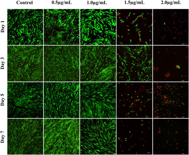
Figure 10. Confocal microscopy images of human fibroblast cells exposed to different concentrations of AgNPs (0.5, 1.0, 1.5, and 2.0 μg per ml) for a week (live cells- green) and dead cells- red) Scale bars at 100 μm. Reprinted with permission from [88]. Copyright (2020).
Download figure:
Standard image High-resolution imageIn another study, the AgNPs showed an excellent antibacterial activity without significant cytotoxicity against human keratinocyte cells. The AgNPs developed through the ethanolic extracts of fenugreek leaves showed antibacterial activity against S. aureus and E. coli bacteria as shown in figure 11. The nanoparticles were further subjected for the cytotoxic studies against HaCaT cells. HaCaT cells are immortalised human keratinocyte cell lines. Through 3-(4, 5-dimethylthiazol-2-yl)-2, 5-diphenyltetrazolium bromide (MTT) assay the cytotoxicity of AgNPs was studied. They exposed the cells with the serial concentrations of silver nanoparticles as depicted in figure 12. They differentiated the cells treated with the lowest and highest concentrations of AgNPs with the control cells for the morphological alterations through microscopy. The AgNPs at 250 μg per ml concentration exerted less cytotoxicity to HaCaT cells as shown in the graphical representation of percentage cell viability. 250μg per ml of concentration of AgNPs showed potential bacterial inhibition. The nanoparticles inhibited the growth of gram positive and gram negative organisms. The zones of inhibitions observed at a concentration of 250 μg per ml was 12 mm for S. aureus, and E. coli displayed the zone of inhibition of 16 mm when treated with 250 μg per ml of AgNPs. Therefore, this method is novel and eco-friendly which results in the formation of sustainable AgNPs with less toxicity to the healthy human cells. This study shows that AgNPs are effective antibacterial materials with no cytotoxic effect on the epithelial cells. Therefore, the nanoparticles are proved non-toxic to epithelial cells [89].
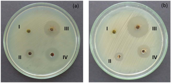
Figure 11. Antibacterial activity of silver nanoparticles at concentration of 250 μg per ml on Staphylococcus aureus (a) and Escherichia coli (b) I. Fenugreek leaves extract, II. Silver nitrate III. Silver nanoparticles and IV. Streptomycin antibiotic. Reprinted with permission from [89]. Copyright (2017).
Download figure:
Standard image High-resolution image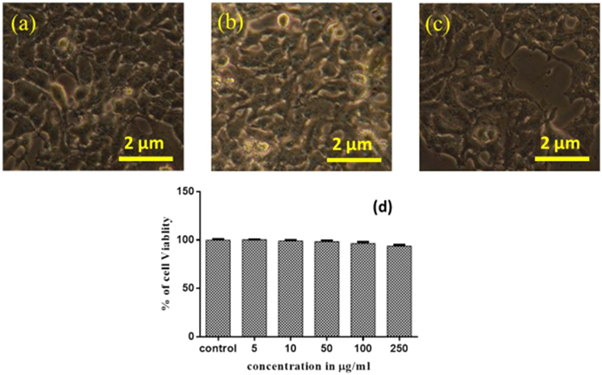
Figure 12. In vitro cytotoxic effect of silver nanoparticles in human keratinocyte cell lines (HaCaT) (a) Control, (b) 5 μg per ml (c) 250 μg per ml of AgNPs treatment and (d) Graphical presentation of percentage cell viability at various concentration of silver nanoparticles. Reprinted with permission from [89]. Copyright (2017).
Download figure:
Standard image High-resolution image
A study of non-antifungal property of nanoparticles is yet to be explored. Agar disk diffusion method [90], Broth micro dilution assays [91], Agar well diffusion method [92], reduction in dry weight determination [93] are some commonly used techniques to study the antifungal activity of nanoparticles.
The nanoparticles can disturb the fungal cell membrane's integrity that results in the inhibition of fungal cells. The cell membrane gets damaged, which leads to the leakage of cellular contents. To the best of our knowledge, the non-antifungal nature of nanoparticles is yet to be reported. The alterations in the redox homeostasis of fungal cells and subsequent oxidative stress result in the membrane damage and imbalance between the osmotic balances of cell. The propidium iodide (PI) staining, real time PCR (RT-PCR), scanning electron microscopy (SEM) techniques can explain the antifungal activity of nanoparticles [94–96]. In the future, these sophisticated techniques can understand the mechanistic approach of antifungal activity of nanoparticles.
5. Future prospects
The significant interactions with the cell membranes, easy translocation can be possible because of the remarkable surface area with a minute size of nanoparticles which results in the distribution of nanoparticles in the human body. Taking into consideration physicochemical and biological properties, it is likely that nanoparticles have unique toxicity mechanisms. There are many research articles available for antibacterial, antifungal and anticancerous activities of various types of nanoparticles. When in vivo applications of these nanoparticles are considered, the particles should not bring about any side effects. Therefore, study of the safety of nanoparticles needs further research. The activity of nanoparticles cannot be same for all cells. The nanoparticles showing the antibacterial activities may or may not be biocompatible for human cells. Therefore, further research regarding the mechanism of activities of nanoparticles in various cell types that is in prokaryotes, eukaryotes, unicellular, multicellular organisms should be carried out. A thorough study of the biocompatible concentrations of nanoparticles for the human system is to be done, to avoid any health hazards to the human healthy cells while using them as a part of therapeutic measure. These nanoparticles will derive various implementations; drug delivery, gene therapy, diagnosis, imaging, and protein, vaccine delivery as a part of therapeutics and diagnosis. When such nanoparticles administered in the body for a treatment purpose, the chronic, long effects of nanoparticles be excluded to offer the safety side for the treatment. Therefore, researchers congregate their attention not only towards fabrication of nanoparticles but also the safety of nanoparticles for the people. The presence of properties non-antibacterial, non-antifungal and non-anticancer makes nanoparticles safer to use for remedy of various pathogenic conditions. Till now only two reports are published on the non-antibacterial and non-anticancerous activities of nanoparticles by Ankamwar et al. [80, 82]. A study of non-antifungal property of nanoparticles is yet to be explored. The nanoparticles ideally with no side effects or else least side effects are to be designed for the biomedical application such as therapeutic and diagnosis.
6. Conclusions
The nanoparticles of non-antibacterial, non-antifungal and non-anticancer properties can be the greatest measures for various therapeutic and diagnostic approaches. The significant applications of nanoparticles as a part of gene delivery, protein delivery, and vaccine delivery would be within reach. These therapeutic applications would not cause any deleterious effects on the microbiome of the living system. The non-specific interactions of the nanoparticles with healthy cells and tissues of the biological system would be diminished with the help of such metallic nanoparticles. Reducing harmful effects on the health is necessary for designing the nanoparticles in such a way that these nanoparticles will not exert the toxicity on the humans. The effects of nanoparticles inside the cells are still not completely understood. Thus, a thorough study on the non-toxic effects of nanoparticles becomes an imperative measure to be employed for various therapeutic applications. The nanoparticles with non-antibacterial, non-antifungal and non-anticancer need to be studied to avoid adverse side effects of nanoparticles on the health. This study plays a significant role when the therapeutic applications are taken into consideration. As researchers focused the attention towards antibacterial, antifungal and anticancer activities of nanoparticles, the non-antibacterial, non-antifungal and non-anticancer properties of nanoparticles eventually bring in forasmuch as a forgotten paradigm.
Acknowledgments
BA extends special gratitude to University Grants Commission-Department of Atomic Energy Consortium for Scientific Research (UGC-DAE CSR), R-5 Shed, Bhabha Atomic Research Centre, Trombay, Mumbai, India (Grant No.UDCSR/MUM/AO/CRS-M-248/2017/1169 dated 14.03.2017) for financial support to Major Research Project. RY thanks Department of Chemistry and Department of Biotechnology, S.P. Pune University for the research work under the supervision of BA.
Data availability statement
All data that support the findings of this study are included within the article (and any supplementary files).
Conflicts of interest
There are no conflicts to declare.