Article

Sustainable Coated Nanostructures Based on Alginate and Electrospun Collagen Loaded with Antimicrobial Agents

by
Ecaterina Matei
1,
Carmen Gaidau
2,
Maria Râpă
1,*,
Laura Mihaela Stefan
3,
Lia-Mara Ditu
4,
Andra Mihaela Predescu
1,
Maria Stanca
2,
Mircea Cristian Pantilimon
1,
Mariana Daniela Berechet
2,
Cristian Predescu
1 and
Anamaria Mosutiu
5
1
Faculty of Materials Science and Engineeing, University POLITEHNICA of Bucharest, 313 Splaiul Independentei, 060042 Bucharest, Romania
2
The National Research & Development Institute for Textiles and Leather—Division Leather and Footwear Research Institute Bucharest, 93 Ion Minulescu Street, 031215 Bucharest, Romania
3
National Institute of Research and Development for Biological Sciences, 296 Splaiul Independenţei, 060031 Bucharest, Romania
4
Faculty of Biology, University of Bucharest, 60101 Bucharest, Romania
5
SPD STAR SRL, Loc. Caseiu No. 136/C, 407155 Cluj, Romania
*
Author to whom correspondence should be addressed.
Coatings 2021, 11(2), 121; https://doi.org/10.3390/coatings11020121
Submission received: 4 January 2021 / Revised: 15 January 2021 / Accepted: 19 January 2021 / Published: 21 January 2021
(This article belongs to the Special Issue Novel Approaches to Hybrid Films or Coatings)

Abstract

:
In this study, sodium alginate film (Alg) was coated with electrospun collagen glue (Col) extracted from rabbit skin waste, loaded with different commercial antimicrobial agents (chitosan, AG425K and ZnONPs) and investigated in terms of morphological, structural and biological properties. The coated nanostructures were characterized using scanning electron microscopy coupled with the energy-dispersive X-ray (SEM/EDS), Attenuated Total Reflectance Fourier-Transform Infrared spectroscopy (ATR FT-IR), and Atomic Force Microscopy (AFM) tests. The cytotoxicity was investigated on murine L929 fibroblasts using 3-[4,5-dimethylthiazol-2-yl]-2,5-diphenyltetrazolium bromide salt (MTT) and lactate dehydrogenase (LDH) assays. Microbiological tests were performed against Staphylococcus aureus ATCC 25923, Escherichia coli ATCC 25922, Pseudomonas aeruginosa ATCC 27853 and Candida albicans ATCC 27853 standard strains. In vitro cell culture tests showed a good cytocompatibility of the coated nanostructured systems, except the sample loaded with ZnONPs, which exhibited a highly cytotoxic effect. Alg-Col-ZnONPs nanostructure inhibited the growth and multiplication of the Staphylococcus aureus ATCC 25923 and Escherichia coli ATCC 25922 bacterial strains. The results of new coated nanostructures may be useful for the development of sustainable biomaterials in a circular economy, with bioactive properties for medical wound dressings.

1. Introduction

Materials with bioactive properties for tissue engineering and long-term antibacterial properties are strongly desired for wound healing. Although many antibiotic-loaded dressings with excellent antibacterial properties have been developed, toxic effects, anaphylactic reactions and the resulting “super bacteria” that occurred due to drug resistance caused growing concern for finding new treatments [1,2,3]. In addition, various metal-based nanoparticles (NP), such as AgNP, ZnO NP and CeO2 NP, with broad-spectrum antibacterial properties and without drug resistance are used as antibacterial agents in the study of wound dressings [4,5]. Metal nanoparticles can be used for wound treatment as inorganic agents, composites or coated nanomaterials formulated using the electrospinning technique [6]. Special properties of nanofibers are explained by their specific surface-area-to-volume ratio and the ability to form porous and interconnected pores [7]. Besides the special surface-to-volume ratio, the nanofibers are fabricated from natural biopolymers, which can mimic the structure of the extra-cellular matrix (ECM). However, the NP toxicity and accumulation of heavy metal ions in the organs of those treated with nanomaterial antibacterial agents cannot be ignored [8]. Thus, finding antibacterial agents with high antibacterial properties as well as proper biomaterials with proven biocompatibility remains a significant challenge for the preparation of new antibacterial wound dressings.
Due to its excellent biocompatibility, sodium alginate (Alg) is one of the most common applicable natural biopolymers for cell encapsulation [9] and tissue engineering [10]. Alginates are polysaccharides obtained from brown cell walls of seaweed in the form of alginic acid [11]. They are composed of β-D-manuronic acid (M) and α-L-guluronic acid (G) sequences and molecular weights in different proportions. It is water-soluble, induces hemostasis and is able to absorb biological fluids [12]. It was reported that the alginate-based dressings can absorb biological fluids up to 20 times their weight [13]. Crosslinking with glutaraldehyde or multivalent ions (usually Ca2+) with blocks of guluronic acid residues from two different chains resulting in a three-dimensional network are preferred [14,15]. Alginate cannot be electrospun itself because of the lack of chain entanglement [16], but compositions based on collagen and alginate for sustainable drug delivery [17,18] and skin dressing [19,20] have been reported.
Collagen is an extracellular matrix protein that plays a major role in tissue regeneration and is one of the most suitable biomaterials for tissue engineering due to its excellent structural properties and biocompatibility [21]. For example, the 3D collagen matrix has been widely used for several platforms in microfluidics, tissue engineering, basic cell culture and so on [22,23]. For decades, many researchers have studied the techniques, physico-chemical and rheological properties, structural and functional properties to obtaining of 3D matrix collagen for the purpose of investigation for practical applications in wound healing, artificial valves, bone fractures, regeneration of skin membrane and tissues [24,25,26,27,28].
Chitosan as a natural polysaccharide recently attracted attention for covering simple alginate in microcapsules due to its excellent biocompatibility [29,30]. Chitosan has the potential to broadcast in the 3D network and to connect to alginate, forming the membrane of the alginate polyelectrolytic complex/chitosan microcapsules [20,31,32].
Within the concept of a circular economy, the use of resources within closed-loop systems represents an approach about the integration of the well-being of the environment with economic activities [33]. For example, the reuse of agro-food waste by-products such as salted egg whites [34], Aspergillus niger prolyl endopeptidase (An-PEP) from fermentation broth [35], and α-Lactalbumin from cow milk whey [36] is beneficial for the reduction of environmental pollution and increase economic value simultaneously. Developing protein-based functional materials derived from the food industry [37,38], which possess especially antioxidant activity and protein-based nanofiber materials for wound healing [39] with the potential to kill bacteria, has also been reported. Rabbit skin can be considered as waste or by-products from the meat industry [40,41] or the leather industry [42]. In this context, the choice of biomaterials with proper bioactive properties for the production of wound healing is a good strategy. Our team has reported the use of indigenous proteins like collagen extracted from bovine leathers, collagen glue from rabbit skins and keratin from wool sheep [43], in combination with different essential oils [44] or antimicrobial agents [45] for the production of electrospun nanofibers for wound medical dressings, in a circular economy. The antimicrobial biohybrid nanofibers wound dressing is a new class of wound dressings taking the advantages to biomimic the extracellular matrix structure, to be biocompatible and biodegradable, and to have a high surface area able to deliver the antimicrobial agents to the infection zones [46]. The goal of this research was to develop sustainable coated nanostructures containing commercial chitosan, AG425K and ZnONPs antimicrobial agents by mixing them with collagen glue extracted from rabbit skin waste and deposited as electrospun nanofibers onto sodium alginate support. The production of these coated nanostructures will be preferred to known products because they do not use native collagen, which is very expensive, or sophisticated compositions containing synthetic polymers with inflammatory potential or toxic organic solvents or antibiotics, which are reported as having adverse effects. In this paper, the morphological (scanning electron microscopy coupled with the energy-dispersive X-ray (SEM/EDS) and Atomic Force Microscopy (AFM)), structural (Attenuated Total Reflectance Fourier-Transform Infrared spectroscopy (ATR-FT-IR)) and biological properties of the coated nanostructures were investigated to find their potential use as suitable biomaterials for the manufacture of 3D matrix for skin regeneration. The biocompatibility of the coated nanostructured systems was performed on L929 fibroblast cells using MTT and LDH assays. Antimicrobial activity of new coated nanostructures systems was assessed by both qualitative and quantitative methods against Staphylococcus aureus ATCC 25923, Escherichia coli ATCC 25922, Pseudomonas aeruginosa ATCC 27853 and Candida albicans ATCC 10231 bacterial strains by using an adapted spot diffusion method and the determination of CFUs/mL.

2. Materials and Methods

2.1. Materials

Sodium alginate (Alg) films with dimensions of 12 × 12 cm2 were prepared by the casting method, in which 1.5% (w/w) sodium alginate (BioChemica, Darmstadt, Germany) was dissolved in distilled water under magnetic stirrer with speed of 600 rpm, at 90 °C for 4 h.
Collagen glue (Col) was extracted from the indigenous rabbit skins [47] by heating the delimed pelt in a water bath for 4 h. It is characterized by 10% dry substance (SR EN ISO 4684:2006), glue strength of 450 g (Texture analyzer, TEX’AN TOUCH 50N, LRami LAMI Rheology Instruments, Champagne au Mont d’Or, France), molecular weight of 15.5 kDa and an electrical conductivity of 820 μS/cm (C1010, Consort, Turnhout, Belgium). The collagen glue was dried by water sublimation at 2 °C and ground into granules or powder using a mortar.
Three commercial antimicrobial agents were used in this study: (1) chitosan [(C6H11O4N)n] high viscosity is described by a viscosity of 1267 MPaxs and a sulfated ash content of 0.2% (Sigma-Aldrich, Darmstadt, Germany); (2) Ag425K, water-based dispersant of titanium dioxide nanoparticles (TiO2 NPs) in the form of anatase, doped with nitrogen and silver nanoparticles (TiPE Nanotechnology Inc., Shanghai, China) (TiO2-N-Ag NPs), with a particle size of 6–8 nm, pH = 7–10, concentrations of 0.72% Ti and 0.86% Ag, with antibacterial, antifungal and antiviral properties, without toxicity (oral LD50 ≥ 10,000 mg/kg); and (3) water-based dispersant of ZnONPs, with a concentration of 20 wt.% ZnONPs, particle size 100 nm (by Transmission Electron Microscopy (TEM) measurement), ≤40 nm (aerodynamic particle sizer, APS), pH = 7.5 ± 1.5 supplied from Sigma Aldrich, Darmstadt, Germany.
All other chemicals used were of analytical grade.

2.2. Preparing of Electrospinning Solution

Collagen glue (Col) dispersion was obtained according to our previous paper [45] by solubilization in distilled water (0.5:1 w/w) and 40% acetic acid.
Col-Chitosan, Col-Ag425K and Col-ZnONPs electrospinning solutions were prepared by mixing of antimicrobial agents in solution form with Col dispersion, as the amounts of solid antimicrobial agents were 0.1 g in the case of ZnONPs and chitosan, and 0.07 g Ti and 0.08 g Ag in the case of Ag425K. Antimicrobial solutions were prepared at room temperature using a magnetic stirrer with an agitation rate of 200 rpm for 2 h once uniform dispersions were achieved. Subsequently, the dispersions were sonicated (HBW Digital Heated Ultrasonic Cleaner, Toolsidee, Waddinxvee, The Netherlands), at 50 W sonication intensity, 24 kHz frequency, at 30 °C for 30 min in order to remove air bubbles.
The physical characteristics, namely the conductivity (C1010, Consort Turnhout, Belgium) and pH (Consort C831 Multiparameter analyzer, Turnhout, Belgium) of the prepared Col-antimicrobial agent solutions, were measured (Table 1).

2.3. Fabrication of Coated Nanostructures

About 20 mL of each resulting solution were introduced in a Teflon syringe with a G21 needle attached to the other end of the uniaxial electrospinning equipment (TL Pro-BM, Tong Li Tech Co., Ltd., Bao An, Shenzhen, China) and deposited onto the Alg films at a 10 cm distance between the needle and collector. The applied positive voltage and flow rate were optimized to the following values: 20.9–22.6 kV and 3 mL/h in the case of Alg-Col and Alg-Col-Ag425K, 14.3 kV and 2 mL/h in the case of Alg-Col-ZnONPs and 24.1 kV and 1 mL/h in the case of Alg-Col-Chitosan. The electrospinning process was performed at 23 ± 2 °C (room temperature) and 53% ± 2% relative humidity. Then, the coated nanostructures were crosslinked by keeping in vapors of a mixture of 0.5% glutaraldehyde and 96% ethanol (1:1 volume ratio) in a desiccator for 48 h. In all methods, the crosslinked samples were used, except SEM analysis.

2.4. Investigation Methods

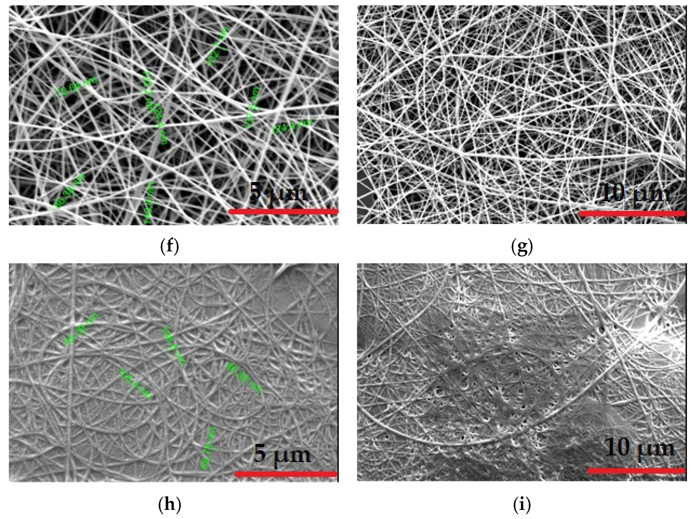
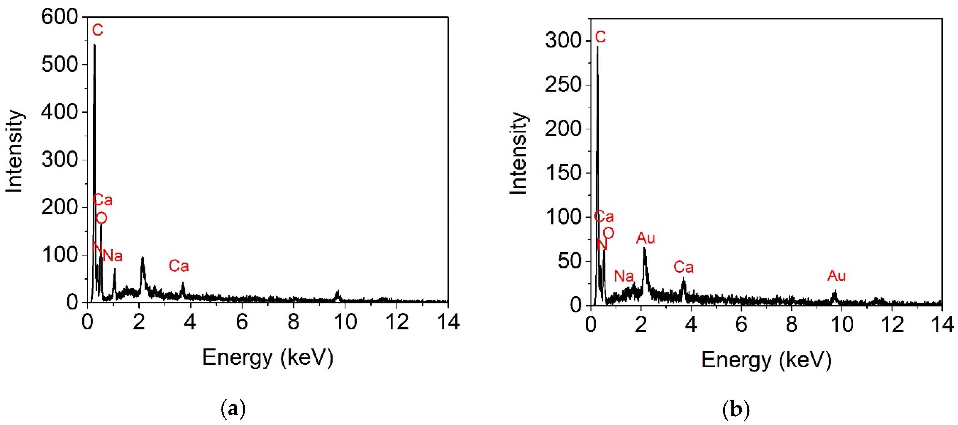
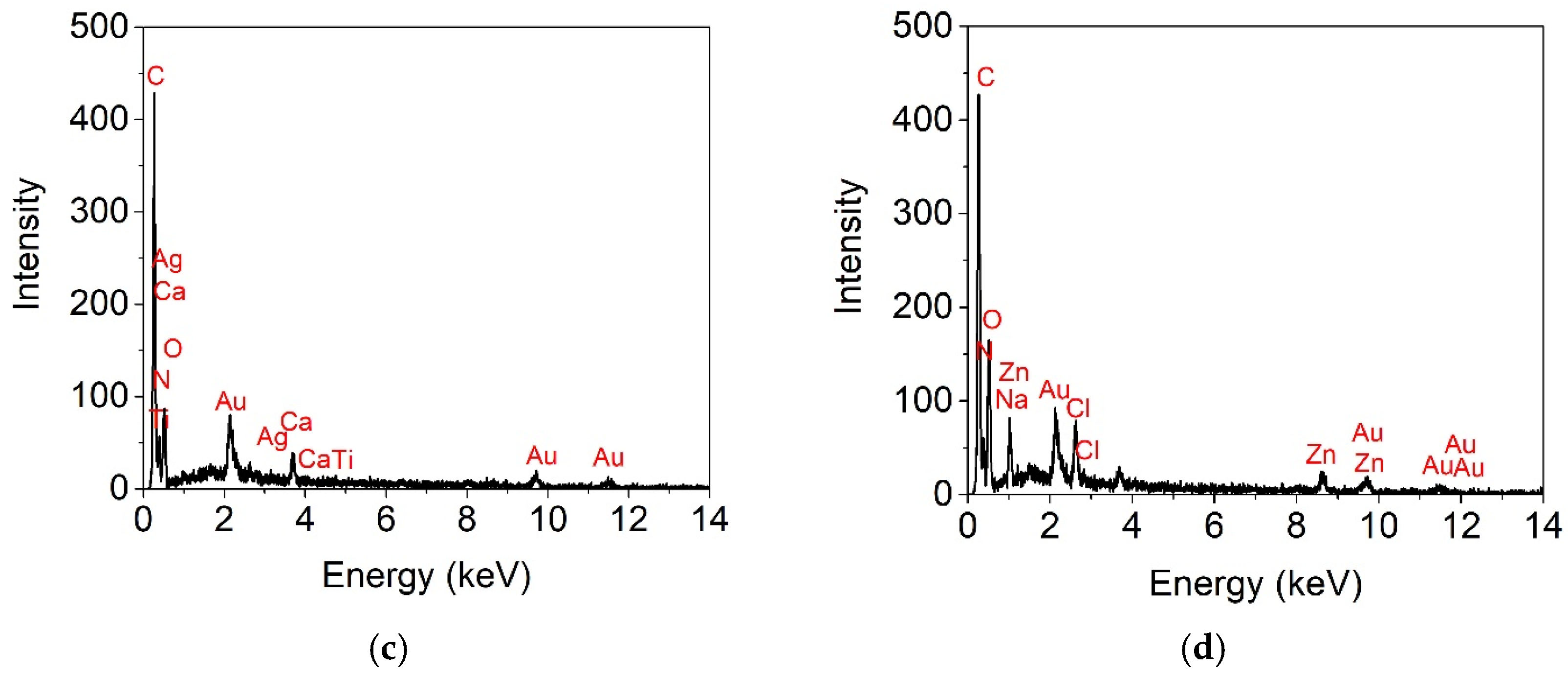
2.4.1. Scanning Electron Microscopy (SEM) Coupled with Energy-Dispersive X-ray Spectroscopy (EDS)

The fiber and diameter size and chemical composition of the coated nanostructures were observed with the help of a scanning electron microscope (SEM), QUANTA 450 FEG (FEI, Eindhoven, The Netherlands) equipped with a field emission gun with 5 kV acceleration voltage and a 1.2 nm resolution X-ray energy-dispersive spectrometer with an acceleration voltage of 30 kV. Before testing, the samples were automatically coated resulting from argon plasma generated by a magnetron and conductive layers of gold using a Q 150R ES sputter coater (Quorum, East Sussex, UK).

2.4.2. Atomic Force Microscopy (AFM)

The surface topography of the coated nanostructures was observed in the tapping mode on an atomic force microscope (AFM) (MultiView 4000SPM/NSOM, Nanonics Imaging LTD, Jerusalem, Israel). SPM images were obtained using the non-contact mode (at a distance in the range of 0.1–10 nm of the sample). The analyzed surface area was 100 m2 with an acquisition speed of 12 ms/point. Root mean square (RMS) was calculated as the root mean square of a surface’s measured microscopic peaks and valleys, while the roughness average (Ra) was the arithmetic average of the absolute values of the roughness profile ordinates. Evaluation of the AFM parameters was achieved by means of WSxM 4.0 software [48].

2.4.3. Fourier-Transform Infrared Spectroscopy—Attenuated Total Reflectance (FT-IR–ATR)

The mid-infrared spectra of electrospun collagen nanofibers loaded with antimicrobial agents and deposited onto Alg film were collected with an INTERSPEC 200-X spectrophotometer (Interspectrum, Tartumaa, Estonia) equipped with an ATR device. The analysis was performed in triplicate by examination the frequency range of 4000–700 cm−1, with 4 cm−1 resolution.

2.4.4. In Vitro Evaluation of Cytotoxicity

In vitro preliminary experiments to assess the potential cytotoxicity of the coated nanostructures were performed on L929 murine fibroblasts, purchased from the European Collection of Authenticated Cell Cultures (ECACC). All samples conditioned as films were cut into disks of 5 mm diameter and sterilized under UV light for 4 h. Cells were grown in Minimum Essential Medium (MEM; Sigma-Aldrich, Steinheim, Germany) supplemented with 10% fetal bovine serum (FBS; Biochrom, Berlin, Germany) and 1% antibiotics (penicillin, streptomycin and neomycin-Sigma-Aldrich, Steinheim, Germany) at 37 °C in a humidified atmosphere with 5% CO2. The cytotoxicity of the samples was evaluated by the direct contact method according to ISO 10993-5 standard. L929 cells were seeded at a density of 5 × 10⁴ cells/mL in 24-well tissue culture plates and incubated for 24 h to allow cell attachment. Then, the culture medium was replaced with fresh medium, samples were added into the wells (1 disk/well) and cells were further maintained in standard conditions for 24 and 48 h, when quantitative MTT 3-[4,5-dimethylthiazol-2-yl]-2,5-diphenyl tetrazolium bromide and LDH assay detecting lactate dehydrogenase activity were performed.
To evaluate the cell viability, cells were incubated with 0.25 mg/mL MTT solution (Sigma-Aldrich, Steinheim, Germany) for 3 h at 37 °C, according to the colorimetric assay described by Mosmann et al. [49]. The insoluble formazan crystals were dissolved with isopropanol, and then the absorbance was measured at 570 nm using the microplate reader Mithras LB 940 (Berthold Technologies, Bad Wildbad, Germany). The amount of formazan was directly correlated to the number of metabolically active cells. The results were expressed as the percentage of viability compared to the control sample (untreated cells), considered 100% viable. Data were presented as the mean of three measurements ± SD.
The cytotoxicity of the electrospun samples was also evaluated by measuring the amount of lactate dehydrogenase released into the culture medium. After 24 and 48 h of treatment, 50 µL of culture media was used to perform the LDH assay using CytoTox96 kit (Promega, Madison, WI, USA), according to the manufacturer’s instructions. The amount of LDH released into the culture medium was recorded at 490 nm using the SPECTROstar® Nano microplate reader (BMG, LABTECH, Ortenberg Germany). The obtained values were directly proportional to the number of cells that lost their cell membrane integrity and, therefore, their viability. Data were presented as the mean of three measurements ± SD.

2.4.5. Antimicrobial Activity

The Staphylococcus aureus ATCC 25923, Escherichia coli ATCC 25922, Pseudomonas aeruginosa ATCC 27853 and Candida albicans ATCC 10231 microbial strains were selected from the collection of microorganisms of the Department of Microbiology, Faculty of Biology, University of Bucharest. To perform the experiment, two successive passages were made by passing the microbial strains on nutritious agar medium and incubating for 24 h at 37 °C. To avoid the impact of contaminants on the experiment, the tested samples were previously sterilized by holding at UV radiation, 30 min on each face.
Qualitative evaluation of the antimicrobial effect was performed by the CLSI adapted spot diffusion method (according to Clinical & Laboratory Standards Institute recommendations, 2020). The microbial suspensions in sterile physiological water, with standard-density 0.5 McFarland (corresponding to 1.5 × 10⁸ CFU/mL), were obtained from 24–48 h bacterial and yeast fresh cultures. The microbial inoculums were uniformly seeded on Muller Hinton agar medium (for bacterial strains) or PDA (potato dextrose agar) (for yeast strain), distributed in Petri dishes, according to CLSI diffusion method (Clinical Laboratory Standard Institute, 2020). Subsequently, the sterile material specimens were disposed on the surface of the inoculated medium and incubated for 24 h/48 h at 37 °C. The sensitivity of microbial strains was assessed by finding the areas of inhibition around the coated nanostructures fragments.
Then, the antimicrobial activity of coated nanostructures samples was quantitatively assessed using Viable Cell Count (VCC) method. The materials were immersed in 1 mL of appropriated broth medium, which was subsequently inoculated with microbial suspension at a final density of 1.5 × 10⁶ CFU/mL. After 24 h of incubation at 37 °C, the microbicide activity of the coated nanostructures samples was evaluated by serial decimal microdilution technique using 96-well microtiter plates. The serial dilutions and plate counts were performed in duplicate, and after incubation at 37 °C for 24 h, the microbial colonies were counted and converted into colony-forming units per milliliter (CFU/mL). Each test was performed in triplicate and repeated on at least three separate occasions.

2.4.6. Statistical Analysis

For biological tests, significant differences between the means of triplicate experiments and the control were determined by using one-way ANOVA statistical analysis (significance difference was noted as * for p < 0.05, and ** for p < 0.01). All data are presented as mean values ± the standard deviations (SD) for biological tests, electrical conductivity and pH measurements.

3. Results

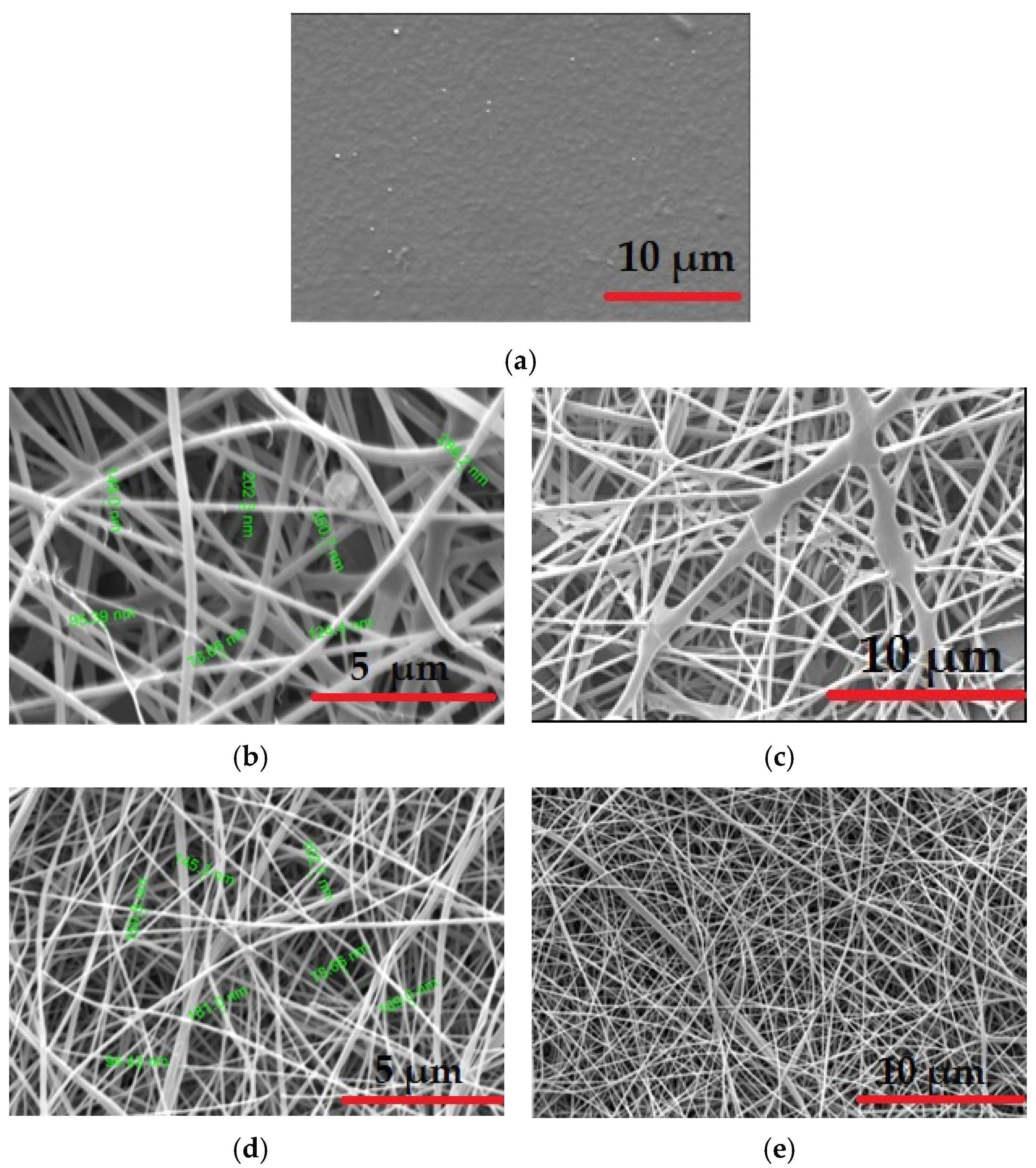
3.1. Scanning Electron Microscopy (SEM) Analysis

The morphology of all coated nanostructures before and after crosslinking was uniform without beads (Figure 1).
Scanning electron microscopy (SEM) image of the Alg film without crosslinking (Figure 1a) shows a smooth and homogeneous surface morphology able to collect the electrospun nanofibers. The prepared solutions for electrospinning as well as the selection of the electrospinning parameters contributed to the obtaining of a relatively wide distribution range for the fiber diameter. These measurements are also due to the nanometric dimension of commercial antimicrobial agents as well as the electrical conductivity of the solutions (Table 1).
According to Figure 1b–i, dense nanofibers covering the Alg films were observed. The dimension of fibers differs depending on composition. Therefore, Alg-Col shows the size fiber distribution in the range of 78.66–350 nm (Figure 1b). The introduction of antimicrobial agent dispersions decreases the size fiber diameter in the range of 72–181 nm, with more uniform fibers produced in the case of Alg-Col-ZnONPs (Figure 1h). This behavior can be explained by the reduction in the viscosity of electrospinning solutions due to the adding of aqueous dispersions [50]. The SEM images of crosslinked samples did not differ significantly from those before crosslinking process.
EDS analysis for the coated nanostructures is presented in Figure 2, while the weight mass of each chemical element from tested compositions is shown in Table 2.
From Table 2, it is observed that nitrogen (N) is found in all coated nanostructures due to the collagen structure. In addition, Alg-Col-Chitosan and Alg-Col-Ag425K samples showed a high amount of N (25.37% and 24.30%), in agreement with the composition of chitosan and Ag425K containing N. The inorganic elements such as Ag, Ti and Zn are found in good correlation with the compositions of antimicrobial dispersions used for electrospinning. Traces of natrium, calcium and chlorine are originated from alginate and collagen glue preparation. Gold presence is due to the antistatic coat used for sample preparation.

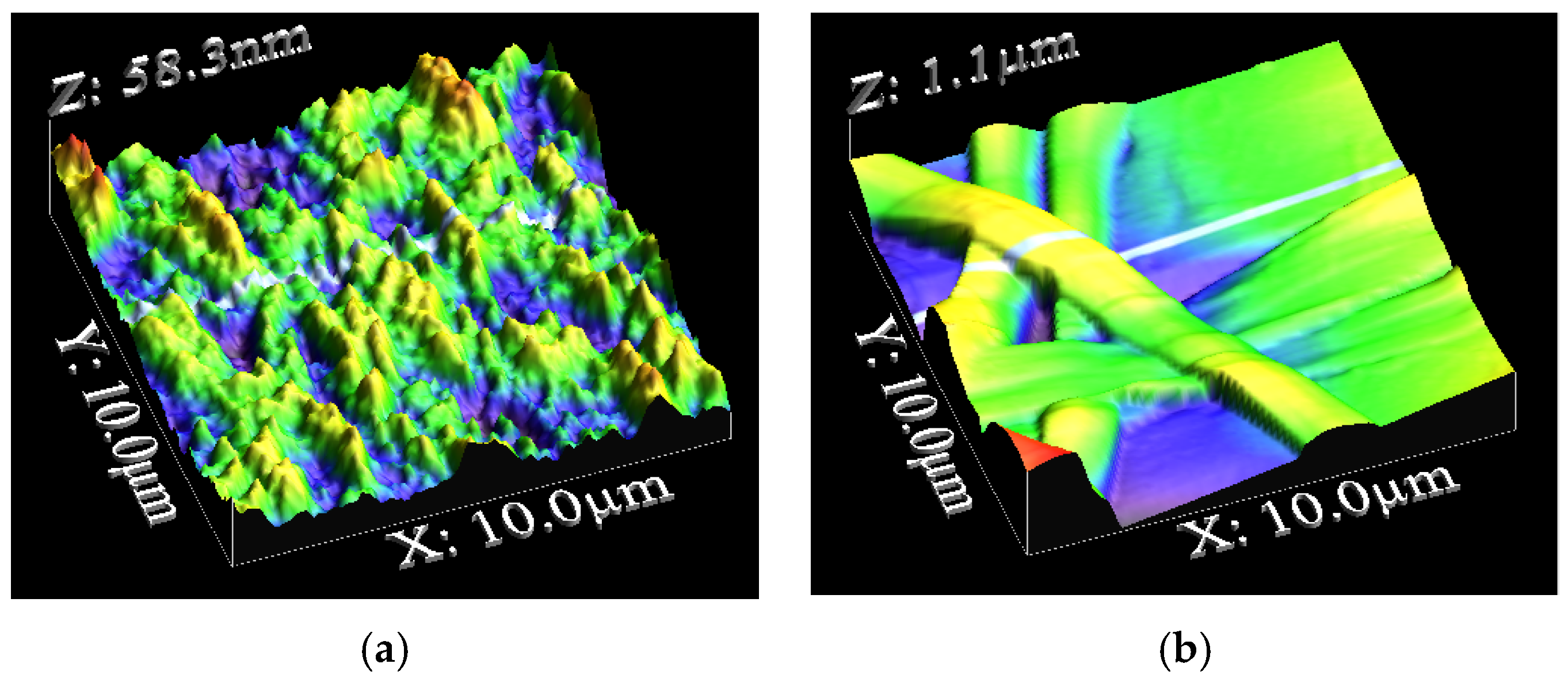
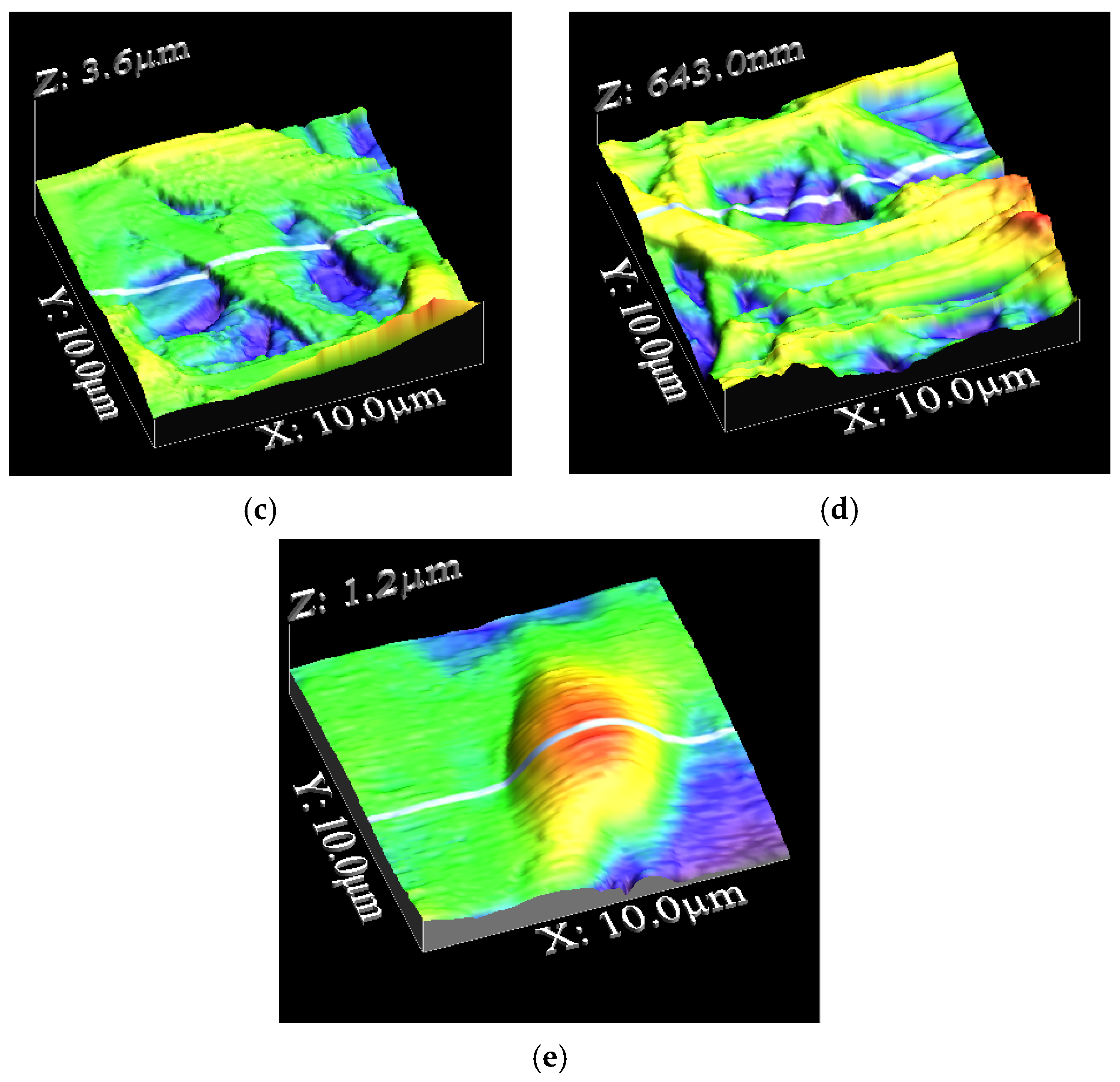
3.2. Atomic Force Microscopy (AFM) Analysis

The surface morphology of the alginate film and coated nanostructures was investigated by AFM analysis (Figure 3).
As shown in Table 3, the coated alginate with collagen (Alg-Col) influences the roughness of Alg film. The Ra value increased for Alg-Col-Chitosan and Alg-Col-ZnONPs samples (191.9 and 148.1 nm) in comparison with the rough surface of Alg-Col (102.9 nm), demonstrating the successful deposition of nanofibers based on collagen with chitosan and ZnONPs antimicrobial agents onto alginate film. The roughness surface of Alg-Col-Ag425K decreased more than that of Alg-Col, probably due to the absorption of small particle size of TiO2NPs doped with nitrogen and AgNPs (size of 6–8 nm) into collagen.

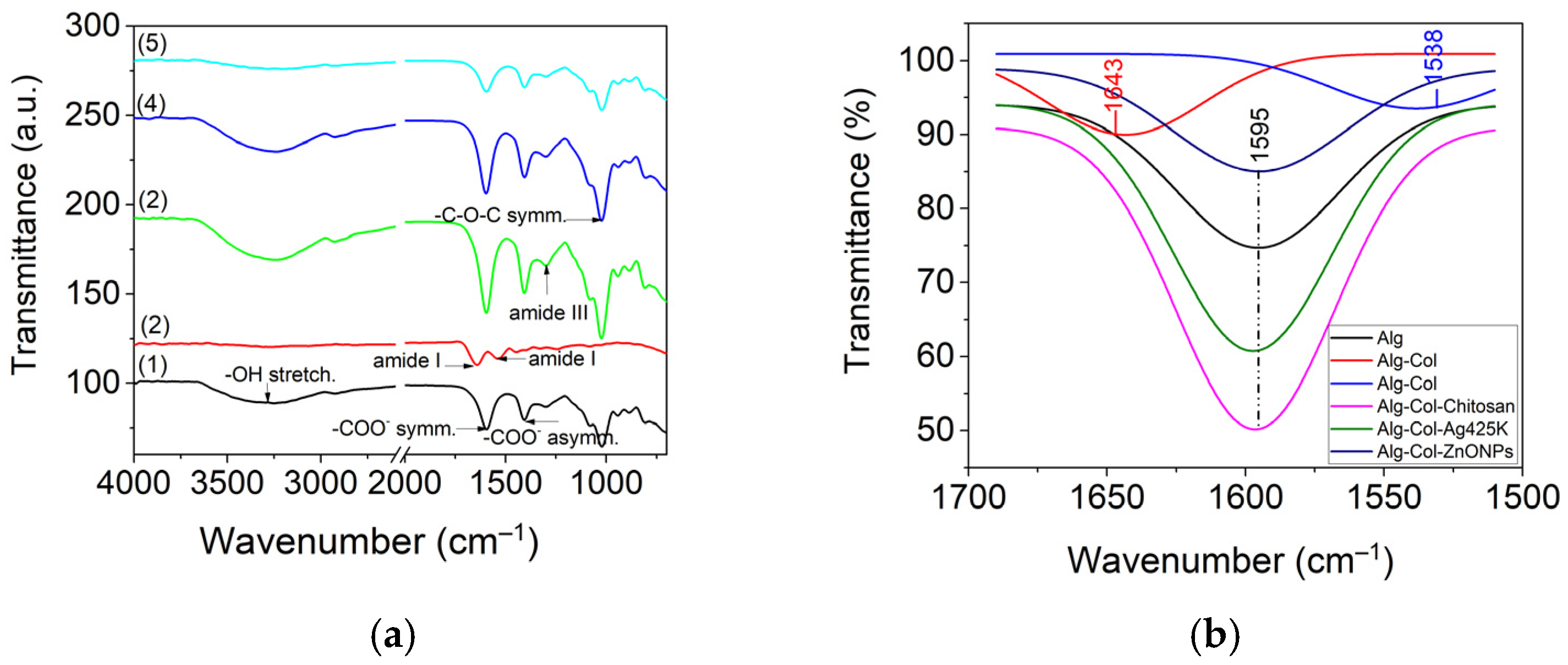
3.3. Fourier-Transform Infrared Spectroscopy—Attenuated Total Reflectance (FT-IR–ATR) Analysis

Figure 4 shows the FT-IR spectra of coated nanostructures from 4000–700 cm−1 range (a) and the deconvolution in the 1700–1500 cm−1 range using the Gauss model (b).
The FTIR spectrum for Alg sample shows characteristic peaks at 1025 cm−1, representing the C–O–C stretching due to the alginate saccharide structure, 3299 cm−1 (OH– stretching) (Figure 4a), and 1595 cm−1 (Figure 4b) and 1405 cm−1 due to the asymmetric and symmetric stretching of carboxylate salt groups, respectively, which were also found in all coated nanostructures [19]. The presence of alginate in the coated nanostructures is also evidenced by the peaks situated around 1031–1030, 1538 cm−1 in Alg-Col, 1596 cm−1 in Alg-Col-Chitosan, 1597 cm−1 in Alg-Col-Ag425K and 1594 cm−1 in Alg-Col-ZnONPs due to amide II (N–H stretching and C–N deformation) [16].
The coated Alg-Col nanostructure shows the absorption peaks at 1205 cm−1 (amide III, stretching vibration of C–H), 1538 cm−1 (amide II band) and 1643 cm−1 associated with the amide I (random coils) (Figure 4b) [51]. The absorption peaks at 1404 and 1449 cm−1 correspond to pyrrolidine ring vibration of hydroxyproline and proline [52]. The coated nanostructures show the peak associated with amide III shifted to high frequencies (1236–1238 cm−1 range) due to the interaction between the antimicrobial agents and collagen. The peaks at 2850 and 2916–2926 cm−1 observed for Alg-Col-ZnONPs and Alg samples are associated with CH3 and CH2 symmetric stretching, respectively. The characteristic peaks of Alg-Col-Chitosan became more intense due to the interaction between the chitosan positively charged and the alginate negatively charged.

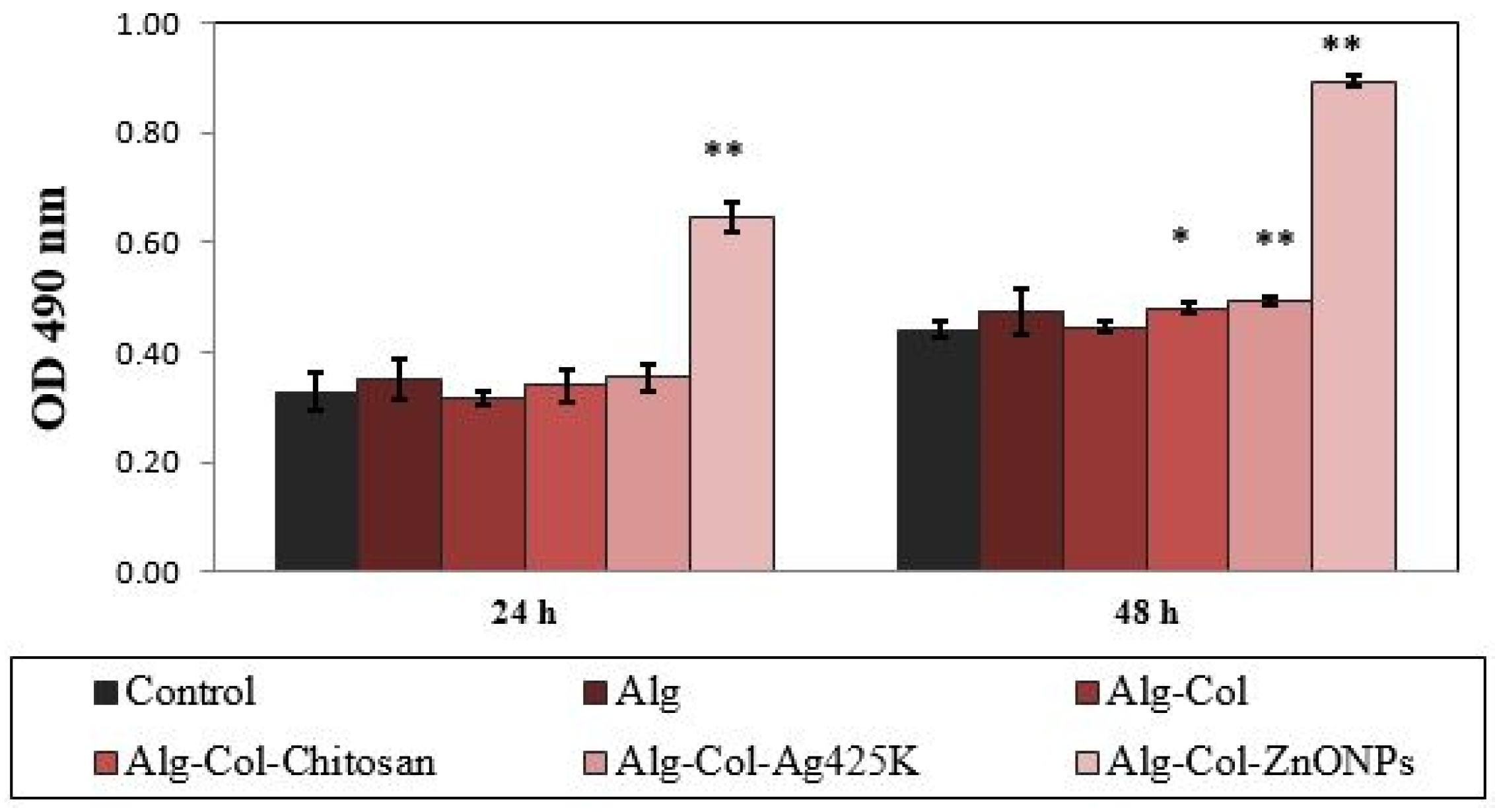
3.4. In Vitro Cytotoxicity Evaluation

The MTT results showed a good cytocompatibility of all tested samples, at both exposure times (24 and 48 h), except for Alg-Col-ZnONPs, which exhibited a severe cytotoxic effect (Figure 5). After 24 h of treatment, the percentages of cell viability ranged between 85.68% for Alg-Col-Ag425K and 97.53% for Alg-Col, while the value for Alg-Col-ZnONPs decreased significantly (12.47%). The statistical differences compared with control were observed at 24 h for Alg (p < 0.01), Alg-Col-Ag425K (p < 0.05) and Alg-Col-ZnONPs (p < 0.01). Although the cell viability slightly decreased after 48 h, the percentages were maintained above 80% (non-cytotoxic effect) for all samples, except Alg-Col-ZnONPs (3.98%) (p < 0.01).
The cell membrane integrity and therefore the degree of cell death were also investigated by measuring the LDH levels secreted into the culture medium. The L929 murine fibroblasts cultivated in the presence of the electrospun materials exhibited low levels of LDH released into the culture medium after 24 and 48 h of treatment, similar to those of the control, except for Alg-Col-ZnONPs, which presented double levels of LDH activity (Figure 6). These results suggested no cytotoxic effect of the coated electrospun alginate and collagen-based materials, except for those containing ZnONPs.

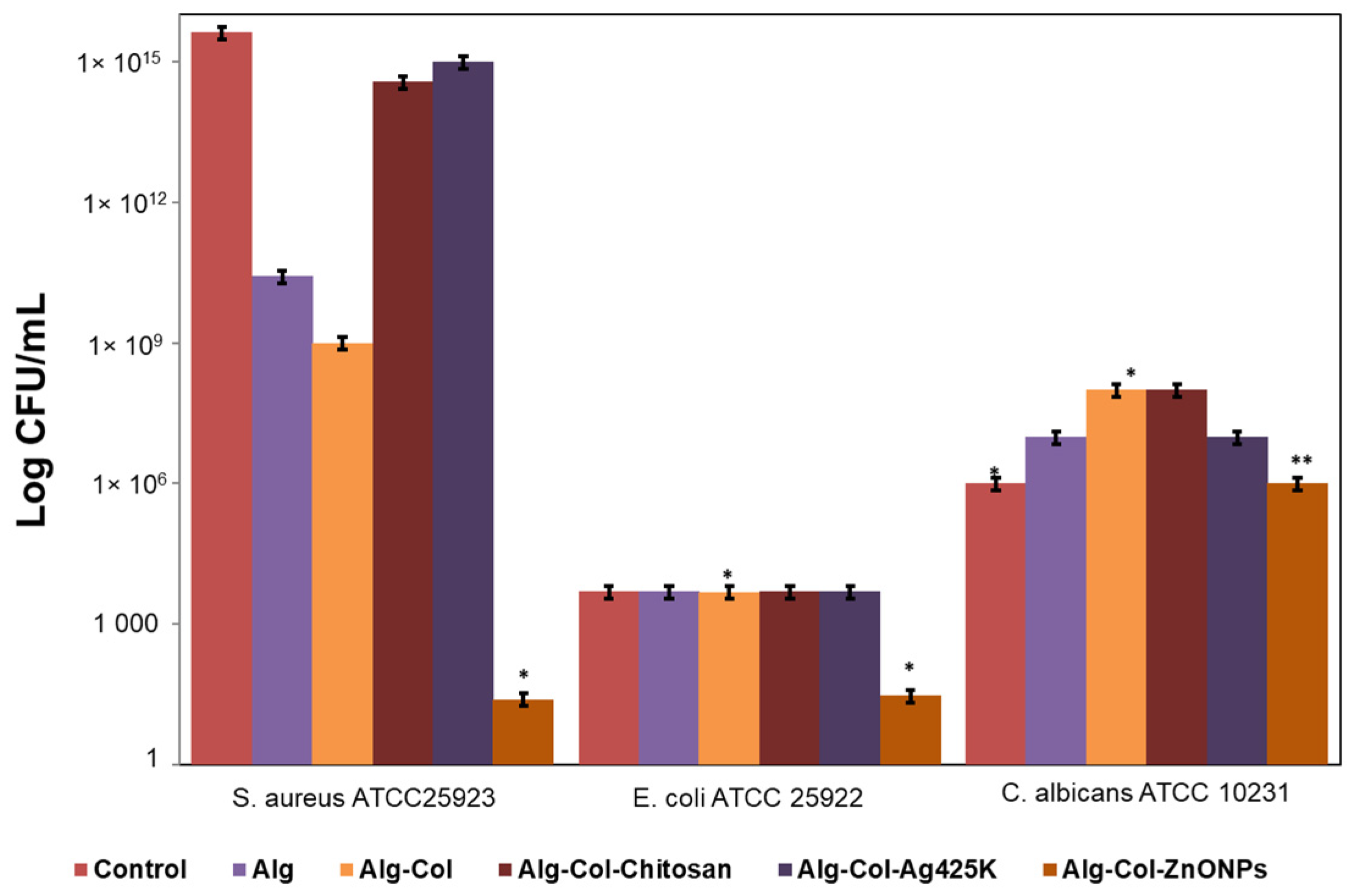
3.5. Antibacterial Assays

The qualitative results showed that a clear growth inhibition area was observed only for the Alg-Col-ZnONPs-coated system tested against Staphylococcus aureus ATCC 25923 and Escherichia coli ATCC 25922, and a less extensive inhibition zone was observed in the case of the Alg-Col toward all the microbial strains. The analyzed samples did not show obvious areas of inhibition against Pseudomonas aeruginosa ATCC 27853 and Candida albicans ATCC 10231 stains. This behavior is due to the accentuated structural instability of the tested coated systems, which were disintegrated shortly after the contact with the surface of the growth media.
For the quantitative assessment of the antimicrobial activity of coated nanostructures samples against Staphylococcus aureus ATCC 25923, Escherichia coli ATCC 25922 and Candida albicans ATCC 10231 standard strains, the Viable Cell Count (VCC) method was performed, and the results are represented in Figure 7.
The antimicrobial activity was exerted by the active compounds released from the tested coated systems against planktonic cells. Evaluating the CFU/mL for planktonic cells, the best effect was also obtained for Alg-Col-ZnONPs nanostructure, which inhibited the growth and multiplication of the Staphylococcus aureus ATCC 25923 (p < 0.05) and Escherichia coli ATCC 25922 (p < 0.05) bacterial strains, with at least 4 logarithmic units in CFU/mL values decrease compared to the growth control (Figure 7). In contrast, the Alg-Col sample did not inhibit the proliferation of Escherichia coli planktonic cells (p < 0.05). In the case of the Candida albicans ATCC 10231 standard strain, the quantitative test was similar to the qualitative test, meaning that the inhibitory effect was not obvious.

4. Discussion

The performance of wound dressings depends on the type of materials/polymers used regarding the specific surface of the contact, porosity or biocompatibility or by the included active substances. In this paper, we prepared sodium alginate films (Alg) and coated them with electrospun nanofibers produced by mixing collagen glue and commercial antimicrobial agents such as chitosan, Ag425K and ZnONPs for the development of potential wound dressings. We used indigenous natural resources based on collagen from rabbit skin glue as an attempt to reduce the environmental pollution, caused by the hazardous leather by-products, in a circular economy. The use of collagen extracted from rabbit skin biowaste in the form of glue has a high potential for the fabrication of biomimetic nanostructures for tissue engineering. Nanofibers obtained by electrospinning technology are able to allow the attachment of cells due to the high surface area of the nanofibers compared to that of the cells. Among other available current technologies, the electrospinning process is non-invasive and does not require the use of coagulation chemicals or high temperatures to obtain nanofibers. The coated nanostructures were characterized in terms of morphology and chemical structure (SEM, EDS, FT-IR), biocompatibility and antimicrobial activities.
Generally, the fiber morphology and topography of the materials can influence cell attachment and proliferation [53]. It was reported that the beaded structures have the lowest cell proliferation rate, ensuring both a less substrate and non-uniformity for cell attachment and growth [54]. We obtained coated nanostructures with uniform morphology, without beads, the nanofibers diameters being between 72 and 181 nm (SEM analysis), closed with the dimension of fibrillar collagen from ECM (50–150 nm) [55]. In our paper, all electrospun nanofibers deposited onto alginate showed a high surface roughness compared with Alg film. This suggests a possible improvement in the cell proliferation and therefore a good biocompatibility. According to the surface roughness of the coated nanostructures, Alg and Alg-Col-Ag425K can be classified as showing nanoroughness (Ra < 100 nm), while Alg-Col, Alg-Col-Chitosan and Alg-Col-ZnONPs show microroughness (Ra in the range of 100 nm–100 μm) [56]. Although the Alg-Col-ZnONPs sample provided a rough surface, it did not pass the cell proliferation test. A similar finding was reported in the case of chitosan/aloe film with controlled delivery of curcumin, showing a rough surface morphology that promotes the regeneration of skin tissues by beneficial interactions with NIH-3T3 cells [57].
The cytotoxic potential of coated nanostructures was evaluated by two quantitative colorimetric assays, which measure the mitochondrial activity (MTT test) and the cell membrane damage (LDH assay). The highest value of cell viability was obtained for Alg-Col (94.27%), followed by Alg-Col-Chitosan with 89.15% (p < 0.01), Alg with 86.98% (p < 0.5) and Alg-Col-Ag425K with 82.58% (p < 0.01), demonstrating a good cytocompatibility. Although the Alg-Col-ZnONPs sample had a high roughness surface (148.1 nm), it was the only one showing an increased degree of cytotoxicity. Our in vitro preliminary results suggested a higher cytocompatibility of electrospun materials based on natural polymers, especially Alg-Col and Alg-Col-Chitosan, compared to those containing inorganic constituents, particularly Ag425K. Our observations were in line with results previously reported, showing enhanced biocompatibility of biomaterials based on naturally derived polymers and their potential use in different fields of tissue engineering. For example, alginate-based chitosan hybrid polymer fibers, containing 0.035% and 0.05% chitosan, promoted in vitro chondrocyte attachment and proliferation [58], whereas higher cell viability and higher expression of specific cartilage gene markers were observed for alginate/collagen bioinks compared to alginate scaffolds [59]. Inorganic materials, such as nanostructured TiO2 or ZnONPs, have been combined with different kinds of polymers in order to produce scaffolds with enhanced biocompatibility and good mechanical properties. Thus, chitin–chitosan/nano TiO2 composite scaffolds, developed using a lyophilization technique for bone tissue engineering, were proved to have no cytotoxic effect on a large array of cell lines, such as osteoblast-like cells (MG-63), fibroblast cells (L929) and human mesenchymal stem cells (hMSCs) [60]. In addition, improved biological properties were also reported for alginate/nanoTiO2 composite scaffolds, suggesting their use for tissue engineering applications [61]. In a recent study, Matei et al. (2020) reported the fabrication of electrospun nanofibers based on collagen glue from rabbit skin (Col) and different antimicrobial agents, such as ZnO NPs, TiO2 NPs doped with nitrogen, Ag NPs and chitosan (CS) in order to be used as nonactive wound dressings. In vitro cell culture tests demonstrated a concentration-dependent cytotoxicity after exposure to L929 murine fibroblasts, with Col/TiO2-N-Ag NPs and Col/CS formulations showing a good biocompatibility on the entire range of tested concentrations (100–1000 µg/mL) and Col/ZnO NPs exhibiting a cytotoxic effect at concentrations higher than 500 µg/mL [45].
The antimicrobial effect of coated nanostructures was examined through both the contact method on solid medium (qualitative test) against Staphylococcus aureus ATCC 25923, Escherichia coli ATCC 25922, Pseudomonas aeruginosa ATCC 27853 and Candida albicans ATCC 10231 bacterial and fungal strains and the quantitative evaluation of CFU/mL. The tests showed clear inhibition zones against Escherichia coli ATCC 25922 and Staphylococcus aureus ATCC 25923 only in the case of Alg-Col-ZnONPs, in good correlation with quantitative tests and fewer inhibition zones in the case of Alg-Col. The other samples were disintegrated when in contact with the culture medium. Similar results were reported in the case of aminated collagen (AC)/oxidized sodium alginate (OSA) hydrogel when a blurry inhibition zone was observed at the contact point with S. aureus [20]. The authors explained that this behavior was due to the high salt concentration of the culture medium, which degrades and overflows the tested hydrogel, hindering the cell growth of S. aureus. In other papers, the beneficial effect of metal oxide nanoparticles combined with biocompatible polymers to produce nanofibers with antimicrobial activity for wound dressing application was reported. For instance, the incorporation of 0.25% and 0.50% (w/w) AgNPs into electrospun chitosan/polyethylene oxide nanofibrous mats intended for wound dressing application led to 100% antibacterial activities against both S. aureus and E. coli bacteria [62]. ZnO NPs added into gelatin nanofibers showed a good antimicrobial effect against S. aureus and E. coli with a significant reduction of bacteria to more than 90% [63]. Although the antimicrobial activity of ZnO NPs is well known, some studies reported the cytotoxicity of ZnO NPs. For example, Cho et al. [64] explained the cytotoxicity of ZnO NPs to be due to their water-soluble ion leading to inhibiting the color of LDH assay. However, more studies for in vitro blood compatibility, angiogenesis to the early stage of wound healing as well as mechanical properties are future research directions needed for the development of new efficient bioactive wound dressing.

5. Conclusions

The novel coated nanostructures based on electrospun collagen–aqueous antimicrobial agents nanofibers deposited onto alginate films were prepared and investigated for assessing these biomaterials as potential candidates for wound medical dressings, thus contributing to environment protection by reducing the quantity of waste from leather industry.
SEM and AFM analyses evidenced the uniform nanofibers with a roughness surface morphology. The characteristic peaks of collagen structure (FT-IR analysis) were found in all coated nanostructures, which evidenced the maintaining of secondary structure from the collagen extracted from rabbit skin waste. In vitro cytotoxicity evaluation indicated a good cytocompatibility for all coated nanostructures, except the one based on ZnONPs. The antibacterial assays suggested that the Alg-Col-ZnONPs-coated nanostructure inhibited the growth of Escherichia coli ATCC 25922 and Staphylococcus aureus ATCC 25923 and decreased with at least 4 logarithmic units in CFU/mL values compared to the growth control. Future studies are needed to clarify the optimum content of antimicrobial agents in the coated nanostructures to ensure the optimum bioactive properties.

Author Contributions

Conceptualization, C.G. and M.R.; data curation, E.M., A.M.P., M.S., and C.P.; formal analysis, L.M.S., L.-M.D., A.M.P., M.D.B., M.C.P., and A.M.; investigation, M.R., L.M.S., L.-M.D., M.D.B., and M.C.P.; methodology, C.G., A.M.P., M.S., and C.P.; project administration, A.M.; software, M.S. and A.M.; supervision, M.R. and C.P.; validation, E.M.; visualization, M.D.B.; writing—original draft, E.M., M.R., L.M.S., and L.-M.D.; writing—review and editing, C.G. All authors have read and agreed to the published version of the manuscript.

Funding

This research was supported by a grant from the Romanian Ministry of Education and Research, CCCDI-UEFISCDI, project no. PN-III-P3-3.5-EUK-2019-0237 within PNCDI III (NonActivPans), Contract 219/23.12.2020 and by National Programme Nucleu, Project No. 25N/2019–19270102 (3D-BIOMAT).

Informed Consent Statement

Not applicable.

Data Availability Statement

This study did not report any data.

Acknowledgments

The FTIR analysis (on INTERSPEC 200-X Spectrophotometer) was possible due to support from the European Regional Development Fund through Competitiveness Operational Program 2014–2020, Priority axis 1, Project No. P_36_611, MySMIS code 107066, and Innovative Technologies for Materials Quality Assurance in Health, Energy and Environmental—Center for Innovative Manufacturing Solutions of Smart Biomaterials and Biomedical Surfaces—INOVABIOMED.

Conflicts of Interest

The authors declare no conflict of interest.

References

  1. Nathan, C.; Cars, O. Antibiotic resistance-problems, progress, and prospects. N. Engl. J. Med. 2014, 371, 1761–1763. [Google Scholar] [CrossRef] [PubMed]
  2. D’Costa, V.M.; King, C.E.; Kalan, L.; Morar, M.; Sung, W.W.L.; Schwarz, C.; Froes, D.; Zazula, G.; Calmels, F.; Debruyne, R.; et al. Antibiotic resistance is ancient. Nature 2011, 477, 457–461. [Google Scholar] [CrossRef] [PubMed]
  3. Jeong, S.-H.; Shin, D.-Y.; Kang, I.-K.; Song, E.-H.; Seong, Y.-J.; Park, J.-U.; Kim, H.-E. Effective wound healing by antibacterial and bioactive calcium-fluoride-containing composite hydrogel dressings prepared using in situ precipitation. ACS Biomater. Sci. Eng. 2018, 4, 2380–2389. [Google Scholar] [CrossRef]
  4. Sener, G.; Hilton, S.A.; Osmond, M.J.; Zgheib, C.; Newsom, J.P.; Dewberry, L.; Singh, S.; Sakthivel, T.S.; Seal, S.; Liechty, K.W.; et al. Injectable, self-healable zwitterionic cryogels with sustained microRNA-cerium oxide nanoparticle release promote accelerated wound healing. Acta Biomater. 2019, 101, 262–272. [Google Scholar] [CrossRef] [PubMed]
  5. Jaiswal, L.; Shankar, S.; Rhim, J.W. Carrageenan-based functional hydrogel film reinforced with sulfur nanoparticles and grapefruit seed extract for wound healing application. Carbohyd. Polym. 2019, 224, 115191. [Google Scholar] [CrossRef]
  6. Mihai, M.M.; Dima, M.B.; Dima, B.; Holban, A.M. Nanomaterials for wound healing and infection control. Materials 2019, 12, 2176. [Google Scholar] [CrossRef] [Green Version]
  7. Jeckson, T.A.; Neo, Y.P.; Sisinthy, S.P.; Gorain, B. Delivery of therapeutics from layer-by-layer electrospun nanofiber matrix for wound healing: An update. J. Pharm. Sci. 2020. [Google Scholar] [CrossRef]
  8. Braydich-Stolle, L.; Hussain, S.; Schlager, J.J.; Hofmann, M.C. In vitro cytotoxicity of nanoparticles in mammalian germline stem cells. Toxicol. Sci. 2005, 88, 412–419. [Google Scholar] [CrossRef] [Green Version]
  9. Wu, Y.; Zhao, Z.; Guan, Y.; Zhang, Y. Galactosylated reversible hydrogels as scaffold for HepG2 spheroid generation. Acta Biomater. 2014, 10, 1965–1974. [Google Scholar] [CrossRef]
  10. Abasalizadeh, F.; Moghaddam, S.V.; Alizadeh, E.; Akbari, E.; Kashani, E.; Fazljou, S.M.B.; Torbati, M.; Akbarzadeh, A. Alginate-based hydrogels as drug delivery vehicles in cancer treatment and their applications in wound dressing and 3D bioprinting. J. Biol. Eng. 2020, 14, 8. [Google Scholar] [CrossRef]
  11. Jönsson, M.; Allahgholi, L.; Sardari, R.R.R.; Hreggviðsson, G.O.; Nordberg Karlsson, E. Extraction and modification of macroalgal polysaccharides for current and next-generation applications. Molecules 2020, 25, 930. [Google Scholar] [CrossRef] [PubMed] [Green Version]
  12. Montalbano, G.; Toumpaniari, S.; Popov, A.; Duan, P.; Chen, J.; Dalgarno, K.; Scott, W.E., III; Ferreira, A.M. Synthesis of bioinspired collagen/alginate/fibrin based hydrogels for soft tissue engineering. Mater. Sci. Eng. C 2018, 91, 236–246. [Google Scholar] [CrossRef] [PubMed]
  13. Kuznetsova, T.A.; Andryukov, B.G.; Besednova, N.N.; Zaporozhets, T.S.; Kalinin, A.V. Marine algae polysaccharides as basis for wound dressings, drug delivery, and tissue engineering: A review. J. Mar. Sci. Eng. 2020, 8, 481. [Google Scholar] [CrossRef]
  14. Kulkarni, A.R.; Soppimath, K.S.; Aralaguppi, M.I.; Aminabhavi, T.M.; Rudzinski, W.E. Preparation of cross-linked sodium alginate microparticles using glutaraldehyde in methanol. Drug Dev. Ind. Pharm. 2000, 26, 1121–1124. [Google Scholar] [CrossRef] [PubMed]
  15. Braccini, I.; Perez, S. Molecular basis of Ca2+-induced gelation in alginates and pectins: The egg-box model revisited. Biomacromolecules 2001, 2, 1089–1096. [Google Scholar] [CrossRef]
  16. Sobhanian, P.; Khorram, M.; Hashemi, S.S.; Mohammadi, A. Development of nanofibrous collagen-grafted poly (vinyl alcohol)/gelatin/alginate scaffolds as potential skin substitute. J. Biol. Macromol. 2019, 130, 977–987. [Google Scholar] [CrossRef] [PubMed]
  17. Wong, F.S.Y.; Tsang, K.K.; Chu, A.M.W.; Chan, B.P.; Yao, K.M.; Lo, A.C.Y. Injectable cell-encapsulating composite alginate-collagen platform with inducible termination switch for safer ocular drug delivery. Biomaterials 2019, 201, 53–67. [Google Scholar] [CrossRef]
  18. Mei, E.; Li, S.; Song, J.; Xing, R.; Li, Z.; Yan, X. Self-assembling collagen/alginate hybrid hydrogels for combinatorial photothermal and immuno tumor therapy. Colloids Surf. A 2019, 577, 570–575. [Google Scholar] [CrossRef]
  19. Iqbal, B.; Muhammad, N.; Jamal, A.; Ahmad, P.; Khan, Z.U.H.; Rahim, A.; Khan, A.S.; Gonfa, G.; Iqbal, J.; Rehman, I.U. An application of ionic liquid for preparation of homogeneous collagen and alginate hydrogels for skin dressing. J. Mol. Liq. 2017, 243, 720–725. [Google Scholar] [CrossRef]
  20. Feng, X.; Zhang, X.; Li, S.; Zheng, Y.; Shi, X.; Li, F.; Guo, S.; Yang, J. Preparation of aminated fish scale collagen and oxidized sodium alginate hybrid hydrogel for enhanced full-thickness wound healing. Int. J. Biol. Macromol. 2020, 164, 626–637. [Google Scholar] [CrossRef]
  21. Karageorgiou, V.; Kaplan, D. Porosity of 3D biomaterial scaffolds and osteogenesis. Biomaterials 2005, 26, 5474–5491. [Google Scholar] [CrossRef] [PubMed]
  22. Rasheena, E.; Jessica, J.B.; Audrey, F.A.; Yang, L. Three-dimensional cell culture systems and their applications in drug discovery and cell-based biosensors. Assay Drug Dev. Technol. 2014, 12, 207–218. [Google Scholar]
  23. Artym, V.V.; Matsumoto, K. Imaging cells in three-dimensional collagen matrix. Curr. Protoc. Cell Biol. 2010, 48, 10.18.1–10.18.20. [Google Scholar] [CrossRef] [Green Version]
  24. Shahin-Shamsabadi, A.; Selvaganapathy, P.R. A rapid biofabrication technique for self-assembled collagen-based multicellular and heterogeneous 3D tissue constructs. Acta Biomater. 2019, 92, 172–183. [Google Scholar] [CrossRef] [PubMed]
  25. Jeevithan, E.; Saravanakumar, K.; Rahman, S.U.; Henrotin, Y.; Regenstein, J.M.; Wu, W.H.; Bao, B. Chitosan-collagen 3D matrix mimics trabecular bone and regulates rankl mediated paracrine cues of differentiated osteoblast and mesenchymal stem cells for bone marrow macrophage-derived osteoclastogenesis. Biomolecules 2019, 9, 173. [Google Scholar] [CrossRef] [Green Version]
  26. Meng, X.; Leslie, P.; Zhang, Y.; Dong, J. Stem cells in a three-dimensional scaffold environment. Springerplus 2014, 3, 80. [Google Scholar] [CrossRef] [Green Version]
  27. Bosi, S.; Rauti, R.; Laishram, J.; Turco, A.; Lonardoni, D.; Nieus, T.; Prato, M.; Scaini, D.; Ballerini, L. From 2D to 3D: Novel nanostructured scaffolds to investigate signalling in reconstructed neuronal networks. Sci. Rep. 2015, 5, 9562. [Google Scholar] [CrossRef] [Green Version]
  28. Ravi, M.; Paramesh, V.; Kaviya, S.R.; Anuradha, E.; Paul Solomon, F.D. 3D cell culture systems: Advantages and applications. J. Cell. Physiol. 2015, 230, 16–26. [Google Scholar] [CrossRef]
  29. Zhang, W.; Zhao, S.; Rao, W.; Snyder, J.; Choi, J.K.; Wang, J.; Khan, I.A.; Saleh, N.B.; Mohler, P.J.; Yu, J. A novel core-shell microcapsule for encapsulation and 3D culture of embryonic stem cells. J. Mater. Chem. B 2013, 1, 1002–1009. [Google Scholar] [CrossRef]
  30. Santos, E.; Zarate, J.; Orive, G.; Hernández, R.M.; Pedraz, J.L. Biomaterials in Cell Microencapsulation. In Therapeutic Applications of Cell Microencapsulation; Pedraz, J.L., Orive, G., Eds.; Springer: New York, NY, USA, 2010; Volume 670. [Google Scholar] [CrossRef]
  31. Aghdam, S.K.; Khoshfetrat, A.B.; Rahbarghazi, R.; Jafarizadeh-Malmiri, H.; Khaksar, M. Collagen modulates functional activity of hepatic cells inside alginate-galactosylated chitosan hydrogel microcapsules. Int. J. Biol. Macromol. 2020, 156, 1270–1278. [Google Scholar] [CrossRef]
  32. Elango, J.; Selvaganapathy, P.R.; Lazzarid, G.; Bao, B.; Wenhui, W. Biomimetic collagen-sodium alginate-titanium oxide (TiO2) 3D matrix supports differentiated periodontal ligament fibroblasts growth for periodontal tissue regeneration. Int. J. Biol. Macromol. 2020, 163, 9–18. [Google Scholar] [CrossRef] [PubMed]
  33. Murray, A.; Skene, K.; Haynes, K. The circular economy: An interdisciplinary exploration of the concept and application in a global context. J. Bus. Ethics 2017, 140, 369–380. [Google Scholar] [CrossRef] [Green Version]
  34. Jiang, B.; Na, J.; Wang, L.; Li, D.; Liu, C.; Feng, Z. Reutilization of food waste: One-step extration, purification and characterization of ovalbumin from salted egg white by aqueous two-phase flotation. Foods 2019, 8, 286. [Google Scholar] [CrossRef] [PubMed] [Green Version]
  35. Jiang, B.; Wang, M.; Wang, X.; Wu, S.; Li, D.; Liu, C.; Feng, Z.; Li, J. Effective separation of prolyl endopeptidase from Aspergillus Niger by aqueous two phase system and its characterization and application. Int. J. Biol. Macromol. 2021, 169, 384–395. [Google Scholar] [CrossRef]
  36. Jiang, B.; Wang, L.; Wang, M.; Wu, S.; Wang, X.; Li, D.; Liu, C.; Feng, Z.; Chi, Y. Direct separation and purification of α-Lactalbumin from cow milk whey by aqueous two-phase flotation of thermo-sensitive polymer/phosphate. J. Sci. Food Agric. 2021. [Google Scholar] [CrossRef]
  37. Jiang, B.; Wang, X.; Wang, L.; Wu, S.; Li, D.; Liu, C.; Feng, Z. Fabrication and characterization of a microemulsion stabilized by integrated phosvitin and gallic acid. J. Agric. Food Chem. 2020, 68, 5437–5447. [Google Scholar] [CrossRef]
  38. Wang, Q.; Liu, W.; Tian, B.; Li, D.; Liu, C.; Jiang, B.; Feng, Z. Preparation and characterization of coating based on protein nanofibers and polyphenol and application for salted duck egg yolks. Foods 2020, 9, 449. [Google Scholar] [CrossRef] [Green Version]
  39. Ahn, S.; Chantre, C.O.; Gannon, A.R.; Lind, J.U.; Campbell, P.H.; Grevesse, T.; O’Connor, B.B.; Parker, K.K. Soy protein/cellulose nanofiber scaffolds mimicking skin extracellular matrix for enhanced wound healing. Adv. Healthc. Mater. 2018, 7, e1701175. [Google Scholar] [CrossRef]
  40. Ma, M.; Ma, L.; Yu, W.; Zhang, X.; Shen, Y.; Zhang, Y. Research on rapid gelatinization of rabbit skin collagen as effect of acid treatment. Food Hydrocoll. 2018, 77, 945–951. [Google Scholar] [CrossRef]
  41. Zhang, X.; Ma, L.; Yu, Y.; Zhou, H.; Guo, T.; Dai, H.; Zhang, Y. Physico-mechanical and antioxidant properties of gelatin film from rabbit skin incorporated with rosemary acid. Food Packag. 2019, 19, 121–130. [Google Scholar] [CrossRef]
  42. Kanuri, W.D.; Onyuka, A.; Tanui, R. An investigation on the properties of rabbit leather from different tannages. IJSRP 2019, 9, 12–17. [Google Scholar]
  43. Râpa, M.; Gaidau, C.; Stefan, L.M.; Matei, E.; Niculescu, M.; Berechet, M.D.; Stanca, M.; Tablet, C.; Tudorache, M.; Gavrila, R.; et al. New nanofibers based on protein by-products with bioactive potential for tissue engineering. Materials 2020, 13, 3149. [Google Scholar] [CrossRef] [PubMed]
  44. Berechet, M.D.; Gaidau, C.; Miletic, A.; Pilic, B.; Râpă, M.; Stanca, M.; Ditu, L.-M.; Constantinescu, R.; Lazea-Stoyanova, A. Bioactive properties of nanofibres based on concentrated collagen hydrolysate loaded with thyme and oregano essential oils. Materials 2020, 13, 1618. [Google Scholar] [CrossRef] [PubMed] [Green Version]
  45. Matei, E.; Gaidau, C.; Râpă, M.; Constantinescu, R.; Savin, S.; Berechet, M.D.; Predescu, A.M.; Berbecaru, A.C.; Coman, G.; Predescu, C. Sustainable rabbit skin glue to produce bioactive nanofibers for nonactive wound dressings. Materials 2020, 13, 5388. [Google Scholar] [CrossRef] [PubMed]
  46. Homaeigohar, S.; Boccaccini, A.R. Antibacterial biohybrid nanofibers for wound dressings. Acta Biomater. 2020, 107, 25–49. [Google Scholar] [CrossRef] [PubMed]
  47. Berechet, M.D.; Niculescu, M.; Gaidau, C.; Ignat, M.; Epure, D.G. Alkaline-Enzymatic hydrolyses of wool waste for different applications. Rev. Chim. Buchar. 2018, 69, 1649–1654. [Google Scholar] [CrossRef]
  48. Horcas, I.; Fernández, R.; Gómez-Rodríguez, J.M.; Colchero, J.; Gómez-Herrero, J.; Baro, A.M. WSXM: A software for scanning probe microscopy and a tool for nanotechnology. Rev. Sci. Instrum. 2007, 78, 013705. [Google Scholar] [CrossRef]
  49. Mosmann, T. Rapid colorimetric assay for cellular growth and survival: Application to proliferation and cytotoxicity assays. J. Immunol. Methods 1983, 65, 55–63. [Google Scholar] [CrossRef]
  50. Mwiiri, F.K.; Brandner, J.M.; Daniels, R. Electrospun bioactive wound dressing containing colloidal dispersions of birch bark dry extract. Pharmaceutics 2020, 12, 770. [Google Scholar] [CrossRef]
  51. Drobota, M.; Gradinaru, L.M.; Vlad, S.; Bargan, A.; Butnaru, M.; Angheloiu, M.; Aflori, M. Preparation and characterization of electrospun collagen based composites for biomedical applications. Materials 2020, 13, 3961. [Google Scholar] [CrossRef]
  52. Felician, F.F.; Yu, R.H.; Li, M.Z.; Li, C.J.; Chen, H.Q.; Jiang, Y.; Tang, T.; Qi, W.Y.; Xu, H.M. The wound healing potential of collagen peptides derived from the jellyfish Rhopilema esculentum. Chin. J. Traumatol. 2019, 22, 12–20. [Google Scholar] [CrossRef] [PubMed]
  53. Guehennec, L.L.; Lopez-Heredia, M.A.; Enkel, B.; Weiss, P.; Amouriq, Y.; Layrolle, P. Osteoblastic cell behaviour on different titanium implant surfaces. Acta Biomat. 2008, 4, 535–543. [Google Scholar] [CrossRef] [PubMed]
  54. Chen, M.; Patra, P.K.; Warner, S.B.; Bhowmick, S. Role of fiber diameter in adhesion and proliferation of NIH 3T3 fibroblast on electrospun polycaprolactone scaffolds. Tissue Eng. 2007, 13, 579–587. [Google Scholar] [CrossRef] [PubMed]
  55. Riesle, J.; Hollander, A.P.; Langer, R.; Freed, L.E.; Vunjak Novakovic, G. Collagen in tissue-engineered cartilage: Types, structure, and crosslinks. J. Cell. Biochem. 1998, 71, 313. [Google Scholar] [CrossRef]
  56. Gonçalves, S.; Dourado, F.; Rodrigues, L.R. Overview on Cell-Biomaterial Interactions. In Advanced Polymers in Medicine; Puoci, F., Ed.; Springer: Cham, Switzerland, 2015; pp. 91–128. [Google Scholar] [CrossRef]
  57. Liu, X.; You, L.; Tarafder, S.; Zou, L.; Fang, Z.; Chen, J.; Lee, C.H.; Zhang, Q. Curcumin releasing chitosan/aloe membrane for skin regeneration. Chem. Eng. J. 2019, 359, 1111–1119. [Google Scholar] [CrossRef]
  58. Iwasaki, N.; Yamane, S.T.; Majima, T.; Kasahara, Y.; Minami, A.; Harada, K.; Nonaka, S.; Maekawa, N.; Tamura, H.; Tokura, S.; et al. Feasibility of polysaccharide hybrid materials for scaffolds in cartilage tissue engineering: Evaluation of chondrocyte adhesion to polyion complex fibers prepared from alginate and chitosan. Biomacromolecules 2004, 10, 828–833. [Google Scholar] [CrossRef]
  59. Yang, X.; Lu, Z.; Wu, H.; Li, W.; Zheng, L.; Zhao, J. Collagen-alginate as bioink for three-dimensional (3D) cell printing based cartilage tissue engineering. Mater. Sci. Eng. C 2018, 83, 195–201. [Google Scholar] [CrossRef]
  60. Jayakumar, R.; Ramachandran, R.; Divyarani, V.V.; Chennazhi, K.P.; Tamura, H.; Nair, S.V. Fabrication of chitin-chitosan/nano TiO2-composite scaffolds for tissue engineering applications. Int. J. Biol. Macromol. 2011, 48, 336–344. [Google Scholar] [CrossRef]
  61. Rani, V.D.; Ramachandran, R.; Chennazhi, K.P.; Tamura, H.; Nair, S.V.; Jayakumar, R. Fabrication of alginate/nanoTiO2 needle composite scaffolds for tissue engineering applications. Carbohydr. Polym. 2011, 83, 858–864. [Google Scholar] [CrossRef]
  62. Kohsari, I.; Shariatinia, Z.; Mahdi, S. Antibacterial electrospun chitosan—polyethylene oxide nanocomposite mats containing bioactive silver nanoparticles. Carbohydr. Polym. 2016, 140, 287–298. [Google Scholar] [CrossRef]
  63. Chen, Y.; Lu, W.; Guo, Y.; Zhu, Y.; Song, Y. Electrospun gelatin fibers surface loaded ZnO particles as a potential biodegradable antibacterial wound dressing. Nanomaterials 2019, 9, 525. [Google Scholar] [CrossRef] [PubMed] [Green Version]
  64. Cho, W.S.; Duffin, R.; Bradley, M.; Megson, I.L.; MacNee, W.; Lee, J.K.; Jeong, J.; Donaldson, K. Predictive value of in vitro assays depends on the mechanism of toxicity of metal oxide nanoparticles. Part. Fibre Toxicol. 2013, 10, 55. [Google Scholar] [CrossRef] [PubMed] [Green Version]
Figure 1. SEM images of the coated nanostructures before (20,000×) and after crosslinking (10,000×) for Alg (a), Alg-Col (b,c), Alg-Col-Chitosan (d,e), Alg-Col-Ag425K (f,g) and Alg-Col-ZnONPs (h,i).
Coatings 11 00121 g001aCoatings 11 00121 g001b
Figure 2. EDS patterns of the coated nanostructures for Alg-Col (a), Alg-Col-Chitosan (b), Alg-Col-Ag425K (c) and Alg-Col-ZnONPs (d).
Coatings 11 00121 g002aCoatings 11 00121 g002b
Figure 3. AFM topography of the coated nanostructures for Alg (a), Alg-Col (b), Alg-Col-Chitosan (c), Alg-Col-Ag425K (d) and Alg-Col-ZnONPs (e).
Coatings 11 00121 g003aCoatings 11 00121 g003b
Figure 4. FT-IR spectra for coated nanostructures, in the range of 4000–700 cm−1 (a), where (1)—Alg, (2)—Alg-Col, (3)—Alg-Col-Chitosan, (4)—Alg-Col-Ag425K and (5)—Alg-Col-ZnONPs, and 1700–1500 cm−1 (b).
Coatings 11 00121 g004
Figure 5. Viability of L929 murine fibroblasts cultivated in the presence of the coated electrospun nanostructures for 24 and 48 h, evaluated by the MTT assay. Samples were reported to the untreated cells (control), considered to have 100% viability. Data were expressed as mean values ± SD (n = 3). * p < 0.05 and ** p < 0.01 compared to the control.
Coatings 11 00121 g005
Figure 6. LDH activity released into the culture medium by L929 murine fibroblasts cultivated in the presence of the coated electrospun nanostructures for 24 and 48 h. Data were expressed as mean values ± SD (n = 3). * p < 0.05 and ** p < 0.01 compared to the control.
Coatings 11 00121 g006
Figure 7. Graphical representation of the logarithmic values of CFU/mL showing the quantification of the antimicrobial activity of the tested materials. Data are expressed as mean values ± SD (n = 3).
Coatings 11 00121 g007
Table 1. Physical characteristics of antimicrobial collagen glue dispersions prior to coating the Alg films by electrospinning process.
PropertyColCol-ChitosanCol-Ag425KCol-ZnONPs
Electrical conductivity at 25 °C (mS/cm)4.40 ± 0.154.73 ± 0.165.18 ± 0.125.96 ± 0.15
pH at 23.4 °C (pH units)3.10 ± 0.12.90 ± 0.13.20 ± 0.13.30 ± 0.1
Table 2. Elements analysis (wt.%) via energy-dispersive X-ray spectrometry (EDS) for coated nanostructures obtained by electrospinning.
ElementAlg-ColAlg-Col-ChitosanAlg-Col-Ag425KAlg-Col-ZnONPs
Carbon (C)48.2145.7946.3444.10
Nitrogen (N)21.8325.3724.3020.12
Oxygen (O)26.4724.0124.7429.53
Natrium (Na)2.990.510.01
Zinc (Zn)1.62
Silver (Ag)0.17
Titanium (Ti)0.18
Calcium (Ca)0.500.990.900.56
Chloride (Cl)0.75
Gold (Au)3.333.373.31
Table 3. RMS and Ra parameters evaluated from AFM analysis for coated nanostructures obtained by electrospinning.
AFM ParametersAlgAlg-ColAlg-Col-ChitosanAlg-Col-Ag425KAlg-Col-ZnONPs
RMS (nm)7.75139.8206.092.6208.8
Ra (nm)6.14102.9191.973.3148.1
Publisher’s Note: MDPI stays neutral with regard to jurisdictional claims in published maps and institutional affiliations.

