Trimethylornithine Membrane Lipids: Discovered in Planctomycetes and Identified in Diverse Environments


Department of Environmental Science, School of Earth and the Environment, Rowan University, Glassboro, NJ 08028, USA
Metabolites 2021, 11(1), 49; https://doi.org/10.3390/metabo11010049
Submission received: 23 November 2020
/
Revised: 3 January 2021
/
Accepted: 5 January 2021
/
Published: 12 January 2021
(This article belongs to the Special Issue Metabolomics of Fatty Acyl Esters of Hydroxy Fatty Acids: Three Diverse Lipid Families)
Abstract
:Intact polar membrane lipids (IPLs) are the building blocks of all cell membranes. There is a wide range of phosphorus-free IPL structures, including amino acid containing IPLs, that can be taxonomically specific. Trimethylornithine membrane lipids (TMOs) were discovered in northern wetland Planctomycete species that were isolated and described in the last decade. The trimethylated terminal nitrogen moiety of the ornithine amino acid in the TMO structure gives the lipid a charged polar head group, similar to certain phospholipids. Since their discovery, TMOs have been identified in various other recently described northern latitude Planctomycete species, and in diverse environments including tundra soil, a boreal eutrophic lake, meso-oligotrophic lakes, and hot springs. The majority of environments or enrichment cultures in which TMOs have been observed include predominately heterotrophic microbial communities involved in the degradation of recalcitrant material and/or low oxygen methanogenic conditions at primarily northern latitudes. Other ecosystems occupied with microbial communities that possess similar metabolic pathways, such as tropical peatlands or coastal salt marshes, may include TMO producing Planctomycetes as well, further allowing these lipids to potentially be used to understand microbial community responses to environmental change in a wide range of systems. The occurrence of TMOs in hot springs indicates that these unique lipids could have broad environmental distribution with different specialized functions. Opportunities also exist to investigate the application of TMOs in microbiome studies, including forensic necrobiomes. Further environmental and microbiome lipidomics research involving TMOs will help reveal the evolution, functions, and applications of these unique membrane lipids.
1. Introduction—Amino Acid Containing Membrane Lipids
Cell membranes are the interface between biological processes and the environment [1]. Intact polar lipids (IPLs) are the constituent components of the cell membrane lipid bilayer, consisting of a polar head group and an apolar core. The diverse molecular structures of IPLs can provide functional and taxonomic information on the microbial community of a given environment [2,3]. Following cell lysis, the head groups of IPLs are rapidly cleaved from the core lipids, on the order of days, thus making IPLs representative of living biomass [4,5]. The structures of IPL head groups are very diverse, including common phosphoglycerol IPLs [6] (e.g., phosphatidylcholine, PC; phosphatidylethanolamine, PE; etc.), and various bacterial IPLs that have polar head groups containing amino acids [7]. The amino acid IPLs phosphatidylserine and homoserine-containing betaine lipids have a glycerol backbone, while other amino acid containing lipids are glycerol free.
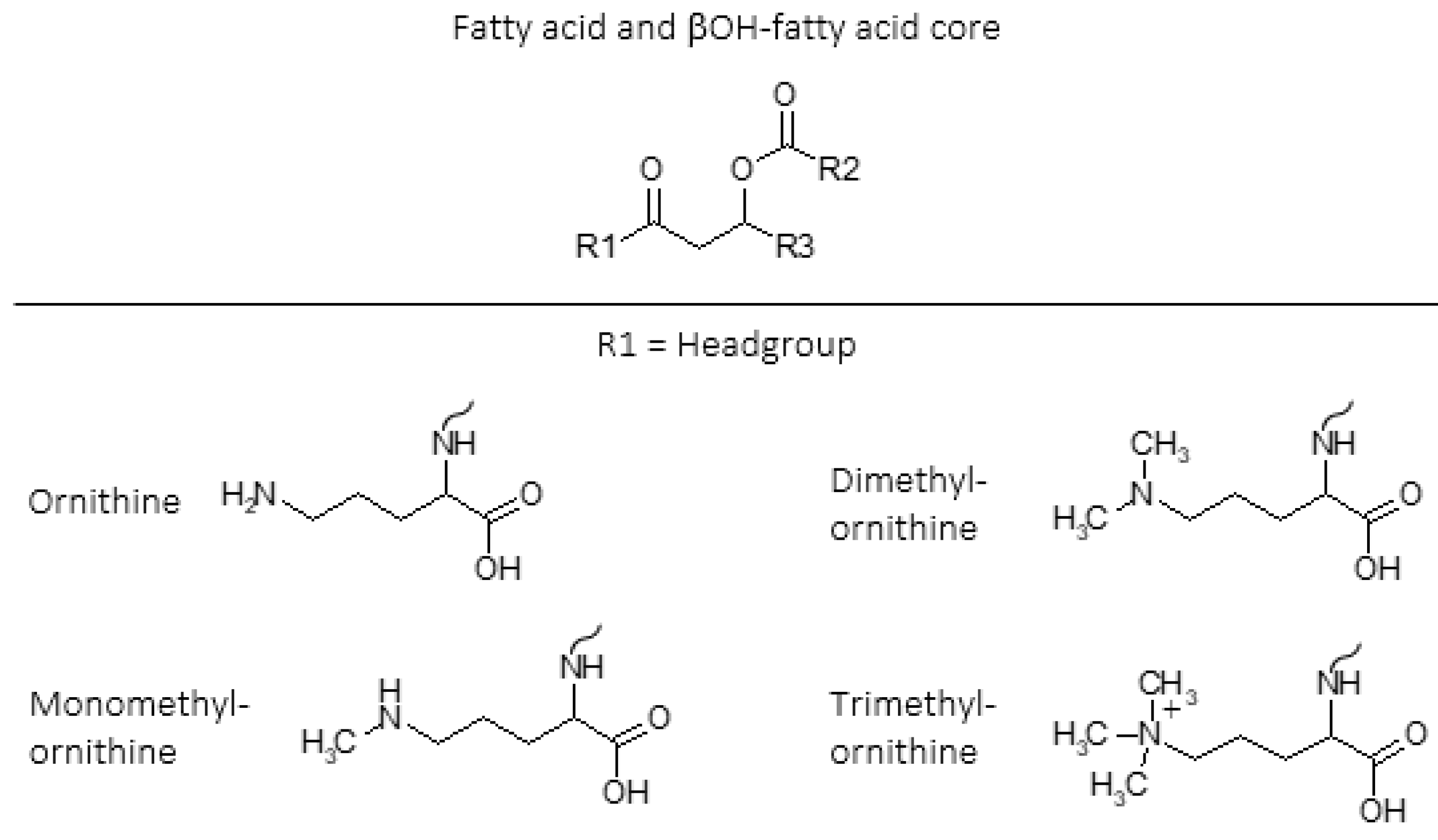
Ornithine lipids (OLs) are a common group of glycerol free amino acid containing lipids in bacteria (Figure 1). Approximately 50% of bacteria whose genomes have been sequenced are predicted to have the capability to synthesize OLs, but the genes necessary to form OLs are absent in archaea and eukaryotes [7,8,9,10]. The OL head group consists of an amino acid that is linked by an amide bond to a β-hydroxy fatty acid, and the β-hydroxy fatty acid is “piggy back” esterified to a fatty acid (Figure 1). Additionally, lysine containing membrane lipids synthesized by Agrobacterium tumefaciens [11] and Pedobacter saltans [12] have the same ester linked fatty acid moiety and amide linked β-OH-fatty acid fatty acid structure that exists in OLs.
Ornithine lipids do not contain phosphorus (Figure 1), and can be produced by certain bacteria as an alternative to phosphoglycerol lipids in response to phosphorus limitation [18,19]. Ornithine lipid head groups and the fatty acid groups can also be hydroxylated in response to different environmental stress conditions [20,21]. It has been proposed that the hydroxylation of OLs increases hydrogen bonding between lipid molecules, thus providing enhanced membrane fluidity and stability [22]. Similarly, it was discovered that lysine lipids (LLs) can be hydroxylated on both the head group and fatty acid chains in response to temperature and pH stress in the soil bacteria Pedobacter saltans [12]. While various other microbial membrane lipids can be produced or modified in response to changing environmental conditions, OLs and other glycerol-free amino acid lipids are commonly observed to be involved in stress response [9].
In 2013, three novel classes of amino acid containing membrane lipids were discovered in multiple Planctomycetes that were isolated from ombrotrophic (receives water only from rain) northern wetlands in European north Russia: mono-, di-, and trimethylated ornithine lipids ([16]; Figure 1). In particular, the unique physical properties of trimethylated ornithine lipids (TMOs) appear to be adapted to the acidic, low nutrient, and anoxic conditions of ombrotrophic northern wetlands. Shortly thereafter, TMOs were identified in various other diverse environments that are often characterized by unique physical or chemical conditions. This review will summarize the discovery of TMOs, their physical and chemical properties, potential functions, and distribution in diverse environments.
2. Methylated Ornithine Lipids Discovered in Northern Wetland Planctomycetes
In recent decades, there has been great interest in the changing conditions of Northern peatlands due to warming temperatures resulting in enhanced decomposition of stored organic carbon and greenhouse gas emissions [23,24,25,26]. Northern peatlands comprise a significant global carbon store, containing roughly one third of global soil organic carbon while only taking up just 3% of the Earth’s land area [27,28]. These low-temperature systems are ombrotrophic, meaning that they are rain fed (only receive water from rain), nutrient poor, and acidic. Microbial communities are the terminal degraders of organic carbon and producers of greenhouse gases from soils and peatlands [29]. Therefore, understanding the wide range of microbial species and communities in Northern peatlands that are involved in carbon degradation and greenhouse gas production is crucial for understanding the impacts of climate change on global terrestrial carbon storage [30,31].
Given that amino acid containing IPLs are often synthesized or modified by bacteria in response to environmental stress conditions, they can be potential biomarkers in rapidly changing ecosystems. The microbiology research group led by Svetlana Dedysh has significantly advanced our understanding of Northern peatland microbial communities, including the discovery of numerous microbial species isolated from these environments [32,33,34,35,36]. The bacterial phylum Planctomycetes were found to be highly abundant in Northern peatlands, and appear to play an important role in the decomposition of the Sphagnum moss-derived vegetation litter that dominates these environments [32,36,37,38].
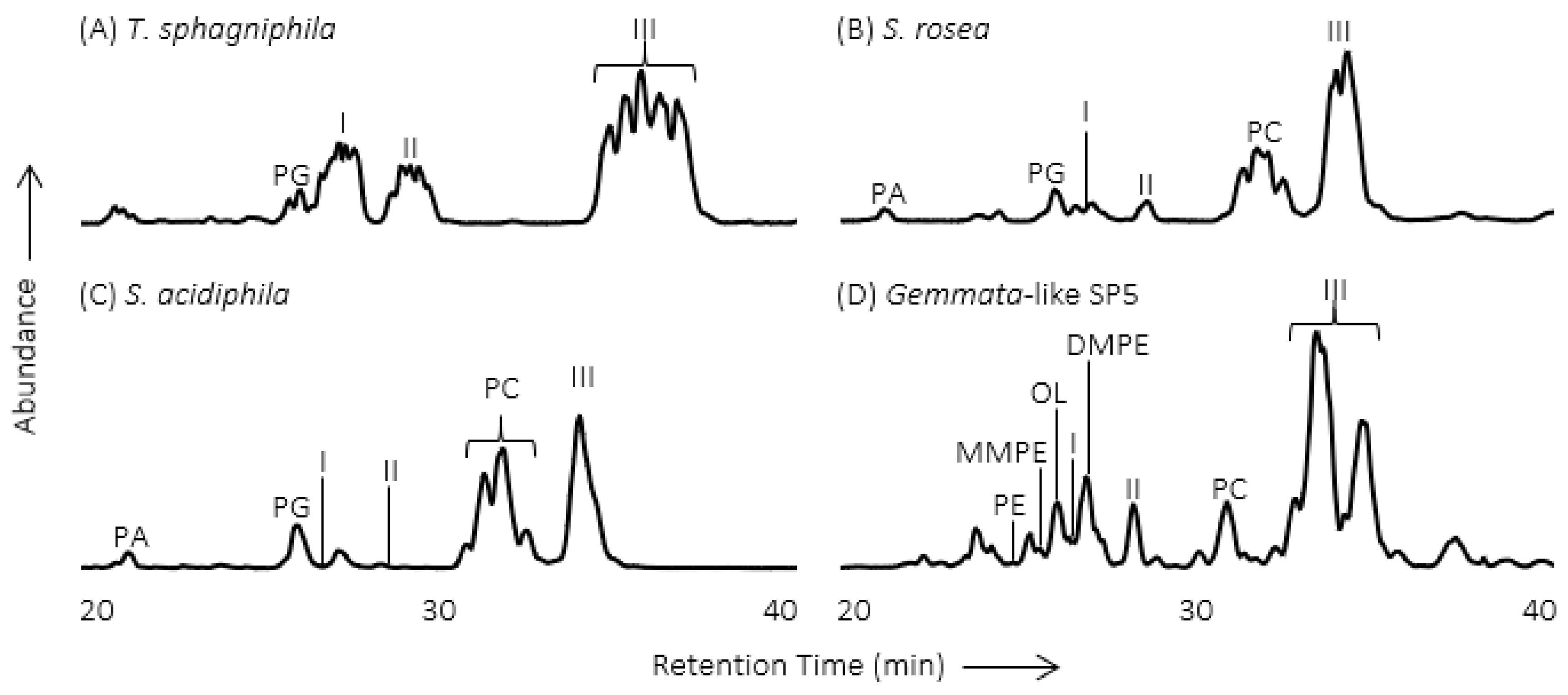
In order to understand how Northern peatland Planctomycetes are suited to live in these unique conditions, and how they may respond to environmental change, the membrane lipids were analyzed from three Planctomycete species (Singulisphaera acidiphila, Singulisphaera rosea, Telmatocola sphagniphila) and one Gemmata-like Planctomycete strain that were all isolated from peatlands of European north Russia [16]. Initial analysis by liquid chromatography-mass spectrometry (LC-MS) resulted in the observation of three unknown IPL classes in each species and strain (Figure 2). The MS fragmentation pattern of the unknown IPLs is similar to OLs, including the sequential loss of a hydroxy fatty acid and a fatty acid, but with a different MS3 product ion m/z value.
The most abundant IPL in T. sphagniphila (peak III from Figure 2A) was purified using LC-MS with fraction collection for further analysis using nuclear magnetic resonance (NMR) and high-resolution mass spectrometry (HRMS). Analysis using both techniques confirmed that the most abundant unknown IPL in T. sphagniphila was an OL that was trimethylated on the terminal nitrogen of the IPL head group (Figure 1; MS fragmentation and NMR results are described in detail by [16]). With this knowledge in hand, HRMS analysis of the lipid extracts from the three Planctomycete species and Gemmata-like strain confirmed the other unknown IPL classes I and II (Figure 2) to be ornithine lipids that were monothylated and dimethylated on the terminal nitrogen of the IPL head group, respectively (Figure 1). Additionally, similar fatty acid distributions between the monomethylornithine, dimethylornithine, and trimethylornithine lipids (MMOs, DMOs, and TMOs) within each species suggest that the three lipid classes are biosynthetically linked. The most abundant core lipids observed in MMOs, DMOs, and TMOs among the analyzed Planctomcyetes were C18:1 and C16:1 for T. sphagniphila, C18:1 and C18:0 for S. rosea, C18:1 and C18:0 for S. acidiphila, and C20:1 and C16:0 for the Gemmata-like strain SP5 [16].
The sequential methylation of the terminal nitrogen among MMOs, DMOs, and TMOs is analogous to the sequential methylation of phosphatidylethanolamine (PE) by the enzyme phosphatidylethanolamine N-methyltransferase, producing monomethylphosphatidylethanol-amine (MMPE), dimethylphosphatidylethanolamine (DMPE), and ultimately the very common IPL phosphatidylcholine (PC) [39,40,41]. The OL N-methyltransferase OlsG (Sinac_1600) gene is responsible for the three-fold methylation of the terminal δ-nitrogen of OL as shown in the TMO producing S. acidiphila [42]. The trimethylated terminal nitrogen of PCs gives them a positively charged quadruple bonded nitrogen, and a cylindrical shape compared to the conical shape of PEs, the un-methylated counterparts of PCs. Due to their polarity and relatively cylindrical shape, PCs spontaneously form lipid bilayers, while the cone shape of PEs causes them to assemble into inverted hexagonal phases. Similarly, the additional three methyl groups on the terminal nitrogen in the head group of TMOs may also give these IPLs a more cylindrical shape and greater polarity than un-methylated ornithine lipids and thus result in greater bilayer stability [16]. It has been proposed that OLs are important for outer membrane stability in Gram-negative bacteria due to their zwitterionic nature [43]. However, the enhanced polarity of the positively charged quadruple bonded nitrogen in TMOs could provide greater membrane stability in the cold acidic conditions of ombrotrophic Northern peatlands without using scarce phosphate in these nutrient limited environments.
3. High Abundance of TMOs at the Oxic/Anoxic Interface of Northern Wetlands
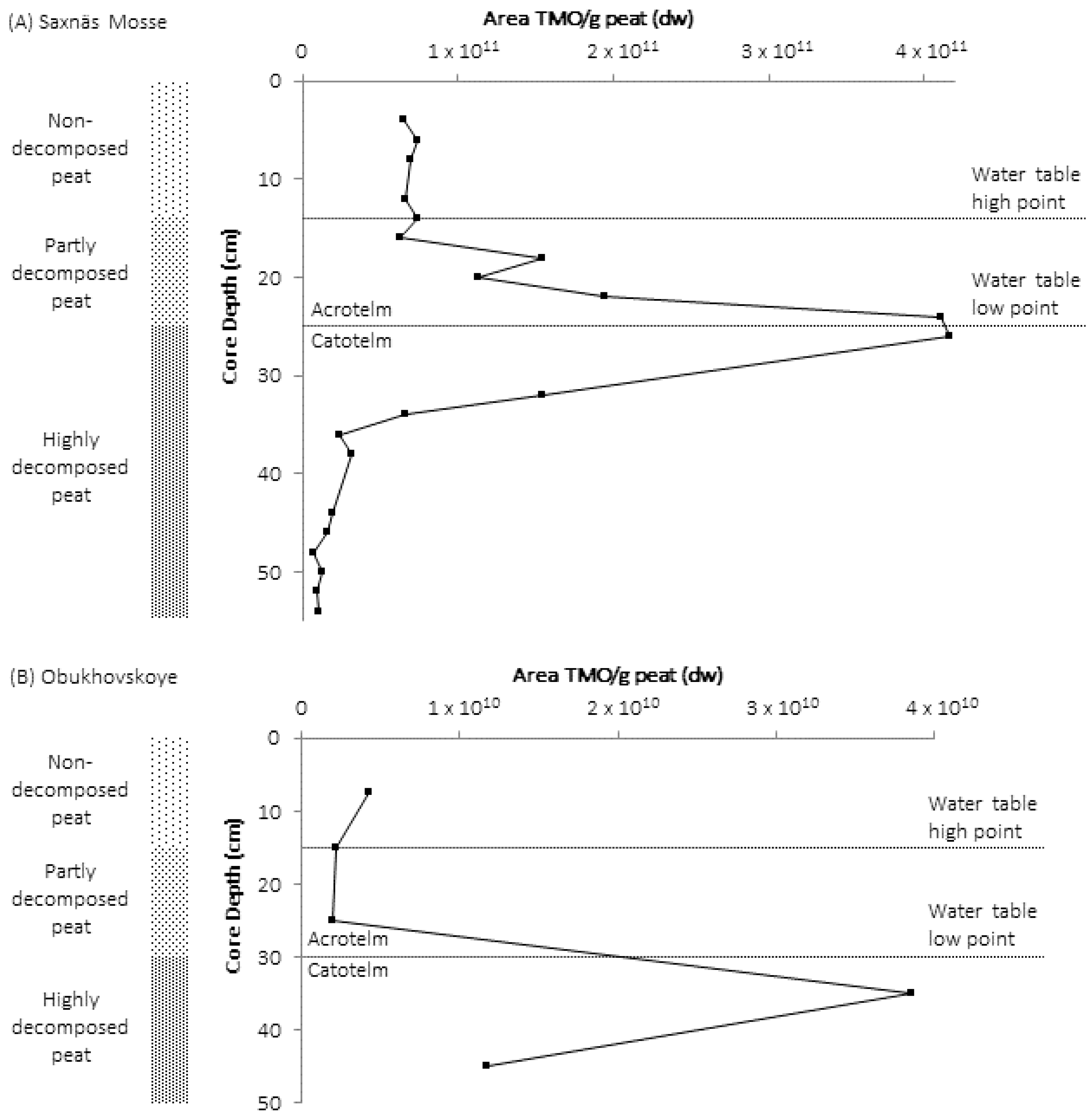
Following the discovery of monomethylornithine, dimethylornithine, and trimethylornithine lipids, a peat core was analyzed from the Obukhovskoye Bog (58°14′ N, 38°12′ E), approximately 200 miles northeast of Moscow, Russia, in order to search for these novel methylated ornithine lipids and observe the importance of Planctomycetes in Northern peatland microbial communities. The peat core had been collected and microbial community of the core had been described in previous studies [34,44]. The peat core was divided into five sections (5 to 10 cm, 10 to 20 cm, 20 to 30 cm, 30 to 40 cm, and 40 to 50 cm) and IPLs were extracted from Obukhoskoye peat using a modified Bligh and Dyer extraction method [45,46]. Intact polar lipid extracts of a peat core collected from the Saxnäs Mosse bog (56°51′20.78 N, 13°27′39.62 E) near the village of Lidhult, southwestern Sweden that were previously collected and analyzed for IPLs [47,48], were analyzed again in order to search for methylated ornithine lipids [49]. The Saxnäs Mosse peat core was extracted for IPLs in 2 cm sections from 0 cm to 54 cm. Additionally, genetic analysis was performed on selected samples from both peat cores in order to confirm the presence Planctomycetes and compare their sequence abundance with other microbial taxa.
Trimethylornithine lipids were present in high abundance throughout the peat core depth profiles of both the Obukhovskoye and Saxnäs Mosse bogs, and the TMO concentration peaked at the oxic/anoxic interface in both cores ([49]; Figure 3). The peak in TMO concentration coincided with the maximum abundance of Planctomycete-specific 16S rRNA gene sequences. Planctomycete gene sequences detected at the oxic/anoxic interface were affiliated with the Isosphaera group, while sequences present in the anoxic peat layers were related to an uncultured Planctomycete group. Pyrosequencing identified Planctomycetes as the major bacterial group at the oxic/anoxic interface in Obukhovskoye peat (54% of total 16S rRNA gene sequence reads), followed by Acidobacteria (19% reads), while in the Saxnäs Mosse peat, Acidobacteria were dominant (46%), and Planctomycetes contributed to 6% of the total reads. The detection of abundant TMO lipids in Planctomycetes isolated from peat bogs [16] and the absence of TMO production by all known cultured species of Acidobacteria suggest that Planctomycetes are the producers of TMOs in the peat bogs.
The distribution of TMO core lipids number of carbons and double bonds changed throughout the Obuhovskoye and Saxnäs Mosse peat cores, which likely reflects the change in Planctomycete community reflected by gene sequences with depth. In the Saxnäs Mosse Bog core, the TMO lipid with C18:1 and βOH-C19:0 core lipids accounted for >60% of total TMOs at the oxic/anoxic interface depth; the TMO with C19:2 and βOH-C18:0 core lipids and the TMO with C18:2 and βOH-C18:0 core lipids were most abundant just above and just below the oxic/anoxic interface; and the TMO with C19:1 and βOH-C16:0 core lipids and the TMO with C18:1 and βOH-C18:0 core lipids were most abundant at the peat surface [49]. In particular, the Isosphaera cluster was the most abundant Planctomycete group at the oxic/anoxic interface. Due to this observation, the Isosphaera-like strain PX4, which was isolated from the Obukhovskoye bog just above the oxic/anoxic interface, was cultured at oxic (7 mg/L) and micro-oxic (1.5 mg/L) conditions. The relative production of TMOs increased in PX4 grown in micro-oxic conditions, including a dramatic increase in concentration of TMO with C19:1 and βOH-C18:0 core lipids. The increase in TMOs and shift in TMO core lipids in micro-oxic conditions, the higher accumulation of TMOs at the oxic/anoxic interface in Northern wetlands, and the change in the Planctomycete community with depth suggest that these IPLs could be synthesized as a response to low oxygen levels and changing redox conditions at the oxic/anoxic interface. Anoxic peat and soils are important environments for the global production of methane [50,51], and TMOs may be an important factor allowing Planctomycetes to be involved in organic matter degradation and subsequent methane production by the microbial community of micro-oxic and anoxic peatlands.
4. Identification of TMOs in Diverse Environments
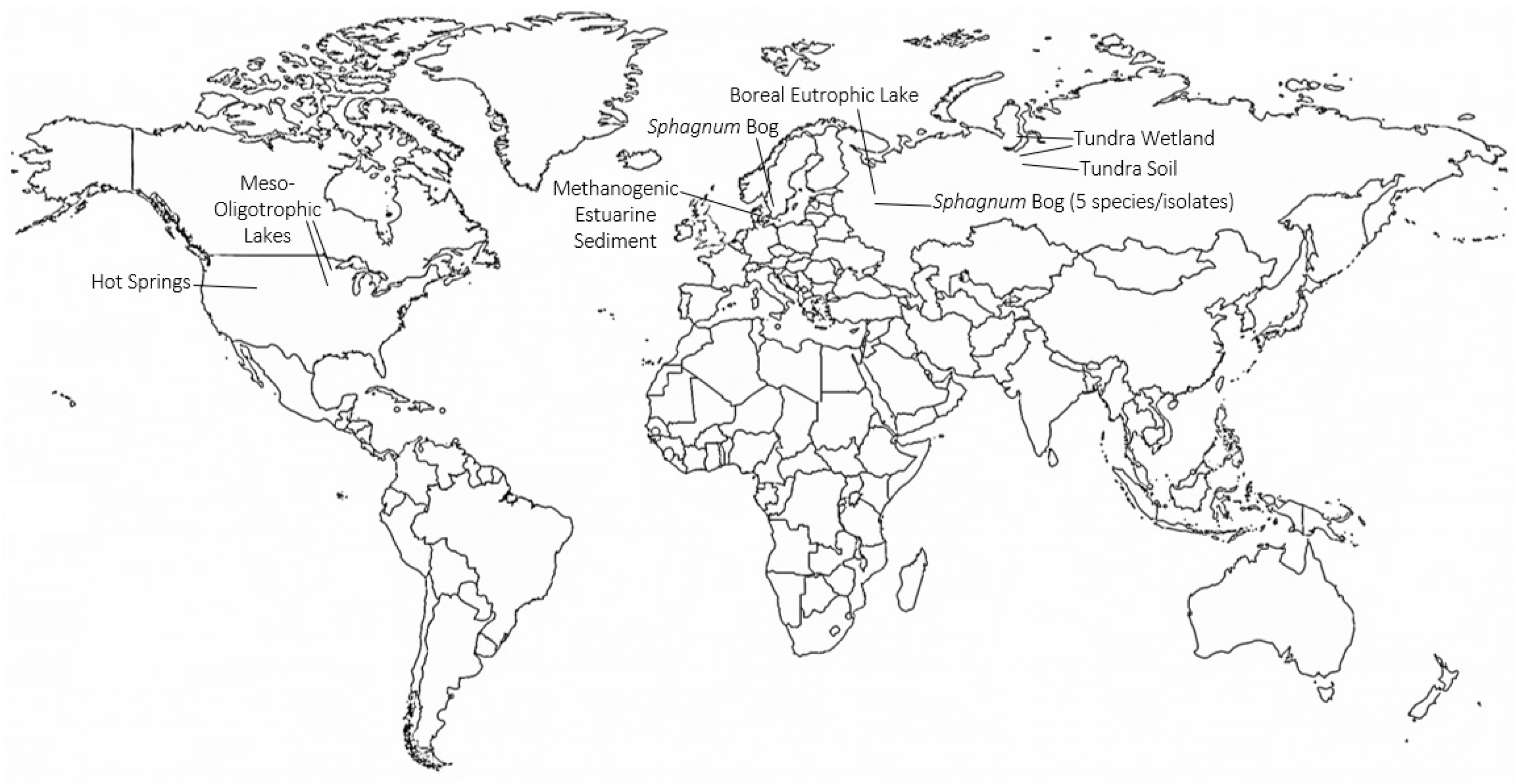
Continued work by the Svetlana Dedysh laboratory to characterize the microbial communities of northern latitude environments has led to the identification of TMOs in various recently described Planctomycete species (Figure 4). Trimethylornithines are the major membrane lipid constituents of the hydrolytic northern tundra wetland Planctomycete Paludisphaera borealis [52], the psychrotolerant Isosphaeraceae Planctomycete from lichen-dominated tundra soils Tundrisphaera lichenicola [53], the Sphagnum peat bog Planctomycete Fimbriiglobus ruber of the proposed family Gemmataceae [54], the freshwater Planctomycete Limnoglobus roseus isolated from a boreal eutrophic lake [55], and the psychrotolerant cellulolytic Gemmataceae Planctomycete from a littoral tundra wetland Frigoriglobus tundricola [56]. The identification of TMOs in Planctomycetes from a northern wetland, tundra soil, peat bog, boreal eutrophic lake, and tundra wetland show that TMOs are more widespread in northern latitude environments than initially thought.
Trimethylornithine lipids have subsequently been identified in a wide range of aquatic environments since their discovery in Northern wetland Planctomycete isolates (Figure 4). An extensive study of trophic state impact on IPL distribution in North American lake surface waters from Minnesota and Iowa found the presence of TMOs, OLs, and betaine lipids in meso-oligotrophic lakes [57]. The higher relative abundance of ornithines and TMOs in the observed meso-oligotrophic lakes was proposed to relate to a higher contribution of heterotrophic bacteria relative to phytoplankton in the lakes, similar to the presence of TMOs in abundant heterotrophic Planctomycetes of Northern peatlands. Trimethylornithines were identified in the oxic, micro-oxic, and anoxic layers of Northern wetland peat [49], therefore, it is consistent to observe TMOs in oxic lake waters. Conversely, TMOs were later identified in enrichment cultures of anoxic methanogenic sediment from Arhus Bay, Denmark [58], akin to the presence of TMOs in anoxic methanogenic peat. Among the enrichment cultures that contained TMOs, the cultures that received sulfate amendments had higher TMO relative abundances than the TMO-containing enrichment culture that did not receive additional sulfate.
Trimethylornithine lipids were also recently observed in microbial communities of multiple Yellowstone National Park hot springs ([59,60]; Figure 4). The fatty acids of some of the hot spring TMOs were hydroxylated [59]. Ornithine lipids were also observed in the hot springs, and various OLs were found to contain hydroxylated fatty acids as well. It has previously been shown that OL and LL fatty acids can be hydroxylated in response to temperature and pH stress [12,20,21]. The hydroxylated fatty acids of Yellowstone TMOs and OLs may also be a stress response to elevated hot spring temperatures in order to create more hydrogen bonding and membrane stability. The high relative abundance of TMOs in the Yellowstone hot spring microbial undermat suggests that these lipids could originate from abundant anoxygenic phototrophic organisms in these layers; however, the authors suggest a chemoheterotrophic origin [60], which aligns much closer with the known metabolic niches of TMO producing microbes. Given that TMOs have only been identified in Planctomycete species, further work characterizing new microbial species in environments where TMOs are observed will help confirm if these lipids are lipid biomarkers for Planctomycetes, or if they are a more taxonomically diverse membrane lipid.
5. TMOs: Specialized Lipids with Potential Broad Distribution
The distribution of TMOs among different types of ecosystems suggests that this class of lipids may have specialized and flexible functions depending on the surrounding environmental conditions or microbial community. The similar structure of the trimethylated terminal nitrogen of TMOs compared to the choline moiety of PCs, without the presence of phosphorus, suggests that TMOs may have a similar membrane structural role to PCs under phosphorus limiting conditions [16]. The increase of relative TMO production in Planctomycete cultures grown under low oxygen conditions [49] and the presence of hydroxylated TMOs in hot spring environments [59] further link TMOs to stress response, similar to the production and modification of other amino acid containing lipids under the nutrient, temperature, or pH stress conditions described above. Additionally, hot spring TMOs are present in high relative abundance at and below the oxic–anoxic interface of the microbial mat, and not present in the oxic upper layer [60]. The increased relative production of TMO lipids in low oxygen cultures, the presence of abundant TMOs in anoxic methanogenic peat, anoxic hot spring microbial mats, and anoxic methanogenic estuarine sediment enrichment cultures further indicate that these lipids may be present in other anoxic and/or methanogenic environments.
In addition to links between TMOs and stress responses, TMOs have been observed in both oxic and anoxic heterotrophic microbial communities in northern peatlands, northern lakes, tundra soils, tundra wetlands, northern eutrophic lakes, and mid-latitude meso-oligotrophic lakes (Figure 4). Future microbial lipidomics studies on aquatic and soil ecosystems at northern and mid-latitudes should include TMOs in their IPL targets to further characterize the microbial community and potential contribution of Planctomycetes in their study systems. Such studies in aquatic and soil ecosystems at lower latitudes will be intriguing for potentially identifying TMOs as well, thus expanding the known geographic distribution of these lipids. Similar to high latitude peatlands, tropical peatlands are important global carbon stores, with variable carbon fluxes in relation to land use and climate change [61,62]. Planctomycetes have been identified in metabolically diverse tropical peat microbial communities [63], suggesting that TMOs could be important microbial membrane constituents in these environments. Coastal salt marshes also contain diverse microbial communities, including Planctomycetes [64], that are involved in the decomposition of recalcitrant organic matter [65,66], that commonly include anoxic and methanogenic sediment zones at depth [67]. The distribution of TMOs and potential modifications of TMOs could inform the role of Planctomycetes and adaptations to lower latitude environments, such as tropical peatlands and coastal salt marshes.
A new area of application for the analysis of TMOs and other IPLs is forensics microbiome research [68,69]. Understanding heterotrophic microbial communities is crucial in forensics studies that investigate changing microbiomes of decaying vertebrate remains. Bacterial community succession analysis of the necrobiome associated with decaying vertebrate remains has revealed a negative relationship for overall taxon richness with increasing decomposition [70], and heterotrophic bacteria have also been observed to increase during decomposition of vertebrate remains [71]. Microbial community succession in forest soil below decomposing human cadavers has shown that Planctomycete sequence abundance decreased during the bloat-active decomposition stage to the advanced decay II stage, but then returned to original relative abundance during the advanced decay III stage [72]. This suggests that the presence of soil Planctomycetes under decayed vertebrate remains could indicate that the advanced decomposition of labile organic matter has taken place allowing Planctomycetes cell numbers to recover. This in agreement with the role of soil Planctomycetes to be involved in the degradation of recalcitrant material after labile material has been consumed [37]. In such a scenario, TMOs and other microbial IPLs could be used to track microbiome changes during carrion decay. Further microbiome and environmental lipidomics research involving TMOs in diverse ecosystems will help reveal the evolution, functions, and applications of these unique membrane lipids.
Funding
Open access publication of this paper was provided by the Rowan University Library Open Access Publishing Fund.
Acknowledgments
I acknowledge Ellen C. Hopmans and Jaap S. Sinninghe Damsté from the Royal Netherlands Institute for Sea Research for their expertise characterizing the molecular structures of monothylornithine, dithylornithine, and trimethylornithine lipids. I also acknowledge the Svetlana Dedysh lab group from the S. N. Winogradsky Institute of Microbiology, Russian Academy of Sciences for their extensive work describing the microbial community in Northern latitude environments.
Conflicts of Interest
The author declares no conflict of interest.
References
- Cooper, G.M. The Cell: A Molecular Approach, 2nd ed.; Sinauer Associates: Sunderland, MA, USA, 2000; ISBN 978-0-87893-106-4. [Google Scholar]
- Sturt, H.F.; Summons, R.E.; Smith, K.; Elvert, M.; Hinrichs, K.-U. Intact polar membrane lipids in prokaryotes and sediments deciphered by high-performance liquid chromatography/electrospray ionization multistage mass spectrometry—New biomarkers for biogeochemistry and microbial ecology. Rapid Commun. Mass Spectrom. 2004, 18, 617–628. [Google Scholar] [CrossRef]
- Schubotz, F.; Wakeham, S.G.; Lipp, J.S.; Fredricks, H.F.; Hinrichs, K.-U. Detection of microbial biomass by intact polar membrane lipid analysis in the water column and surface sediments of the Black Sea. Environ. Microbiol. 2009, 11, 2720–2734. [Google Scholar] [CrossRef] [PubMed]
- White, D.C.; Davis, W.M.; Nickels, J.S.; King, J.D.; Bobbie, R.J. Determination of the sedimentary microbial biomass by extractible lipid phosphate. Oecologia 1979, 40, 51–62. [Google Scholar] [CrossRef] [PubMed]
- Harvey, H.R.; Fallon, R.D.; Patton, J.S. The effect of organic matter and oxygen on the degradation of bacterial membrane lipids in marine sediments. Geochim. Cosmochim. Acta 1986, 50, 795–804. [Google Scholar] [CrossRef]
- Cevc, G. (Ed.) Phospholipids Handbook; CRC Press: Boca Raton, FL, USA, 1993. [Google Scholar]
- Geiger, O.; González-Silva, N.; López-Lara, I.M.; Sohlenkamp, C. Amino acid-containing membrane lipids in bacteria. Prog. Lipid Res. 2010, 49, 46–60. [Google Scholar] [CrossRef]
- López-Lara, I.M.; Sohlenkamp, C.; Geiger, O. Membrane Lipids in Plant-Associated Bacteria: Their Biosyntheses and Possible Functions. Mol. Plant-Microbe Interact. 2003, 16, 567–579. [Google Scholar] [CrossRef]
- Vences-Guzmán, M.Á.; Geiger, O.; Sohlenkamp, C. Ornithine lipids and their structural modifications: From A to E and beyond. FEMS Microbiol. Lett. 2012, 335, 1–10. [Google Scholar] [CrossRef] [Green Version]
- Vences-Guzmán, M.Á.; Guan, Z.; Escobedo-Hinojosa, W.I.; Bermúdez-Barrientos, J.R.; Geiger, O.; Sohlenkamp, C. Discovery of a bifunctional acyltransferase responsible for ornithine lipid synthesis in Serratia proteamaculans. Environ. Microbiol. 2015, 17, 1487–1496. [Google Scholar] [CrossRef]
- Tahara, Y.; Yamada, Y.; Kondo, K. A New Lysine-containing Lipid Isolated from Agrobacterium tumefaciens. Agric. Biol. Chem. 1976, 40, 1449–1450. [Google Scholar] [CrossRef] [Green Version]
- Moore, E.K.; Hopmans, E.C.; Rijpstra, W.I.C.; Sanchez Andrea, I.; Villanueva, L.; Wienk, H.; Schoutsen, F.; Stams, A.; Sinninghe Damste, J. Lysine and novel hydroxylysine lipids in soil bacteria: Amino acid membrane lipid response to temperature and pH in Pseudopedobacter saltans. Front. Microbiol. 2015, 6, 637. [Google Scholar] [CrossRef] [Green Version]
- Hilker, D.R.; Gross, M.L.; Knocke, H.W.; Shively, J.M. The interpretation of the mass spectrum of an ornithine-containing lipid from Thiobacillus thiooxidans. Biomed. Mass Spectrom. 1978, 5, 64–71. [Google Scholar] [CrossRef] [PubMed]
- Cerny, R.L.; Tomer, K.B.; Gross, M.L. Desorption ionization combined with tandem mass spectrometry: Advantages for investigating complex lipids, disaccharides and organometallic complexes. Org. Mass Spectrom. 1986, 21, 655–660. [Google Scholar] [CrossRef]
- Zhang, X.; Ferguson-Miller, S.M.; Reid, G.E. Characterization of ornithine and glutamine lipids extracted from cell membranes of Rhodobacter sphaeroides. J. Am. Soc. Mass Spectrom. 2009, 20, 198–212. [Google Scholar] [CrossRef] [PubMed] [Green Version]
- Moore, E.K.; Hopmans, E.C.; Rijpstra, W.I.C.; Villanueva, L.; Dedysh, S.N.; Kulichevskaya, I.S.; Wienk, H.; Schoutsen, F.; Damsté, J.S.S. Novel Mono-, Di-, and Trimethylornithine Membrane Lipids in Northern Wetland Planctomycetes. Appl. Environ. Microbiol. 2013, 79, 6874–6884. [Google Scholar] [CrossRef] [Green Version]
- Moore, E.K.; Hopmans, E.C.; Rijpstra, W.I.C.; Villanueva, L.; Damsté, J.S.S. Elucidation and identification of amino acid containing membrane lipids using liquid chromatography/high-resolution mass spectrometry. Rapid Commun. Mass Spectrom. 2016, 30, 739–750. [Google Scholar] [CrossRef] [Green Version]
- Weissenmayer, B.; Gao, J.-L.; López-Lara, I.M.; Geiger, O. Identification of a gene required for the biosynthesis of ornithine-derived lipids. Mol. Microbiol. 2002, 45, 721–733. [Google Scholar] [CrossRef]
- Gao, J.-L.; Weissenmayer, B.; Taylor, A.M.; Thomas-Oates, J.; López-Lara, I.M.; Geiger, O. Identification of a gene required for the formation of lyso-ornithine lipid, an intermediate in the biosynthesis of ornithine-containing lipids. Mol. Microbiol. 2004, 53, 1757–1770. [Google Scholar] [CrossRef]
- Vences-Guzmán, M.Á.; Guan, Z.; Ormeño-Orrillo, E.; González-Silva, N.; López-Lara, I.M.; Martínez-Romero, E.; Geiger, O.; Sohlenkamp, C. Hydroxylated ornithine lipids increase stress tolerance in Rhizobium tropici CIAT899. Mol. Microbiol. 2011, 79, 1496–1514. [Google Scholar] [CrossRef] [Green Version]
- González-Silva, N.; López-Lara, I.M.; Reyes-Lamothe, R.; Taylor, A.M.; Sumpton, D.; Thomas-Oates, J.; Geiger, O. The Dioxygenase-Encoding olsD Gene from Burkholderia cenocepacia Causes the Hydroxylation of the Amide-Linked Fatty Acyl Moiety of Ornithine-Containing Membrane Lipids. Biochemistry 2011, 50, 6396–6408. [Google Scholar] [CrossRef]
- Gibbons, H.S.; Lin, S.; Cotter, R.J.; Raetz, C.R.H. Oxygen Requirement for the Biosynthesis of theS-2-Hydroxymyristate Moiety in Salmonella typhimurium Lipid a function of LpxO, a new Fe2+/α-Ketoglutarate-Dependent Dioxygenase Homologue. J. Biol. Chem. 2000, 275, 32940–32949. [Google Scholar] [CrossRef] [Green Version]
- Kirschbaum, M.U.F. The temperature dependence of soil organic matter decomposition, and the effect of global warming on soil organic C storage. Soil Biol. Biochem. 1995, 27, 753–760. [Google Scholar] [CrossRef]
- Biasi, C.; Rusalimova, O.; Meyer, H.; Kaiser, C.; Wanek, W.; Barsukov, P.; Junger, H.; Richter, A. Temperature-dependent shift from labile to recalcitrant carbon sources of arctic heterotrophs. Rapid Commun. Mass Spectrom. 2005, 19, 1401–1408. [Google Scholar] [CrossRef] [PubMed]
- Dorrepaal, E.; Toet, S.; van Logtestijn, R.S.P.; Swart, E.; van de Weg, M.J.; Callaghan, T.V.; Aerts, R. Carbon respiration from subsurface peat accelerated by climate warming in the subarctic. Nature 2009, 460, 616–619. [Google Scholar] [CrossRef]
- Lai, D.Y.F. Methane Dynamics in Northern Peatlands: A Review. Pedosphere 2009, 19, 409–421. [Google Scholar] [CrossRef]
- Gorham, E. Northern Peatlands: Role in the Carbon Cycle and Probable Responses to Climatic Warming. Ecol. Appl. 1991, 1, 182–195. [Google Scholar] [CrossRef]
- Bain, C.G.; Bonn, A.; Stoneman, R.; Chapman, S.; Coupar, A.; Evans, M.; Gearey, B.; Howat, M.; Joosten, H.; Keenleyside, C.; et al. IUCN UK Commission of Inquiry on Peatlands; IUCN UK Peatland Programme: Edinburgh, UK, 2011. [Google Scholar]
- Sundh, I.; Nilsson, M.; Granberg, G.; Svensson, B.H. Depth distribution of microbial production and oxidation of methane in northern boreal peatlands. Microb. Ecol. 1994, 27, 253–265. [Google Scholar] [CrossRef]
- Basiliko, N.; Henry, K.; Gupta, V.; Moore, T.; Driscoll, B.; Dunfield, P. Controls on bacterial and archaeal community structure and greenhouse gas production in natural, mined, and restored Canadian peatlands. Front. Microbiol. 2013, 4. [Google Scholar] [CrossRef] [Green Version]
- McCalley, C.K.; Woodcroft, B.J.; Hodgkins, S.B.; Wehr, R.A.; Kim, E.-H.; Mondav, R.; Crill, P.M.; Chanton, J.P.; Rich, V.I.; Tyson, G.W.; et al. Methane dynamics regulated by microbial community response to permafrost thaw. Nature 2014, 514, 478–481. [Google Scholar] [CrossRef] [Green Version]
- Kulichevskaya, I.S.; Pankratov, T.A.; Dedysh, S.N. Detection of representatives of the Planctomycetes in Sphagnum peat bogs by molecular and cultivation approaches. Microbiology 2006, 75, 329–335. [Google Scholar] [CrossRef]
- Dedysh, S.N. Cultivating Uncultured Bacteria from Northern Wetlands: Knowledge Gained and Remaining Gaps. Front. Microbiol. 2011, 2. [Google Scholar] [CrossRef] [Green Version]
- Serkebaeva, Y.M.; Kim, Y.; Liesack, W.; Dedysh, S.N. Pyrosequencing-Based Assessment of the Bacteria Diversity in Surface and Subsurface Peat Layers of a Northern Wetland, with Focus on Poorly Studied Phyla and Candidate Divisions. PLoS ONE 2013, 8, e63994. [Google Scholar] [CrossRef] [Green Version]
- Dedysh, S.N.; Damsté, J.S.S. Acidobacteria. In eLS; American Cancer Society: Atlanta, GA, USA, 2018; pp. 1–10. ISBN 978-0-470-01590-2. [Google Scholar]
- Dedysh, S.N.; Ivanova, A.A. Planctomycetes in boreal and subarctic wetlands: Diversity patterns and potential ecological functions. FEMS Microbiol. Ecol. 2019, 95. [Google Scholar] [CrossRef] [PubMed] [Green Version]
- Kulichevskaia, I.S.; Belova, S.E.; Kevbrin, V.V.; Dedysh, S.N.; Zavarzin, G.A. Analysis of the bacterial community developing in the course of Sphagnum moss decomposition. Mikrobiologiia 2007, 76, 702–710. [Google Scholar] [CrossRef]
- Dedysh, S.N.; Ivanova, A.O. Abundance, Diversity, and Depth Distribution of Planctomycetes in Acidic Northern Wetlands. Front. Microbiol. 2012, 3. [Google Scholar] [CrossRef] [Green Version]
- Bremer, J.; Greenberg, D.M. Methyl transfering enzyme system of microsomes in the biosynthesis of lecithin (phosphatidylcholine). Biochim. Biophys. Acta 1961, 46, 205–216. [Google Scholar] [CrossRef]
- Yamashita, S.; Oshima, A.; Nikawa, J.; Hosaka, K. Regulation of the phosphatidylethanolamine methylation pathway in Saccharomyces cerevisiae. Eur. J. Biochem. 1982, 128, 589–595. [Google Scholar] [CrossRef] [PubMed]
- Gaynor, P.M.; Gill, T.; Toutenhoofd, S.; Summers, E.F.; McGraw, P.; Homann, M.J.; Henry, S.A.; Carman, G.M. Regulation of phosphatidylethanolamine methyltransferase and phospholipid methyltransferase by phospholipid precursors in Saccharomyces cerevisiae. Biochim. Biophys. Acta Gene Struct. Expr. 1991, 1090, 326–332. [Google Scholar] [CrossRef]
- Escobedo-Hinojosa, W.I.; Vences-Guzmán, M.Á.; Schubotz, F.; Sandoval-Calderón, M.; Summons, R.E.; López-Lara, I.M.; Geiger, O.; Sohlenkamp, C. OlsG (Sinac_1600) Is an Ornithine LipidN-Methyltransferase from the PlanctomyceteSingulisphaera acidiphila. J. Biol. Chem. 2015, 290, 15102–15111. [Google Scholar] [CrossRef] [Green Version]
- Freer, E.; Moreno, E.; Moriyón, I.; Pizarro-Cerdá, J.; Weintraub, A.; Gorvel, J.P. Brucella-Salmonella lipopolysaccharide chimeras are less permeable to hydrophobic probes and more sensitive to cationic peptides and EDTA than are their native Brucella sp. counterparts. J. Bacteriol. 1996, 178, 5867–5876. [Google Scholar] [CrossRef] [Green Version]
- Pankratov, T.A.; Ivanova, A.O.; Dedysh, S.N.; Liesack, W. Bacterial populations and environmental factors controlling cellulose degradation in an acidic Sphagnum peat. Environ. Microbiol. 2011, 13, 1800–1814. [Google Scholar] [CrossRef]
- Bligh, E.G.; Dyer, W.J. A rapid method of total lipid extraction and purification. Can. J. Biochem. Physiol. 1959, 37, 911–917. [Google Scholar] [CrossRef] [PubMed] [Green Version]
- Rütters, H.; Sass, H.; Cypionka, H.; Rullkötter, J. Phospholipid analysis as a tool to study complex microbial communities in marine sediments. J. Microbiol. Methods 2002, 48, 149–160. [Google Scholar] [CrossRef]
- Weijers, J.W.H.; Schouten, S.; van der Linden, M.; van Geel, B.; Sinninghe Damsté, J.S. Water table related variations in the abundance of intact archaeal membrane lipids in a Swedish peat bog. FEMS Microbiol. Lett. 2004, 239, 51–56. [Google Scholar] [CrossRef] [PubMed]
- Peterse, F.; Hopmans, E.C.; Schouten, S.; Mets, A.; Rijpstra, W.I.C.; Sinninghe Damsté, J.S. Identification and distribution of intact polar branched tetraether lipids in peat and soil. Org. Geochem. 2011, 42, 1007–1015. [Google Scholar] [CrossRef]
- Moore, E.K.; Villanueva, L.; Hopmans, E.C.; Rijpstra, W.I.C.; Mets, A.; Dedysh, S.N.; Damsté, J.S.S. Abundant Trimethylornithine Lipids and Specific Gene Sequences Are Indicative of Planctomycete Importance at the Oxic/Anoxic Interface in Sphagnum-Dominated Northern Wetlands. Appl. Environ. Microbiol. 2015, 81, 6333–6344. [Google Scholar] [CrossRef] [Green Version]
- Roslev, P.; King, G.M. Regulation of methane oxidation in a freshwater wetland by water table changes and anoxia. FEMS Microbiol. Ecol. 1996, 19, 105–115. [Google Scholar] [CrossRef]
- Angel, R.; Claus, P.; Conrad, R. Methanogenic archaea are globally ubiquitous in aerated soils and become active under wet anoxic conditions. ISME J. 2012, 6, 847–862. [Google Scholar] [CrossRef] [Green Version]
- Kulichevskaya, I.S.; Ivanova, A.A.; Suzina, N.E.; Rijpstra, W.I.C.; Sinninghe Damsté, J.S.; Dedysh, S.N. Paludisphaera borealis gen. nov., sp. nov., a hydrolytic planctomycete from northern wetlands, and proposal of Isosphaeraceae fam. nov. Int. J. Syst. Evol. Microbiol. 2016, 66, 837–844. [Google Scholar] [CrossRef]
- Kulichevskaya, I.S.; Ivanova, A.A.; Detkova, E.N.; Rijpstra, W.I.C.; Sinninghe Damsté, J.S.; Dedysh, S.N. Tundrisphaera lichenicola gen. nov., sp. nov., a psychrotolerant representative of the family Isosphaeraceae from lichen-dominated tundra soils. Int. J. Syst. Evol. Microbiol. 2017, 67, 3583–3589. [Google Scholar] [CrossRef]
- Kulichevskaya, I.S.; Ivanova, A.A.; Baulina, O.I.; Rijpstra, W.I.C.; Sinninghe Damsté, J.S.; Dedysh, S.N. Fimbriiglobus ruber gen. nov., sp. nov., a Gemmata-like planctomycete from Sphagnum peat bog and the proposal of Gemmataceae fam. nov. Int. J. Syst. Evol. Microbiol. 2017, 67, 218–224. [Google Scholar] [CrossRef]
- Kulichevskaya, I.S.; Naumoff, D.G.; Miroshnikov, K.K.; Ivanova, A.A.; Philippov, D.A.; Hakobyan, A.; Rijpstra, W.I.C.; Damsté, J.S.S.; Liesack, W.; Dedysh, S.N. Limnoglobus roseus gen. nov., sp. nov., a novel freshwater planctomycete with a giant genome from the family Gemmataceae. Int. J. Syst. Evol. Microbiol. 2020, 70, 1240–1249. [Google Scholar] [CrossRef] [PubMed]
- Kulichevskaya, I.S.; Ivanova, A.A.; Naumoff, D.G.; Beletsky, A.V.; Rijpstra, W.I.C.; Sinninghe Damsté, J.S.; Mardanov, A.V.; Ravin, N.V.; Dedysh, S.N. Frigoriglobus tundricola gen. nov., sp. nov., a psychrotolerant cellulolytic planctomycete of the family Gemmataceae from a littoral tundra wetland. Syst. Appl. Microbiol. 2020, 43, 126129. [Google Scholar] [CrossRef] [PubMed]
- Bale, N.J.; Hopmans, E.C.; Schoon, P.L.; Kluijver, A.d.; Downing, J.A.; Middelburg, J.J.; Damsté, J.S.S.; Schouten, S. Impact of trophic state on the distribution of intact polar lipids in surface waters of lakes. Limnol. Oceanogr. 2016, 61, 1065–1077. [Google Scholar] [CrossRef] [Green Version]
- Ozuolmez, D.; Moore, E.K.; Hopmans, E.C.; Sinninghe Damsté, J.S.; Stams, A.J.M.; Plugge, C.M. Butyrate Conversion by Sulfate-Reducing and Methanogenic Communities from Anoxic Sediments of Aarhus Bay, Denmark. Microorganisms 2020, 8, 606. [Google Scholar] [CrossRef]
- Boyer, G.M.; Schubotz, F.; Summons, R.E.; Woods, J.; Shock, E.L. Carbon Oxidation State in Microbial Polar Lipids Suggests Adaptation to Hot Spring Temperature and Redox Gradients. Front. Microbiol. 2020, 11, 229. [Google Scholar] [CrossRef] [Green Version]
- Wörmer, L.; Gajendra, N.; Schubotz, F.; Matys, E.D.; Evans, T.W.; Summons, R.E.; Hinrichs, K.-U. A micrometer-scale snapshot on phototroph spatial distributions: mass spectrometry imaging of microbial mats in Octopus Spring, Yellowstone National Park. Geobiology 2020, 18, 742–759. [Google Scholar] [CrossRef]
- Jauhiainen, J.; Takahashi, H.; Heikkinen, J.E.P.; Martikainen, P.J.; Vasander, H. Carbon fluxes from a tropical peat swamp forest floor. Glob. Chang. Biol. 2005, 11, 1788–1797. [Google Scholar] [CrossRef]
- Hirano, T.; Segah, H.; Kusin, K.; Limin, S.; Takahashi, H.; Osaki, M. Effects of disturbances on the carbon balance of tropical peat swamp forests. Glob. Chang. Biol. 2012, 18, 3410–3422. [Google Scholar] [CrossRef]
- Kanokratana, P.; Uengwetwanit, T.; Rattanachomsri, U.; Bunterngsook, B.; Nimchua, T.; Tangphatsornruang, S.; Plengvidhya, V.; Champreda, V.; Eurwilaichitr, L. Insights into the phylogeny and metabolic potential of a primary tropical peat swamp forest microbial community by metagenomic analysis. Microb. Ecol. 2011, 61, 518–528. [Google Scholar] [CrossRef]
- Bowen, J.L.; Morrison, H.G.; Hobbie, J.E.; Sogin, M.L. Salt marsh sediment diversity: A test of the variability of the rare biosphere among environmental replicates. ISME J. 2012, 6, 2014–2023. [Google Scholar] [CrossRef] [Green Version]
- Osburn, C.L.; Mikan, M.P.; Etheridge, J.R.; Burchell, M.R.; Birgand, F. Seasonal variation in the quality of dissolved and particulate organic matter exchanged between a salt marsh and its adjacent estuary. J. Geophys. Res. Biogeosciences 2015, 120, 1430–1449. [Google Scholar] [CrossRef]
- Sun, H.; Jiang, J.; Cui, L.; Feng, W.; Wang, Y.; Zhang, J. Soil organic carbon stabilization mechanisms in a subtropical mangrove and salt marsh ecosystems. Sci. Total Environ. 2019, 673, 502–510. [Google Scholar] [CrossRef] [PubMed]
- Oremland, R.S.; Marsh, L.M.; Polcin, S. Methane production and simultaneous sulphate reduction in anoxic, salt marsh sediments. Nature 1982, 296, 143–145. [Google Scholar] [CrossRef]
- Metcalf, J.L.; Xu, Z.Z.; Bouslimani, A.; Dorrestein, P.; Carter, D.O.; Knight, R. Microbiome Tools for Forensic Science. Trends Biotechnol. 2017, 35, 814–823. [Google Scholar] [CrossRef]
- Langley, N.R.; Wood, P.; Herling, P.; Steadman, D.W. Forensic Postmortem Interval Estimation from Skeletal Muscle Tissue: A Lipidomics Approach. Forensic Anthropol. 2019, 2, 152–157. [Google Scholar] [CrossRef]
- Pechal, J.L.; Crippen, T.L.; Benbow, M.E.; Tarone, A.M.; Dowd, S.; Tomberlin, J.K. The potential use of bacterial community succession in forensics as described by high throughput metagenomic sequencing. Int. J. Legal Med. 2014, 128, 193–205. [Google Scholar] [CrossRef]
- Howard, G.T.; Duos, B.; Watson-Horzelski, E.J. Characterization of the soil microbial community associated with the decomposition of a swine carcass. Int. Biodeterior. Biodegrad. 2010, 64, 300–304. [Google Scholar] [CrossRef]
- Cobaugh, K.L.; Schaeffer, S.M.; DeBruyn, J.M. Functional and Structural Succession of Soil Microbial Communities below Decomposing Human Cadavers. PLoS ONE 2015, 10, e0130201. [Google Scholar] [CrossRef]
Figure 1.
Intact polar lipid (IPL) structures of ornithine (OL), monomethylornithine (MMO), dimethylornithine (DMO), and trimethylornithine (TMO) lipids including fatty acid and βOH-fatty acid core lipids. R1 = headgroup; R2, R3 = alkane/alkene chains. Ornithine (OL; [13,14,15]); monomethylornithine (MMO; [16]); dimethylornithine (DMO; [16]); trimethylornithine (TMO; [16]). Figure adapted from [17].

Figure 2.
High-performance liquid chromatography-mass spectrometry (HPLC-MS) chromatograms (m/z 400 to 2000) of Bligh and Dyer lipid extracts from Planctomycete cultures. (A) T. sphagniphila; (B) S. rosea; (C) S. acidiphila; (D) Gemmata-like strain SP5. PG = phosphatidylglycerol; PA = phosphatidic acid; PC = phosphatidylcholine; PE = phosphatidylethanolamine; MMPE = monomethyl-phosphatidylethanolamine; DMPE = dimethyl-phosphatidylethanolamine; OL = ornithine lipid; I = monomethylornithine; II = dimethylornithine; III = trimethylornithine. Figure adapted from [16].

Figure 3.
Relative abundances of trimethylornithine IPL (TMO) from HPLC-MS analysis down core in Saxnäs Mosse peat (A) and Obukhovskoye peat (B). dw, dry weight. Figure adapted from [49].
Figure 3.
Relative abundances of trimethylornithine IPL (TMO) from HPLC-MS analysis down core in Saxnäs Mosse peat (A) and Obukhovskoye peat (B). dw, dry weight. Figure adapted from [49].

Figure 4.
Global map showing locations and ecosystems where trimethylornithine lipids have been identified. Global map made with mapchart.net.
Figure 4.
Global map showing locations and ecosystems where trimethylornithine lipids have been identified. Global map made with mapchart.net.

Publisher’s Note: MDPI stays neutral with regard to jurisdictional claims in published maps and institutional affiliations. |
© 2021 by the author. Licensee MDPI, Basel, Switzerland. This article is an open access article distributed under the terms and conditions of the Creative Commons Attribution (CC BY) license (http://creativecommons.org/licenses/by/4.0/).
Share and Cite
MDPI and ACS Style
Moore, E.K. Trimethylornithine Membrane Lipids: Discovered in Planctomycetes and Identified in Diverse Environments. Metabolites 2021, 11, 49. https://doi.org/10.3390/metabo11010049
AMA Style
Moore EK. Trimethylornithine Membrane Lipids: Discovered in Planctomycetes and Identified in Diverse Environments. Metabolites. 2021; 11(1):49. https://doi.org/10.3390/metabo11010049
Chicago/Turabian StyleMoore, Eli K. 2021. "Trimethylornithine Membrane Lipids: Discovered in Planctomycetes and Identified in Diverse Environments" Metabolites 11, no. 1: 49. https://doi.org/10.3390/metabo11010049
Note that from the first issue of 2016, this journal uses article numbers instead of page numbers. See further details here.