Abnormal Levels of Metal Micronutrients and Autism Spectrum Disorder: A Perspective Review
 Supriya Behl
 Sunil Mehta
 Mukesh K. Pandey
- 1Department of Pediatric and Adolescent Medicine, Mayo Clinic, Rochester, MN, United States
- 2Department of Radiology, Mayo Clinic, Rochester, MN, United States
The aim of the present review is to summarize the prevalence of abnormal levels of various metal micronutrients including copper (Cu), iron (Fe), magnesium (Mg), zinc (Zn), and selenium (Se) in Autism Spectrum Disorder (ASD) using hair, nail and serum samples. A correlation of selected abnormal metal ions with known neurodevelopmental processes using Gene Ontology (GO) term was also conducted. Data included in this review are derived from ASD clinical studies performed globally. Metal ion disparity data is also analyzed and discussed based on gender (Male/Female) to establish any gender dependent correlation. Finally, a rational perspective and possible path to better understand the role of metal micronutrients in ASD is suggested.
Introduction
Autism Spectrum Disorder: Epidemiology and Etiology
Autism spectrum disorder (ASD) is a complex condition characterized by impaired social communication and restricted, repetitive behaviors (Lord et al., 2018). Globally, it is estimated that 0.76% of children have ASD. The United States of America (USA) prevalence rates are higher at 1.9%, and have increased since 2000 (Maenner et al., 2020). The USA prevalence varies by race and ethnicity: prevalence in Hispanic children is approximately 17% lower than that of white children. Approximately one third of children with ASD also have an intellectual disability.
ASD is a heterogeneous condition which is likely caused by multiple environmental and genetic factors, and many risk factors have been identified. In a recent literature review of 67 environmental risk factors and 52 biomarkers, the most convincing risk factors for ASD were maternal factors before or during pregnancy. These included maternal age (≥35 years), maternal chronic and gestational hypertension, maternal overweight pre-pregnancy or during pregnancy, maternal pre-eclampsia, pre-pregnancy maternal antidepressant use (Kim et al., 2019), and maternal immune activation (Bilbo et al., 2018). The gut microbiome is also implicated in ASD symptomatology. In fact, gut microbiome plays an integral role in immune reactions and inflammation. One hypothesis is that various antigens induce peripheral immunoreactions through the GI tract, which subsequently alters CNS activity (Cristiano et al., 2018). Further, mouse models of maternal immune activation results in ASD-like behaviors as well as decreased gastrointestinal tract permeability. Because of this, diet modulations have been suggested as a therapeutic option for ASD (Rosenfeld, 2015). Other likely environmental risk factors for ASD include to paternal age (>45 years) (Gabis et al., 2010; Simard et al., 2019), and family history of autoimmune diseases (Wu et al., 2015) such as psoriasis (Croen et al., 2019), rheumatoid arthritis (Rom et al., 2018), placenta morphology (Straughen et al., 2017), and type 1 diabetes (Atladottir et al., 2009).
Since early twin studies in the late 1980’s, a strong genetic influence is seen for ASD. A recent literature review of all twin studies on ASD found a 0.98 correlation between monozygotic twins and an overall heritability estimate of approximately 64–91% (Tick et al., 2016). Today, over 900 autism susceptibility genes have been reported (Banerjee-Basu and Packer, 2010), indicating the condition’s heterogeneous nature. Both common single nucleotide polymorphisms (SNPs) and more rare copy number variations (CNVs) likely play a role in ASD incidence. A study looking at common SNPs across the genome found that ASD may be caused by a collective effort of many small effect SNPs rather than one or a few genes (Klei et al., 2012). CNVs in ASD are common on nine of 23 chromosomes (Bergbaum and Ogilvie, 2016), and have been reported on every chromosome. While most genetic variations are inherited in ASD, approximately 10% of ASD can be attributed to de novo genetic variations (Sebat et al., 2007).
The Role of Metals in ASD
There is a growing interest in the role of metals in ASD incidence. The increasing environmental dissemination of toxic metals and chemicals are likely a cause for ASD, among other neurodevelopmental disorders (Grandjean and Landrigan, 2014). Typical ASD characteristics such as intellectual disability and language problems have been associated with exposure of toxic metals prior to or during pregnancy. In a review of almost 100 studies, 74% suggest that physiological mercury levels are a risk factor for ASD (Kern et al., 2016), likely due to autoimmune activation, oxidative stress, and subsequent neuroinflammation. Aluminum is found at high levels in both white and gray matter of ASD patients, and is hypothesized to be able to cross the blood-brain barriers and taken up by microglial cells (Mold et al., 2018). Further, animal studies examining neurodevelopmental effects of metals found changes in brain structures also implicated in ASD (Arora et al., 2017).
Aside from heavy and toxic metals, which actively disrupt neurodevelopmental processes, dyshomeostasis of metal micronutrients may also be involved in ASD etiology. Zinc, copper, selenium, iron, and magnesium levels have been associated with ASD incidence.
Zinc (Zn)
Zinc is the second most abundant element in the human body and is involved in a plethora of cellular functions. Approximately 2,800 proteins or 10% of the human proteome may bind zinc in vivo (Andreini et al., 2006). Zinc is particularly involved in glutamatergic transmission (i.e., GABA pathway) during embryonic and childhood development. Deficiency of zinc in mice models leads to altered neural tube closure (Li et al., 2018) and ASD-related behavior such as weakened vocalization and social behavior through SHANK proteins, a family of postsynaptic scaffolding proteins (Grabrucker et al., 2014). When dietary zinc is added, non-ASD behavior is restored in SHANK3-mutant mice (Fourie et al., 2018). Brain zinc levels are 10-fold higher than serum Zinc levels, indicating a larger role of Zinc in neurodevelopment (Portbury and Adlard, 2017). Zinc is more abundant in neuronal-rich areas, and is important in neuronal modulation, synaptic plasticity, learning, and memory (Qi and Liu, 2019). Of note, current literature link zinc to ASD in the context of the central nervous system, and therefore a lot remains unknown within the context of the peripheral nervous system. Novel pharmacological therapies for brain injury target levels of free zinc in the brain to restore homeostasis, indicating the significance of zinc homeostasis in the brain (Frederickson et al., 2005).
Zinc is also involved in the gut-brain interaction, and many ASD patients also have gastrointestinal symptoms. Maternal zinc levels are likely a factor in fetal gut formation and therefore the gut-brain interaction in ASD (Vela et al., 2015). Prenatal zinc levels also influence the morphology of placenta (Wilson et al., 2017), and placental function is notable in ASD (Straughen et al., 2017).
There is a well-known link between autism and the immune system (Meltzer and Van de Water, 2017; Hughes et al., 2018), to which zinc plays an integral role in both innate and adaptive immunity, such as monocytes, natural killer, T-, and B-cells (Shankar and Prasad, 1998).
There is considerable evidence for an association between zinc deficiency and ASD (Yasuda and Tsutsui, 2013; Li et al., 2014; Goyal et al., 2019). Zinc-binding genes associated with ASD are up-regulated in all neurodevelopmental stages (Supplementary Table S1). In a study examining 1,967 children with ASD, almost 30% had low zinc concentration in hair samples (Yasuda et al., 2011). Another small study found lower zinc levels in saliva of autistic children when compared to healthy controls (Deshpande et al., 2019). Zinc levels may also be correlated to severity of ASD presentation (Guo et al., 2018). It is important to note, however, that significant variance is observed when comparing zinc from hair and nails (Giuseppe De Palma et al., 2011; Lakshmi Priya and Geetha, 2011), suggesting that serum may be a better source for zinc measurement. When serum was evaluated in 78 children with autism, 71.8% of children had zinc levels either in the lowest 10% or below the reference range (Faber et al., 2009).
Zinc levels may also be affected by geographic-specific factors (Table 1); studies in Ireland (Sweetman et al., 2019) and Brazil (Saldanha Tschinkel et al., 2018) found zinc levels in ASD children to be equivalent to that of healthy controls. One study in Oman found higher levels of zinc in ASD patients than in controls (Al-Farsi et al., 2013). Geographical differences may be attributed to differences in social determinants of health such as nutrition, economic status, and associated illnesses. Zinc deficiency in infants is prevalent in countries with malnourishment, and is globally a recognized public health issue (Ackland and Michalczyk, 2016). Geographic-specific differences may also be due to sample size and age variability between studies.
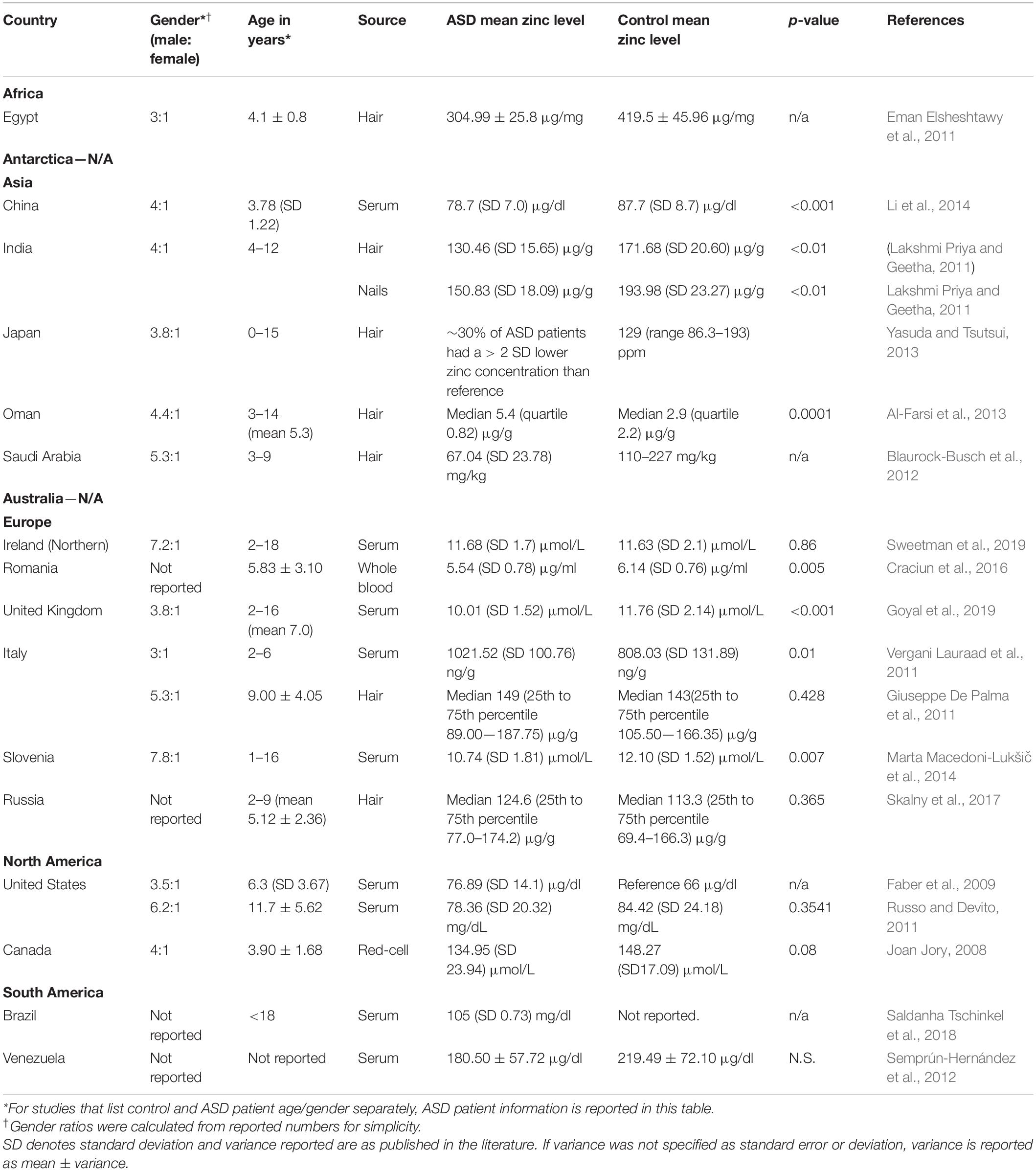
Table 1. Geographical differences in physiological zinc levels.
Copper (Cu)
Copper also has important roles in the human body, and is involved in cell growth, among many others. Copper is involved in reactions connected to neurological diseases, and dyshomeostasis of copper has been seen in disorders such as Parkinson’s, Alzheimer’s, and Huntington’s Diseases (Faber et al., 2009). Further, copper is integral in several autism-related biological processes, such as immunity (Kelley et al., 1995) and placental development. Copper levels are typically higher than average in ASD patients.
In 78 children with ASD, 15.4% had higher copper levels than the reference range, and 30.8% were in the highest 10% of the copper reference range (Faber et al., 2009). In another study, mean serum copper levels were significantly higher in ASD children than in healthy controls (Li et al., 2014). A third study of 79 autistic individuals found a similar pattern, in which autistic and Pervasive Developmental Disorder-Not Otherwise Specified (PDD-NOS) patients had significantly higher plasma levels of copper (Russo and Devito, 2011). Increase in physiological copper levels may also correlate with increasing severity of ASD (Lakshmi Priya and Geetha, 2011). Copper levels in ASD patients do not seem to vary by geographical region (Table 2); regardless of region, copper levels vary in comparison to controls between studies.
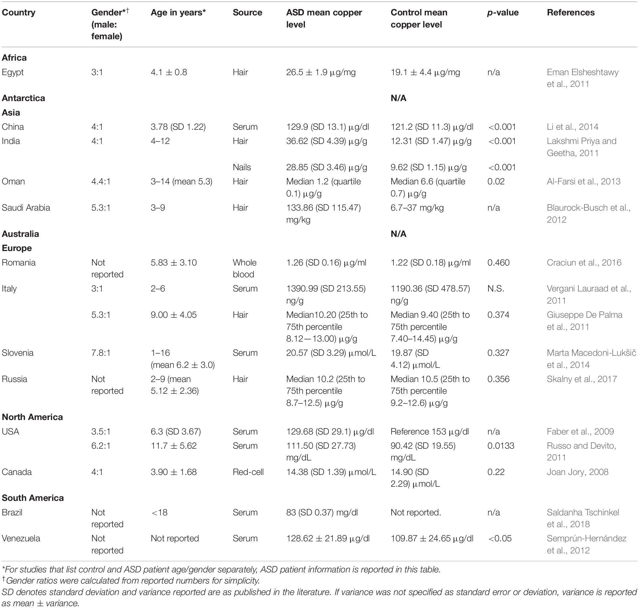
Table 2. Geographical differences in physiological copper levels.
Interestingly, copper and zinc play competing roles physiologically, such that an increase in copper leads to zinc deficiency (Grabrucker, 2012). The zinc/copper ratio has therefore been examined in the ASD setting. Patients and children with ASD tend to have lower zinc/copper ratios than controls (Bjorklund, 2013), even if differences are not seen in copper levels alone (Craciun et al., 2016). Such differences are not known to be sex-dependent (Faber et al., 2009), though copper levels alone may differ by sex due to oral contraceptive use (Babic et al., 2013). One study found that the zinc/copper ratio could be used as a diagnostic biomarker (Li et al., 2014). Zinc/copper cycles may play a role in ASD occurrence, and the rhythmicity of these cycles can be used as a diagnostic tool to classify ASD (Curtin et al., 2018).
Selenium (Se)
Selenium and selenium-dependent proteins are essential in brain development and managing oxidative damage in the brain, and it has been suggested that dyshomeostasis in selenium may be associated with ASD incidence (Raymond et al., 2014). In a recent literature review of 10 studies comparing hair trace element levels in ASD and controls, four of them found a significant difference in Selenium levels. However, two found a significant increase in Selenium levels in children with ASD, and two found a significant decrease (Tinkov et al., 2019). Another meta-analysis found no significant differences in mean hair or erythrocyte selenium concentrations among 12 studies (Saghazadeh et al., 2017). Though dyshomeostasis is likely involved in ASD incidence, the contradictory data indicate a need for a more comprehensive study evaluating Selenium levels in ASD patients (Anatoly et al., 2018).
Iron (Fe)
Iron is the most abundant trace element in the body (Wood and Sperling, 2019). Iron deficiency anemia is a major health concern in both developed and developing countries and can result in inadequate cellular function at a young age (Bener et al., 2017). Iron is involved in several neurodevelopmental processes, such as transmitter synthesis, myelin production, and synaptogenesis, and deficiency leads to malfunction of these processes (Pivina et al., 2019). Subsequently, iron deficiency is associated with developmental delay (McCann and Ames, 2007) and likely in ASD. Deficiency of iron has been seen in ASD children when compared to controls (Bener et al., 2017). While one meta-analysis in 2017 found lower iron levels in ASD patients than controls (Saghazadeh et al., 2017), another meta-analysis in 2018 found no differences in peripheral iron levels in ASD children (Tseng et al., 2018). Iron deficiency may be associated with ASD symptoms and particularly correlates with severity of emotional and behavioral problems (Saghazadeh et al., 2017).
Magnesium (Mg)
Magnesium is involved in basic cellular processes such as nucleic acid formation and energy metabolism. In neurodevelopment, magnesium regulates glutamate-activated channels in neuronal membranes, a process highly correlated with ASD pathogenesis (Saghazadeh et al., 2017). Magnesium has been seen as deficient in children with ASD (Lakshmi Priya and Geetha, 2011). In combination with vitamin B6, magnesium has been argued as a potential nutritional intervention for ASD. However, several systematic reviews from the late 1990’s to early 2000’s found no substantial evidence for magnesium and vitamin B6 as treatment for ASD (Karhu et al., 2019). Since then, not many studies have examined the role of magnesium in ASD. A more recent review, however, found a significant magnesium deficiency in ASD patients, and suggests monitoring of magnesium status in patients with ASD (Saghazadeh et al., 2017).
Association Between Metal Micronutrients and Biological Processes
Iron, zinc, and copper have been considered as essential metal nutrients for neurodevelopment processes. Iron is required for the enzyme ribonucleotide reductase that regulates the central nervous system. Iron also plays a role in myelin synthesis (Prado and Dewey, 2014). Zinc is needed for cell division because of its role in DNA synthesis. Zinc is also required for modulation of postsynaptic plasticity, NMDA receptors for glutamate and inhibits GABA receptor activation (Prado and Dewey, 2014). For example, mouse models indicate alterations in zinc levels lead to differences in both synaptic plasticity and neurogenesis (Nam et al., 2017). When mothers are deprived of zinc early in pregnancy or after birth, rodent pups exhibit impaired DNA synthesis and improperly incorporate of thymine in brain DNA (Sandstead, 1985). Zinc deficiency in early development also results in morphologic brain defects including the hippocampus, a part of the brain most notably significant in working memory.
Along with folic acid and vitamin A, copper is needed for the formation of the neural plate and neural tube very early in the development (Prado and Dewey, 2014). Copper is also a cofactor of dopamine-β-hydroxylase, peptidyl-a-monooxygenase and many other enzymes which are involved in vital central nervous system processes (Lutsenko et al., 2010; Telianidis et al., 2013). Copper is particularly involved in neurotransmitter synthesis and neuromodulation (Scheiber et al., 2014). Subsequently, several neurodegenerative diseases associate with copper dyshomeostasis. Other essential trace metals, such as Mn, Mo, and trace elements such as Se ions also play a critical role in neurodevelopment. Metal ion contents also vary with age (Lutsenko et al., 2010; Xu et al., 2012). Deficiency or dyshomeostasis of any of these metal ions will affect the neurodevelopmental process and may not be corrected even after the repletion of these metal ions (Lutsenko et al., 2010). Considering the importance of metal ions as integral part of various metalloproteins and enzymes, perturbation of metal ions homeostasis either through dietary deficiencies or via genetic alterations in metalloproteins and enzymes can have detrimental effect on neurodevelopment. There are many zinc-binding genes within each neurodevelopmental pathway (Supplementary Table S1), providing an example of metal micronutrients’ role in neurodevelopment.
Sex and Gender Differences in ASD
ASD incidence is four times higher in males than in females (Baird et al., 2006; Baio et al., 2018), and ASD phenotypes present differently between sexes (Grove et al., 2017). Females may possess a protective factor for ASD, due to their earlier development of language (Dickerson et al., 2017). However, among individuals with a high intellectual capacity, females out-number males by a ratio of 11:1. When ASD patients do not have physical or cerebral abnormalities, this ratio has been seen at as high as 23:1 (Schaafsma and Pfaff, 2014).
ASD sex differences can be explained by many mechanisms. As ASD has a genetic component, there may be ASD-associated genes on the X and Y sex chromosomes. In a 2014 study examining children with an extra X chromosome (i.e., Klinefelter syndrome and Trisomy X), levels of social dysfunction and autism symptoms were higher than those of controls. Social anxiety in children with an extra X chromosome was at a higher level than ASD patients (van Rijn et al., 2014). Genes on each sex chromosome can be up- or down-regulated through a number of cellular mechanisms, all which may impact gene expression.
ASD differences may also be explained by hormonal mechanisms. One study found a significant difference in hormone levels between ASD patients and controls (Geier and Geier, 2006). Further, genes relating to sex steroids are associated with autistic phenotypes (Chakrabarti et al., 2009). Hormonal dyshomeostasis leading to ASD-like symptoms may be due to prenatal gonadal hormones: Prenatal and amniotic testosterone has been seen to correlate with ASD-similar behaviors, in both females and males (Schaafsma and Pfaff, 2014).
Previous studies show that levels of metal micronutrients differ by sex. For instance, copper levels are increased in women using oral contraceptives (Babic et al., 2013). The testosterone to estrogen conversion is regulated by a zinc-dependent protein. Zinc deficiency may therefore affect young girls and boys differently (Grabrucker et al., 2016). In a recent literature review of metal levels in ASD, a strong argument was made for the need to stratify results by sex. Many studies had been published on a plethora of metals and markers of exposure in hair, urine, blood, teeth, fingernails, and air pollution. However, only three studies were found to have reported on sex differences in metal levels. Further, results for each metal were conflicting; suggesting that stratification for sex in these studies may lead to more accurate findings. In the present review, all studies had a higher number of boys than girls studied, which is representative of the overall ASD population. Only one study evaluated sex differences in metal levels. Three studies did not report the gender distribution of their cohort. There are sex differences in metal exposures, metabolism, and physiological storage, which may explain possible metal differences between sexes (Faber et al., 2009; Dickerson et al., 2017).
Perspective
There is a growing interest in the role of metal micronutrients in ASD. Metal micronutrients, essential to a large majority of biological processes, are recommended as therapeutic options for children with ASD (Cristiano et al., 2018). Particularly, zinc and copper, including the zinc: copper ratio, are evidently involved in ASD incidence and severity. The role of other metals, such as selenium, in ASD is less clear. Selenium is indeed crucial to neurological function, and it is implicated in neurological disorders (Pillai et al., 2014; Dominiak et al., 2016) such as Alzheimer’s (Solovyev et al., 2018), suggesting that it may also play a role in ASD. There is, however, conflicting evidence indicating the presence or absence of selenium in ASD patients. There remains a need for a more comprehensive look at selenium levels in ASD patients.
Many studies examining metal micronutrient levels in ASD patients did so with nail or hair trace levels, which is an arguable source for such analyses. Very few have compared hair, nail, and plasma levels of metal micronutrients. We recommend further investigations of the accuracy and reliability of using each of these sources in the ASD setting.
Finally, we recommend and encourage future analyses of metal micronutrients in ASD to be stratified by sex and age. ASD prevalence rates greatly differ by sex, and several hypotheses have been made to explain such differences. However, only three studies have been identified to stratify their results by sex when examining metals in ASD. Two of these three studies found significant sex differences in metal concentrations. Because of the inherent sex differences in ASD incidence and presentation, it is evident that there is a need for sex stratification in metal analyses in ASD. Further, age ranges should be considered when designing future studies regarding metal micronutrients in ASD. Throughout the course of the lifespan, clinical requirements differ (current clinical reference ranges are indicated in Supplementary Table S3). Therefore, future studies should delineate age brackets of participants.
Based on the summarized findings on the role of metal micronutrients in ASD, we also recommend analyzing the metal micronutrients in all diagnosed ASD children to broaden the understanding of association of metal micronutrients in ASD. Future studies should be conducted comprehensively, as small sample sizes have led to inconsistent results in the literature.
Conclusion
Autism spectrum disorder (ASD) is a complex condition with a combination of both genetic and environmental factors involved in each case. While toxic metals and ASD incidence are widely characterized, much is to be understood of metal micronutrients in ASD. Zinc and copper play evident roles in neurodevelopment. The extents to which they play roles in ASD likely differ by geographic location. Of all genes involved in ASD incidence, 134 are zinc-binding. Within these, several ASD-related and metal-binding genes can be mapped to specific neurodevelopmental processes through a GO analysis.
There is a lot that is still unknown regarding metal micronutrients and ASD. Specifically, while selenium clearly plays a role in neurodevelopment, the literature shows conflicting evidence for selenium in ASD incidence. Further, studies often use one of hair, nail, or plasma as their source to compare concentrations, but clear differences have been seen in the three, suggesting comprehensive studies with multiple sources. Last, there remains a critical need to stratify metal micronutrient results by sex and age, considering the significant sex and age differences in all other aspects of ASD.
Data Availability Statement
The original contributions presented in the study are included in the article/Supplementary Materials, further inquiries can be directed to the corresponding author/s.
Author Contributions
SB wrote the review article under the guidance of MP and SM. MP and SM reviewed and edited the article. All authors have actively participated in completion of this manuscript.
Conflict of Interest
The authors declare that the research was conducted in the absence of any commercial or financial relationships that could be construed as a potential conflict of interest.
Acknowledgments
We would like to thank the Children’s Research Center, Transform to Practice, the Department of Psychiatry and the Department of Radiology Mayo Clinic, Rochester, Minnesota, for their financial support.
Supplementary Material
The Supplementary Material for this article can be found online at: https://www.frontiersin.org/articles/10.3389/fnmol.2020.586209/full#supplementary-material
References
Ackland, M. L., and Michalczyk, A. A. (2016). Zinc and infant nutrition. Arch. Biochem. Biophys. 611, 51–57. doi: 10.1016/j.abb.2016.06.011
Al-Farsi, Y. M., Waly, M. I., Al-Sharbati, M. M., Al-Shafaee, M. A., Al-Farsi, O. A., Al-Khaduri, M. M., et al. (2013). Levels of heavy metals and essential minerals in hair samples of children with autism in Oman: a case-control study. Biol. Trace Elem. Res. 151, 181–186. doi: 10.1007/s12011-012-9553-z
Anatoly, V., Skalny, M. G., Skalnaya, G. B., Gritsenko, V. A., Aaseth, J., and Tinkov, A. A. (2018). “Selenium and autism spectrum disorder,” in Selenium Molecular and Integrative Toxicology, ed. B. Michalke (Cham: Springer), 193–210.
Andreini, C., Banci, L., Bertini, I., and Rosato, A. (2006). Counting the zinc-proteins encoded in the human genome. J. Proteome Res. 5, 196–201. doi: 10.1021/pr050361j
Arora, M., Reichenberg, A., Willfors, C., Austin, C., Gennings, C., Berggren, S., et al. (2017). Fetal and postnatal metal dysregulation in autism. Nat. Commun. 8:15493. doi: 10.1038/ncomms15493
Atladottir, H. O., Pedersen, M. G., Thorsen, P., Mortensen, P. B., Deleuran, B., Eaton, W. W., et al. (2009). Association of family history of autoimmune diseases and autism spectrum disorders. Pediatrics 124, 687–694. doi: 10.1542/peds.2008-2445
Babic, Z., Tariba, B., Kovacic, J., Pizent, A., Varnai, V. M., and Macan, J. (2013). Relevance of serum copper elevation induced by oral contraceptives: a meta-analysis. Contraception 87, 790–800. doi: 10.1016/j.contraception.2012.10.006
Baio, J., Wiggins, L., Christensen, D. L., Maenner, M. J., Daniels, J., Warren, Z., et al. (2018). Prevalence of autism spectrum disorder among children aged 8 Years — autism and developmental disabilities monitoring network, 11 Sites, United States, 2014. MMWR Surveill Summ. 67, 1–23. doi: 10.15585/mmwr.ss6802a1
Baird, G., Simonoff, E., Pickles, A., Chandler, S., Loucas, T., Meldrum, D., et al. (2006). Prevalence of disorders of the autism spectrum in a population cohort of children in South Thames: the Special Needs and Autism Project (SNAP). Lancet 368, 210–215. doi: 10.1016/S0140-6736(06)69041-7
Banerjee-Basu, S., and Packer, A. S. F. A. R. I. (2010). Gene: an evolving database for the autism research community. Dis. Model. Mech. 3, 133–135. doi: 10.1242/dmm.005439
Bener, A., Khattab, A. O., Bhugra, D., and Hoffmann, G. F. (2017). Iron and vitamin D levels among autism spectrum disorders children. Ann. Afr. Med. 16, 186–191. doi: 10.4103/aam.aam_17_17
Bergbaum, A., and Ogilvie, C. M. (2016). Autism and chromosome abnormalities-a review. Clin. Anat. 29, 620–627. doi: 10.1002/ca.22719
Bilbo, S. D., Block, C. L., Bolton, J. L., Hanamsagar, R., and Tran, P. K. (2018). Beyond infection – maternal immune activation by environmental factors, microglial development, and relevance for autism spectrum disorders. Exp. Neurol. 299, 241–251. doi: 10.1016/j.expneurol.2017.07.002
Bjorklund, G. (2013). The role of zinc and copper in autism spectrum disorders. Acta Neurobiol. Exp. 73, 225–236.
Blaurock-Busch, E., Amin, O. R., Dessoki, H. H., and Rabah, T. (2012). Toxic metals and essential elements in hair and severity of symptoms among children with autism. Maedica 7, 38–48.
Chakrabarti, B., Dudbridge, F., Kent, L., Wheelwright, S., Hill-Cawthorne, G., Allison, C., et al. (2009). Genes related to sex steroids, neural growth, and social-emotional behavior are associated with autistic traits, empathy, and Asperger syndrome. Autism Res. 2, 157–177. doi: 10.1002/aur.80
Craciun, E. C., Bjorklund, G., Tinkov, A. A., Urbina, M. A., Skalny, A. V., Rad, F., et al. (2016). Evaluation of whole blood zinc and copper levels in children with autism spectrum disorder. Metab. Brain Dis. 31, 887–890. doi: 10.1007/s11011-016-9823-0
Cristiano, C., Lama, A., Lembo, F., Mollica, M. P., Calignano, A., and Mattace Raso, G. (2018). Interplay between peripheral and central inflammation in autism spectrum disorders: possible nutritional and therapeutic strategies. Front. Physiol. 9:184. doi: 10.3389/fphys.2018.00184
Croen, L. A., Qian, Y., Ashwood, P., Daniels, J. L., Fallin, D., Schendel, D., et al. (2019). Family history of immune conditions and autism spectrum and developmental disorders: Findings from the study to explore early development. Autism Res. 12, 123–135. doi: 10.1002/aur.1979
Curtin, P., Austin, C., Curtin, A., Gennings, C., Arora, M., Tammimies, K., et al. (2018). Dynamical features in fetal and postnatal zinc-copper metabolic cycles predict the emergence of autism spectrum disorder. Sci. Adv. 4:eaat1293. doi: 10.1126/sciadv.aat1293
Deshpande, R. R., Dungarwal, P. P., Bagde, K. K., Thakur, P. S., Gajjar, P. M., and Kamath, A. P. (2019). Comparative evaluation of salivary zinc concentration in autistic and healthy children in mixed dentition age group-pilot study. Indian J. Dent. Res. 30, 43–46. doi: 10.4103/ijdr.IJDR_728_16
Dickerson, A. S., Rotem, R. S., Christian, M. A., Nguyen, V. T., and Specht, A. J. (2017). Potential sex differences relative to autism spectrum disorder and metals. Curr. Environ. Health Rep. 4, 405–414. doi: 10.1007/s40572-017-0164-x
Dominiak, A., Wilkaniec, A., Wroczynski, P., and Adamczyk, A. (2016). Selenium in the therapy of neurological diseases. Where is it going? Curr. Neuropharmacol. 14, 282–299. doi: 10.2174/1570159x14666151223100011
Eman Elsheshtawy, S. T., Sherra, K., Atallah, S., and Elkasaby, R. (2011). Study of some biomarkers in hair of children with autism. Middle East Curr. Psychiatry 18, 6–10. doi: 10.1097/01.xme.0000392842.64112.64
Faber, S., Zinn, G. M., Kern, J. C. II, and Kingston, H. M. (2009). The plasma zinc/serum copper ratio as a biomarker in children with autism spectrum disorders. Biomarkers 14, 171–180. doi: 10.1080/13547500902783747
Fourie, C., Vyas, Y., Lee, K., Jung, Y., Garner, C. C., and Montgomery, J. M. (2018). Dietary zinc supplementation prevents autism related behaviors and striatal synaptic dysfunction in Shank3 Exon 13-16 mutant mice. Front. Cell Neurosci. 12:374. doi: 10.3389/fncel.2018.00374
Frederickson, C. J., Koh, J. Y., and Bush, A. I. (2005). The neurobiology of zinc in health and disease. Nat. Rev. Neurosci. 6, 449–462. doi: 10.1038/nrn1671
Gabis, L., Raz, R., and Kesner-Baruch, Y. (2010). Paternal age in autism spectrum disorders and ADHD. Pediatr. Neurol. 43, 300–302. doi: 10.1016/j.pediatrneurol.2010.05.022
Geier, D. A., and Geier, M. R. (2006). A clinical and laboratory evaluation of methionine cycle-transsulfuration and androgen pathway markers in children with autistic disorders. Horm. Res. 66, 182–188. doi: 10.1159/000094467
Giuseppe De Palma, S. C., Franco, A., Brighenti, M., and Apostoli, P. (2011). Lack of correlation between metallic elements analyzed in hair by ICP-MS and Autism. J. Autism Dev. Disord. 42, 342–353. doi: 10.1007/s10803-011-1245-6
Goyal, D. K., Neil, J., Simmons, S. D., Mansab, F., Benjamin, S., Pitfield, V., et al. (2019). Zinc deficiency in autism: a controlled study. Insights Biomed. 4:12. doi: 10.36648/2572-5610.4.3.63
Grabrucker, A. M. (2012). Environmental factors in autism. Front. Psychiatry. 3:118. doi: 10.3389/fpsyt.2012.00118
Grabrucker, S., Boeckers, T. M., and Grabrucker, A. M. (2016). Gender dependent evaluation of autism like behavior in mice exposed to prenatal zinc deficiency. Front. Behav. Neurosci. 10:37. doi: 10.3389/fnbeh.2016.00037
Grabrucker, S., Jannetti, L., Eckert, M., Gaub, S., Chhabra, R., Pfaender, S., et al. (2014). Zinc deficiency dysregulates the synaptic ProSAP/Shank scaffold and might contribute to autism spectrum disorders. Brain 137(Pt 1), 137–152. doi: 10.1093/brain/awt303
Grandjean, P., and Landrigan, P. J. (2014). Neurobehavioural effects of developmental toxicity. Lancet Neurol. 13, 330–338. doi: 10.1016/S1474-4422(13)70278-3
Grove, R., Hoekstra, R. A., Wierda, M., and Begeer, S. (2017). Exploring sex differences in autistic traits: a factor analytic study of adults with autism. Autism 21, 760–768. doi: 10.1177/1362361316667283
Guo, M., Li, L., Zhang, Q., Chen, L., Dai, Y., Liu, L., et al. (2018). Vitamin and mineral status of children with autism spectrum disorder in Hainan Province of China: associations with symptoms. Nutr. Neurosci. 23, 803–810. doi: 10.1080/1028415X.2018.1558762
Hughes, H. K., Mills Ko, E., Rose, D., and Ashwood, P. (2018). Immune dysfunction and autoimmunity as pathological mechanisms in autism spectrum disorders. Front. Cell Neurosci. 12:405. doi: 10.3389/fncel.2018.00405
Joan Jory, W. R. M. (2008). Red-cell trace minerals in children with autism. Am. J. Biochem. Biotechnol. 4, 101–104. doi: 10.3844/ajbbsp.2008.101.104
Karhu, E., Zukerman, R., Eshraghi, R. S., Mittal, J., Deth, R. C., Castejon, A. M., et al. (2019). Nutritional interventions for autism spectrum disorder. Nutr. Rev. 78, 515–531. doi: 10.1093/nutrit/nuz092
Kelley, D. S., Daudu, P. A., Taylor, P. C., Mackey, B. E., and Turnlund, J. R. (1995). Effects of low-copper diets on human immune response. Am. J. Clin. Nutr. 62, 412–416. doi: 10.1093/ajcn/62.2.412
Kern, J. K., Geier, D. A., Sykes, L. K., Haley, B. E., and Geier, M. R. (2016). The relationship between mercury and autism: a comprehensive review and discussion. J. Trace Elem. Med. Biol. 37, 8–24. doi: 10.1016/j.jtemb.2016.06.002
Kim, J. Y., Son, M. J., Son, C. Y., Radua, J., Eisenhut, M., Gressier, F., et al. (2019). Environmental risk factors and biomarkers for autism spectrum disorder: an umbrella review of the evidence. Lancet Psychiatry 6, 590–600. doi: 10.1016/S2215-0366(19)30181-6
Klei, L., Sanders, S. J., Murtha, M. T., Hus, V., Lowe, J. K., Willsey, A. J., et al. (2012). Common genetic variants, acting additively, are a major source of risk for autism. Mol. Autism 3:9. doi: 10.1186/2040-2392-3-9
Lakshmi Priya, M. D., and Geetha, A. (2011). Level of trace elements (copper, zinc, magnesium and selenium) and toxic elements (lead and mercury) in the hair and nail of children with autism. Biol. Trace Elem. Res. 142, 148–158. doi: 10.1007/s12011-010-8766-2
Li, H., Zhang, J., and Niswander, L. (2018). Zinc deficiency causes neural tube defects through attenuation of p53 ubiquitylation. Development 145:dev169797. doi: 10.1242/dev.169797
Li, S. O., Wang, J. L., Bjorklund, G., Zhao, W. N., and Yin, C. H. (2014). Serum copper and zinc levels in individuals with autism spectrum disorders. Neuroreport 25, 1216–1220. doi: 10.1097/WNR.0000000000000251
Lord, C., Elsabbagh, M., Baird, G., and Veenstra-Vanderweele, J. (2018). Autism spectrum disorder. Lancet 392, 508–520. doi: 10.1016/S0140-6736(18)31129-2
Lutsenko, S., Bhattacharjee, A., and Hubbard, A. L. (2010). Copper handling machinery of the brain. Metallomics 2, 596–608. doi: 10.1039/c0mt00006j
Maenner, M. J., Shaw, K. A., Baio, J., Washington, A., Patrick, M., DiRienzo, M., et al. (2020). Prevalence of autism spectrum disorder among children aged 8 years – autism and developmental disabilities monitoring network, 11 Sites, United States, 2016. MMWR Surveill Summ. 69, 1–12. doi: 10.15585/mmwr.ss6904a1
Marta Macedoni-Lukšič, D. G., Bjørklund, G., Oražem, J., Kodrič, J., Lešnik-Musek, P., Zupančič, M., et al. (2014). Levels of metals in the blood and specific porphyrins in the urine in children with autism spectrum disorders. Biol. Trace Elem. Res. 163, 2–10. doi: 10.1007/s12011-014-0121-6
McCann, J. C., and Ames, B. N. (2007). An overview of evidence for a causal relation between iron deficiency during development and deficits in cognitive or behavioral function. Am. J. Clin. Nutr. 85, 931–945. doi: 10.1093/ajcn/85.4.931
Meltzer, A., and Van de Water, J. (2017). The role of the immune system in autism spectrum disorder. Neuropsychopharmacology 42, 284–298. doi: 10.1038/npp.2016.158
Mold, M., Umar, D., King, A., and Exley, C. (2018). Aluminium in brain tissue in autism. J. Trace Elem. Med. Biol. 46, 76–82. doi: 10.1016/j.jtemb.2017.11.012
Nam, S. M., Kim, J. W., Kwon, H. J., Yoo, D. Y., Jung, H. Y., Kim, D. W., et al. (2017). Differential effects of low- and high-dose zinc supplementation on synaptic plasticity and neurogenesis in the hippocampus of control and high-fat diet-fed mice. Neurochem. Res. 42, 3149–3159. doi: 10.1007/s11064-017-2353-2
Pillai, R., Uyehara-Lock, J. H., and Bellinger, F. P. (2014). Selenium and selenoprotein function in brain disorders. IUBMB Life 66, 229–239. doi: 10.1002/iub.1262
Pivina, L., Semenova, Y., Dosa, M. D., Dauletyarova, M., and Bjorklund, G. (2019). Iron deficiency, cognitive functions, and neurobehavioral disorders in children. J. Mol. Neurosci. 68, 1–10. doi: 10.1007/s12031-019-01276-1
Portbury, S. D., and Adlard, P. A. (2017). Zinc signal in brain diseases. Int. J. Mol. Sci. 18:2506. doi: 10.3390/ijms18122506
Prado, E. L., and Dewey, K. G. (2014). Nutrition and brain development in early life. Nutr. Rev. 72, 267–284. doi: 10.1111/nure.12102
Qi, Z., and Liu, K. J. (2019). The interaction of zinc and the blood-brain barrier under physiological and ischemic conditions. Toxicol. Appl Pharmacol. 364, 114–119. doi: 10.1016/j.taap.2018.12.018
Raymond, L. J., Deth, R. C., and Ralston, N. V. (2014). Potential role of selenoenzymes and antioxidant metabolism in relation to autism etiology and pathology. Autism Res. Treat. 2014:164938. doi: 10.1155/2014/164938
Rom, A. L., Wu, C. S., Olsen, J., Jawaheer, D., Hetland, M. L., and Morch, L. S. (2018). Parental rheumatoid arthritis and autism spectrum disorders in offspring: a danish nationwide cohort study. J. Am. Acad. Child Adolesc. Psychiatry 57, 28–32.e1. doi: 10.1016/j.jaac.2017.10.002
Rosenfeld, C. S. (2015). Microbiome disturbances and autism spectrum disorders. Drug Metab. Dispos. 43, 1557–1571. doi: 10.1124/dmd.115.063826
Russo, A. J., and Devito, R. (2011). Analysis of copper and zinc plasma concentration and the efficacy of zinc therapy in individuals with Asperger’s syndrome, pervasive developmental disorder not otherwise specified (PDD-NOS) and autism. Biomark. Insights 6, 127–133. doi: 10.4137/BMI.S7286
Saghazadeh, A., Ahangari, N., Hendi, K., Saleh, F., and Rezaei, N. (2017). Status of essential elements in autism spectrum disorder: systematic review and meta-analysis. Rev. Neurosci. 28, 783–809. doi: 10.1515/revneuro-2017-0015
Saldanha Tschinkel, P. F., Bjorklund, G., Conon, L. Z. Z., Chirumbolo, S., and Nascimento, V. A. (2018). Plasma concentrations of the trace elements copper, zinc and selenium in Brazilian children with autism spectrum disorder. Biomed. Pharmacother. 106, 605–609. doi: 10.1016/j.biopha.2018.06.174
Sandstead, H. H. (1985). W.O. Atwater memorial lecture. Zinc: essentiality for brain development and function. Nutr. Rev. 43, 129–137. doi: 10.1111/j.1753-4887.1985.tb06889.x
Schaafsma, S. M., and Pfaff, D. W. (2014). Etiologies underlying sex differences in autism spectrum disorders. Front. Neuroendocrinol. 35:255–271. doi: 10.1016/j.yfrne.2014.03.006
Scheiber, I. F., Mercer, J. F., and Dringen, R. (2014). Metabolism and functions of copper in brain. Prog. Neurobiol. 116, 33–57. doi: 10.1016/j.pneurobio.2014.01.002
Sebat, J., Lakshmi, B., Malhotra, D., Troge, J., Lese-Martin, C., Walsh, T., et al. (2007). Strong association of de novo copy number mutations with autism. Science 316, 445–449. doi: 10.1126/science.1138659
Semprún-Hernández, N., Bohorques, A., Bravo, A., Bohórquez, R. C., Hernandez, F., Maury-Sintjago, E., et al. (2012). Copper, zinc, calcium and magnesium profiles in subjects with autistic disorder according to their functioning level. Trace Elem. Electr. 29, 1–5. doi: 10.5414/TEX01188
Shankar, A. H., and Prasad, A. S. (1998). Zinc and immune function: the biological basis of altered resistance to infection. Am. J. Clin. Nutr. 68, 447S–463S. doi: 10.1093/ajcn/68.2.447S
Simard, M., Laprise, C., and Girard, S. L. (2019). Impact of paternal age at conception on human health. Clin. Chem. 65, 146–152. doi: 10.1373/clinchem.2018.294421
Skalny, A. V., Simashkova, N. V., Klyushnik, T. P., Grabeklis, A. R., Bjorklund, G., Skalnaya, M. G., et al. (2017). Hair toxic and essential trace elements in children with autism spectrum disorder. Metab. Brain Dis. 32, 195–202. doi: 10.1007/s11011-016-9899-6
Solovyev, N., Drobyshev, E., Bjorklund, G., Dubrovskii, Y., Lysiuk, R., and Rayman, M. P. (2018). Selenium, selenoprotein P, and Alzheimer’s disease: is there a link? Free Radic. Biol. Med. 127, 124–133. doi: 10.1016/j.freeradbiomed.2018.02.030
Straughen, J. K., Misra, D. P., Divine, G., Shah, R., Perez, G., VanHorn, S., et al. (2017). The association between placental histopathology and autism spectrum disorder. Placenta 57, 183–188. doi: 10.1016/j.placenta.2017.07.006
Sweetman, D. U., O’Donnell, S. M., Lalor, A., Grant, T., and Greaney, H. (2019). Zinc and vitamin A deficiency in a cohort of children with autism spectrum disorder. Child Care Health Dev. 45, 380–386. doi: 10.1111/cch.12655
Telianidis, J., Hung, Y. H., Materia, S., and Fontaine, S. L. (2013). Role of the P-Type ATPases, ATP7A and ATP7B in brain copper homeostasis. Front. Aging Neurosci. 5:44. doi: 10.3389/fnagi.2013.00044
Tick, B., Bolton, P., Happe, F., Rutter, M., and Rijsdijk, F. (2016). Heritability of autism spectrum disorders: a meta-analysis of twin studies. J. Child Psychol. Psychiatry. 57, 585–595. doi: 10.1111/jcpp.12499
Tinkov, A. A., Skalnaya, M. G., Simashkova, N. V., Klyushnik, T. P., Skalnaya, A. A., Bjorklund, G., et al. (2019). Association between catatonia and levels of hair and serum trace elements and minerals in autism spectrum disorder. Biomed. Pharmacother. 109, 174–180. doi: 10.1016/j.biopha.2018.10.051
Tseng, P. T., Cheng, Y. S., Chen, Y. W., Stubbs, B., Whiteley, P., Carvalho, A. F., et al. (2018). Peripheral iron levels in children with autism spectrum disorders vs controls: a systematic review and meta-analysis. Nutr. Res. 50, 44–52. doi: 10.1016/j.nutres.2017.11.004
van Rijn, S., Stockmann, L., Borghgraef, M., Bruining, H., van Ravenswaaij-Arts, C., Govaerts, L., et al. (2014). The social behavioral phenotype in boys and girls with an extra X chromosome (Klinefelter syndrome and Trisomy X): a comparison with autism spectrum disorder. J. Autism Dev. Disord. 44, 310–320. doi: 10.1007/s10803-013-1860-5
Vela, G., Stark, P., Socha, M., Sauer, A. K., Hagmeyer, S., and Grabrucker, A. M. (2015). Zinc in gut-brain interaction in autism and neurological disorders. Neural Plast. 2015:972791. doi: 10.1155/2015/972791
Vergani Lauraad, L. C., Paolae, R., Luisae, A. M., Shytia, G., Edvigeb, V., Giuseppec, M., et al. (2011). Metals, metallothioneins and oxidative stress in blood of autistic children. Res. Autism Spectr. Disord. 5, 286–293. doi: 10.1016/j.rasd.2010.04.010
Wilson, R. L., Leemaqz, S. Y., Goh, Z., McAninch, D., Jankovic-Karasoulos, T., Leghi, G. E., et al. (2017). Zinc is a critical regulator of placental morphogenesis and maternal hemodynamics during pregnancy in mice. Sci. Rep. 7:15137. doi: 10.1038/s41598-017-15085-2
Wood, S. K., and Sperling, R. (2019). Pediatric screening: development, anemia, and lead. Prim. Care 46, 69–84. doi: 10.1016/j.pop.2018.10.008
Wu, S., Ding, Y., Wu, F., Li, R., Xie, G., Hou, J., et al. (2015). Family history of autoimmune diseases is associated with an increased risk of autism in children: a systematic review and meta-analysis. Neurosci. Biobehav. Rev. 55, 322–332. doi: 10.1016/j.neubiorev.2015.05.004
Xu, J., Jia, Z., Knutson, M. D., and Leeuwenburgh, C. (2012). Impaired iron status in aging research. Int. J. Mol. Sci. 13, 2368–2386. doi: 10.3390/ijms13022368
Yasuda, H., and Tsutsui, T. (2013). Assessment of infantile mineral imbalances in autism spectrum disorders (ASDs). Int. J. Environ. Res. Public Health 10, 6027–6043. doi: 10.3390/ijerph10116027
Keywords: metal micronutrients, biometals, copper, iron, magnesium, zinc, selenium and autism spectrum disorder
Citation: Behl S, Mehta S and Pandey MK (2020) Abnormal Levels of Metal Micronutrients and Autism Spectrum Disorder: A Perspective Review. Front. Mol. Neurosci. 13:586209. doi: 10.3389/fnmol.2020.586209
Received: 22 July 2020; Accepted: 20 October 2020;
Published: 10 December 2020.
Edited by:
Barbara Bardoni, UMR7275 Institut de pharmacologie moléculaire et cellulaire (IPMC), FranceReviewed by:
Rebecca Eshraghi, University of Miami, United StatesManabu Makinodan, Nara Medical University, Japan
Copyright © 2020 Behl, Mehta and Pandey. This is an open-access article distributed under the terms of the Creative Commons Attribution License (CC BY). The use, distribution or reproduction in other forums is permitted, provided the original author(s) and the copyright owner(s) are credited and that the original publication in this journal is cited, in accordance with accepted academic practice. No use, distribution or reproduction is permitted which does not comply with these terms.
*Correspondence: Sunil Mehta, Mehta.Sunil@mayo.edu; Mukesh K. Pandey, Pandey.Mukesh@mayo.edu