Article

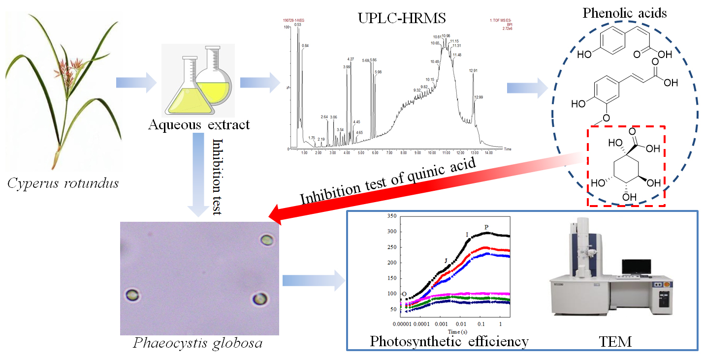
Algicidal Activity of Cyperus rotundus Aqueous Extracts Reflected by Photosynthetic Efficiency and Cell Integrity of Harmful Algae Phaeocystis globosa

1
Department of Ecology, College of Life Science and Technology, Jinan University, Guangzhou 510632, China
2
South China Institute of Environmental Science, Ministry of Ecology and Environment, Guangzhou 510535, China
3
Institute of Groundwater and Earth Sciences, Jinan University, Guangzhou 510632, China
*
Author to whom correspondence should be addressed.
These authors contributed equally to this work.
Water 2020, 12(11), 3256; https://doi.org/10.3390/w12113256
Submission received: 17 September 2020 / Revised: 13 November 2020 / Accepted: 16 November 2020 / Published: 20 November 2020
(This article belongs to the Section Oceans and Coastal Zones)

Abstract

:
Phaeocystis globosa is regarded as a notoriously harmful algal bloom species. Suppressing harmful algae using algicidal substances extracted from plants is considered an effective method. The physiological and biochemical processes of P. globosa were explored by exposure to different concentrations of aqueous extracts of Cyperus rotundus. All treatments indicated various inhibitory effects on the algal growth compared to the control samples without adding extracts. At 48 h, the 4, 8, and 16 mg/mL treatment groups showed a significant inhibitory effect, consistent with a decrease in the chlorophyll-a content and photosynthetic efficiency. The images of the transmission electron microscope (TEM) further confirmed that a subset of the cells in the treatment groups exhibited morphological anomalies. The algicidal active substances were mainly identified as phenolic acids containing maximal content of quinic acid in aqueous extracts according to the results of ultra-high-performance liquid chromatography-tandem time-of-flight mass spectrometer (UPLC-HRMS). The 50% anti-algal effect concentration of quinic acid was 22 mg/L at 96 h (EC50–96h). Thus, the phenolic acids might be considered as major inhibitors of the growth of P. globosa. These results demonstrated that the aqueous extracts of C. rotundus could potentially control the growth of P. globosa.

Graphical Abstract

1. Introduction

Algal blooms are natural phenomena in the aquatic ecosystem and are designated as harmful algal blooms (HABs) with detrimental effects on ecosystems. The anthropogenic activities, including agricultural and industrial sewage discharge, the development of ports, and the over-expansion of aquaculture, have resulted in eutrophication and intensified the occurrence of HABs worldwide [1]. Some HABs species gain superiority in resource competition for mass proliferation, while leading to hypoxia or the mortality of marine organisms due to high biomass and oxygen depletion due to their self-decomposition [2]. Toxic HABs species may produce toxicants that are absorbed by aquatic organisms, especially shellfish, ultimately threatening human life [3].
Phaeocystis globosa (Prymnesiophyceae) is a dense bloom-forming, broad temperature tolerance, and broad salinity tolerance HABs species [4]. It leads to massive mortality of fishes, and numerous giant colonies block the supplement of cooling water of nuclear power plants, causing great harm to marine ecology, aquaculture, and nuclear power safety [5,6]. In 2002, nearly 90% of the animal and plant species in tidal reefs of Phan Ri Bay (Binh Thuan Province, southern central Viet Nam) were seriously damaged during the P. globosa bloom, causing substantial economic losses [7]. Thus, frequent global outbreaks of P. globosa in the ocean have gained extensive attention in the last decades [8,9,10,11]. Phaeocystis globosa has a complex polymorphic life cycle involving diploid colonial cells and diploid and haploid flagellates belonging to different life cycles that can adapt to different environments [8,12]. The colonial cells have high growth rates and can resist small predators [13], while the flagellates reproduce asexually and adapt to conditions of limited nutrients [14]. During the disintegration of P. globosa, the vesicles of the cells burst and white foam floats on the surface of the water [15,16]. Seuront et al. [17] found that the seawater viscosity increased continually during bloom and declined subsequently. Previous studies reported several measures to inhibit the growth of P. globosa, such as Chinese traditional herbs and herb-modified clays [18], mangroves [19] and coastal plants [20]. Therefore, P. globosa is considered as an excellent species to study the environmental stress on its growth due to its wide adaptability, life cycle, dominance in the microalgae community, and ecological hazards [21].
The long-term strategy for controlling HABs is to reduce nutrient inputs. However, the usage of physical, chemical, and biological methods rapidly and effectively suppresses the growth of HABs [22,23,24]. The natural secondary metabolites extracted from different plant tissues are under intensive focus. Previous studies reported several algicidal substances extracted from seagrass [25], riparian plants [26], and terrestrial plants [27]. Owing to the adaptive capacities of some HAB species, the ecological safety of non-targeted microalgae or aquatic animals, and the resources of plants, exploring more plants is crucial. Cyperus rotundus, commonly known as purple nutsedge or nutgrass, belongs to the Cyperaceae family and is believed to be native to the Indian subcontinent. Cyperus rotundus is widely distributed in tropical, subtropical and temperate regions, as well as coastal areas. It is an invasive weed that competes strongly with the adjacent plants because of its allelopathic ability [28]. For instance, the allelopathic chemicals released from C. rotundus could reduce the crop yields and replanting problems in the orchards [29,30]. Despite the negative effect, most studies have focused on the pharmaceutical functions of its rhizomes and tubers mainly based on the component of essential or volatile oils obtained by hydrodistillation [31,32,33]. Instead of pharmacological activities, the function of C. rotundus aqueous extracts gained less attention [34].
In previous decades, P. globosa blooms have frequently been presented in the South China Sea [35,36]. Consequently, the application of aqueous extracts of C. rotundus to inhibit the proliferation of HABs is a novel methodological attempt. The present study aimed to (1) evaluate the algal growth of short-term exposure to C. rotundus extracts, (2) elucidate the microalgae potential physiological response of extracts by measuring the chlorophyll-a content, photosynthetic efficiency and changes in the intracellular microstructure of P. globosa involved in stress tolerance and (3) analyze the main components in C. rotundus aqueous extracts, identify the potential algicidal substances and test the algicidal effect of single phenolic acid by using standard substances of quinic acid.

2. Materials and Methods

2.1. Phaeocystis Globosa Cultures

The strain of P. globosa was isolated by the capillary pipette method [37] from Junk Bay, Hongkong in 1999. The identification of the partial sequence of 18S rDNA indicated that the strain was P. globosa [38].
Natural seawater (salinity of 10‰) was collected from Pearl River Estuary in south China. Filtered seawater enriched with f/2 medium without silicate [39] was used as the culture medium for algal culture. Before the experiments, the culture conditions were maintained at 23 ± 0.1 °C under a 12:12 h (light:dark) photoperiod cycle and an irradiance of 120 μmol/m2/s. The tested microalgae, consisting of haploid flagellates, were cultured to the exponential phase. Then, the culture was inoculated in the subsequent experiments.

2.2. Cyperus Rotundus Plant Sampling and Treatments

Fresh and healthy C. rotundus whole-plants except for rhizomes and tubers (including leaves, stalks and inflorescence) were collected in October 2018 from the coastal tidal belt of the Qi’ao island, Guangdong Province, China (22°25′ N, 113°37′ E). The collected samples were cleaned with filtered seawater containing 1% of the surfactant to remove surface epiphytes, following which they were dipped in filtered seawater containing chloramphenicol (50 ppm) for 3 min to obliterate residual epiphytic cells and bacteria. After rinsing in filtered seawater, the samples were heated at 105 °C for 30 min and dried to a constant weight at 60 °C. The dry samples were cut into pieces and ground into powder by a pulverizer.
An equivalent of 16 g samples was weighed for extraction with 1 L seawater filtered by a 0.22-μm nylon mesh, followed by extraction with ultrasonic waves overnight at room temperature. The extracted solution was passed through the filter again to obtain a stock filtrate at a concentration of 16 mg/mL C. rotundus dried plant extracts.

2.3. Co-Cultures of C. rotundus Extracts and P. globosa

The stock filtrate was diluted to a series of concentrations and added to each group (e.g., the 16 mg/mL treatment group contained 98 mL stock filtrate and 1 mL 32 mg/mL C. rotundus aqueous extracts). A volume of 1 mL algae suspension was added to the above working solution (final algal concentration was 2.5 × 10⁵ cells/mL) with a final concentration gradient of 1, 2, 4, 8 and 16 mg/mL C. rotundus dried plant extracts. The culture medium was considered as the control. All the experiments were carried out in three replicates. The cell density was estimated by flow cytometer at 0, 12, 24, 48, 72 and 96 h, respectively.

2.4. Measurement of Photosynthetic Efficiency and Chlorophyll-a Content

The performance of photosystem II (PSII) was determined by the Handy Plant Efficiency Analyzer apparatus (Handy PEA, Hansatech Instruments, Pentney, UK). The samples were adapted to the darkness for 20 min at room temperature before measurement. The fast phase gave rise to the “O-J-I-P” (O: initial fluorescence value, J: fluorescence value in 2 ms, I: fluorescence value in 30 ms, P: maximum fluorescence value) fluorescence transients within the recordation of 3 s (Table 1). The fluorescence transients were used to measure the photochemical quantum yield of PSII and the electron transport properties within PSII. The selected parameters of fluorescence transients (including Fv/Fm, ABS/RC, TR0/RC, ET0/RC, DI0/RC, and PI) were calculated from the original date using the formula based on previous studies [40,41,42]. The photosynthetic apparatus was altered under stress conditions, as assessed by the JIP-test (the fluorescence analysis based on the O-J-I-P curve, for reviews, see [40,41,42,43]). Chlorophyll-a content was measured spectrophotometrically after extraction in 90% acetone at 4 °C overnight as described by Lin et al. [44].

2.5. TEM Analysis of the Cell Ultrastructure

The algae solution of the control and the experimental groups (1 and 4 mg/mL) was collected after 48 h and fixed in 0.1 M PBS containing 2.5% glutaraldehyde (v/v) at 4 °C for 2–4 h. The cells were harvested and 0.1 M PBS was added containing 1% osmium tetroxide for post-fixation at room temperature for 2 h. Then, the cells were dehydrated with a graded alcohol series (50%, 70%, 80%, 90%, 95% and 100%) for 15 min each, followed by 100% acetone (15 min × 3). The samples were embedded in the 812 embedding medium, sliced at 60–80 nm thickness with an ultramicrotome (Lecia EM UC7, Vienna, Austria) and stained with uranium-lead stains (2% uranyl acetate and 2% lead citrate). Finally, the cell ultrastructure was observed using TEM with 0.144-nm lattice resolution and 0.33-nm point resolution (Hitachi 7700, Tokyo, Japan).

2.6. Analysis of the Main Compounds of C. rotundus Extracts

The main compounds of C. rotundus extracts were analyzed using ultra-high-performance liquid chromatography (Waters Acquity UPLC H-Class, Milford, MA, USA) tandem time-of-flight mass spectrometer (Waters Xevo G2-S tof, Milford, MA, USA) (UPLC-HRMS). The chromatographic column Acquity UPLC BEH C18, 2.1 mm × 50 mm, 1.7 μm was used to separate compounds. The column temperature was set at 45 °C, the injection volume was 1 μL, and the flow rate was 0.40 mL/min. The mobile phase comprised acetonitrile (A) and water containing 0.1% formic acid (B). The gradient elution procedures were as follows: 0–2 min, 3–10% (A); 2–6 min, 10–50% (A); 6–8 min, 50–80% (A); 8–11 min, 80–95% (A); 11–12 min, 95% (A); 12–13 min, 95–3% (A); 13–15 min, 3% (A). Electron spray ionization was used for the high-resolution mass spectrum (HRMS) in positive ion and negative ion mode, respectively. The temperature of the ion source was 120 °C. The other MS parameters were set as follows: the capillary voltage was 2.5 kV, the cone voltage was 25 V, the desolvation gas temperature was 400 °C, the desolvation gas flow was 1000 L/h, and the scanning pattern was MSE. The chemical information of different components was downloaded from the National Center for Biotechnology Information, the U.S. National Library of Medicine (https://pubchem.ncbi.nlm.nih.gov, Bethesda, MD, USA).

2.7. Reagents and Algal Cultures

To understand the dose-response of algicidal effect, 1.6 g standard substances of quinic acid (>98%, Sigma-Aldrich Shanghai Trading Co., Ltd., Shanghai, China) were solubilized in 1 L culture medium to prepare a stock solution. Then, the stock solution, culture medium and 1 mL algae suspension were added into flasks (250 mL) and diluted to 100 mL (final algal concentration was 2.5 × 10⁵ cells/mL) at final concentrations of quinic acid solution: 0.05, 0.1, 0.2, 0.4, 0.8, and 1.6 mg/mL. The experimental conditions were the same as the co-culture of C. rotundus extracts and P. globosa.

2.8. Statistical Analyses

The cell density was estimated after homogenization using a flow cytometer (BD Accuri C6, Franklin Lakes, NJ, USA). The inhibition rate of C. rotundus aqueous extracts (IRCr) or quinic acid (IRqa) on algal growth was estimated by the inhibition rate of two algicidal substances using the following formula:
I R ( % ) = ( 1 N N 0 ) × 100
where N 0 represents the cell density in the control groups and N represents the cell density in the treatment groups.
The differences among the groups were assessed by one-way analysis of variance (ANOVA) analysis, followed by Duncan’s test (p < 0.05) [45]. EC50 was estimated by the Probit analysis based on the IR values. The graphs were constructed using Origin 9.0 (Origin Lab Corporation, Microcal, MA, USA).

3. Results

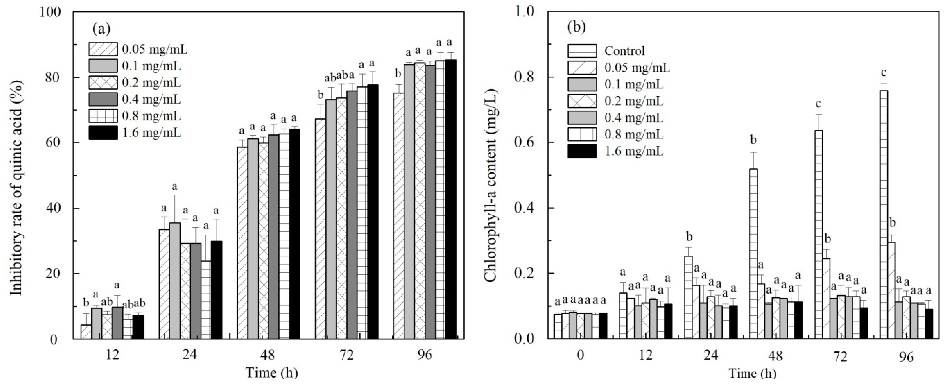
3.1. Effect of C. rotundus Extracts on Algal Growth and Chlorophyll-a Content

The treatment groups with adding C. rotundus aqueous extracts exhibited various inhibitions on the growth of P. globosa during the experimental period. The concentration of 4 mg/mL exhibited the highest IRCr of algal growth at 12 and 24 h of exposure. The IRCr of 73.15%, 72.80%, and 81.22% in the treatment groups of 4, 8, and 16 mg/mL, respectively, indicated significantly higher IRCr than that in the 1 and 2 mg/mL treatment groups (p < 0.05). These effects were noted at 48 h, and the IRCr increased continuously in the following 48 h. The 16 mg/mL treatment group showed a profound inhibition in the growth of P. globosa compared to the other treatment groups and reached the maximum IRCr (98.54%) at 96 h (Figure 1a).
At the beginning of the 24 h, the chlorophyll-a content of the control and all treatment groups increased over time, albeit without a significant difference among these groups at the same sampling time (p > 0.05). Following 48 h of exposure to aqueous extracts, the chlorophyll-a content of the concentrations of 4, 8, and 16 mg/mL was significantly lower than that in the control and the 1 and 2 mg/mL groups (p < 0.05). The control groups reached the maximum content (1.29 mg/L) at 96 h, which was significantly higher than that in the other treatment groups (p < 0.05) (Figure 1b).

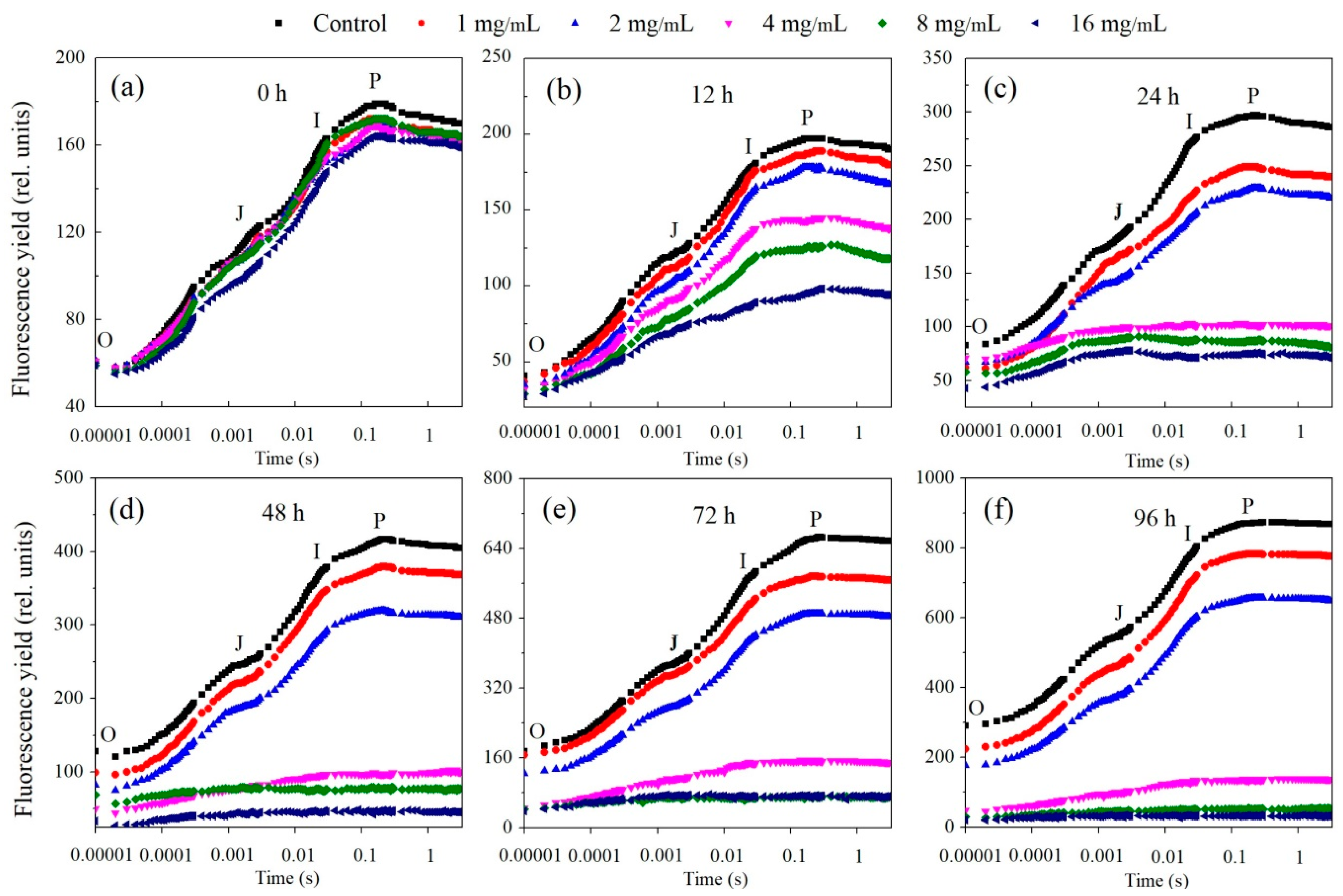
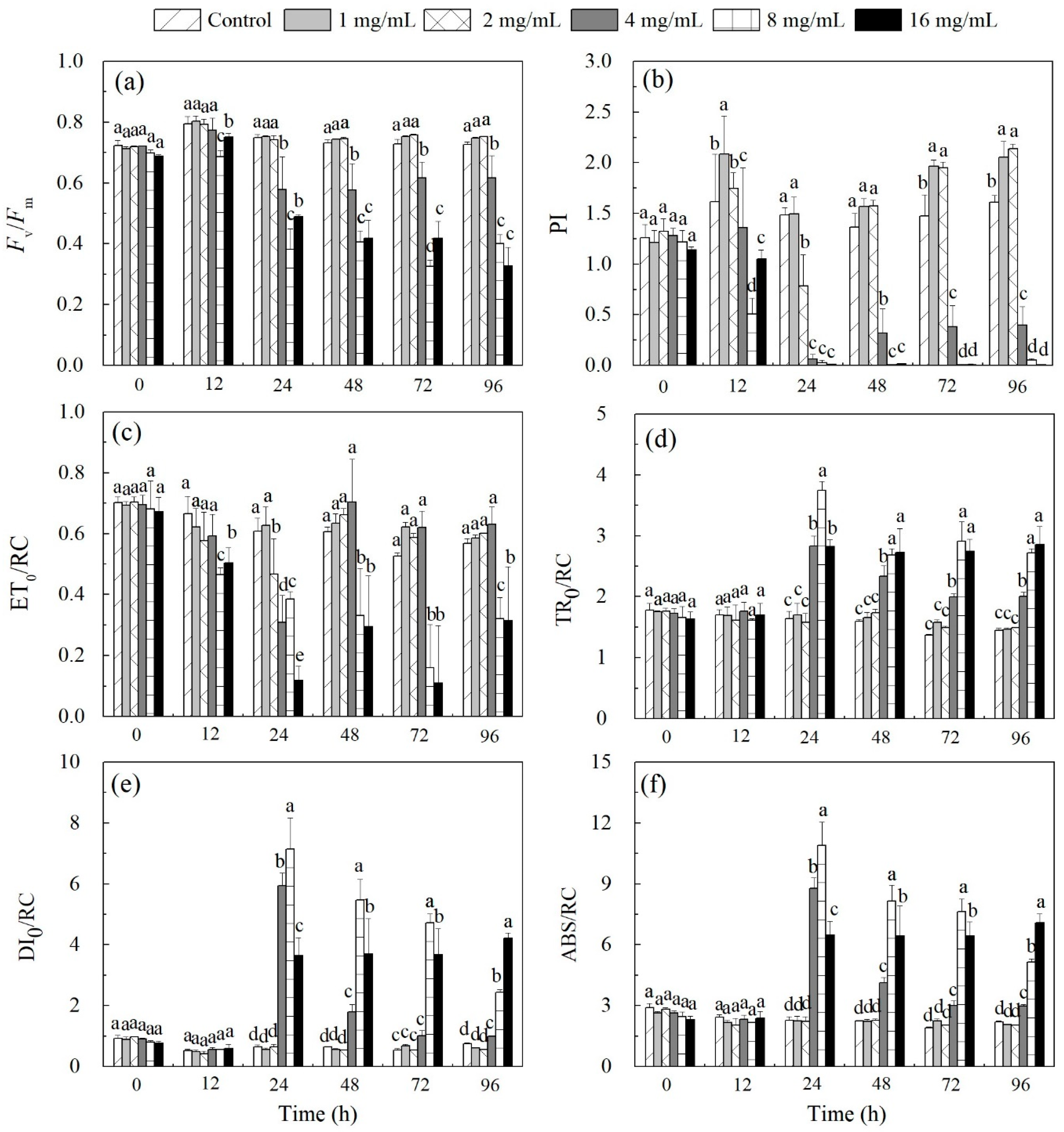
3.2. Inhibition of Algal Photosynthetic Efficiency

The fluorescence intensity of the control groups and the 1 and 2 mg/mL treatment groups increased in a time-dependent manner and that in the control groups grew higher than the two treatment groups during the experimental period. The fluorescence intensity of the control and all treatment groups increased variably at 12 h, whereas that of the 4, 8, and 16 mg/mL treatment groups was lower than that of the control and the 1 and 2 mg/mL treatment groups. After 24 h of cultivation, the O-J, J-I, and I-P phases of the treatment groups (4, 8, and 16 mg/mL) were indistinguishable (Figure 2).
The Fv/Fm values of the control and low treatment groups (1 and 2 mg/mL) were about 0.7 during the experiment, whereas that of the other treatments decreased to <0.6 following 24-h exposure. The Fv/Fm values of 8 and 16 mg/mL continued to decline in the following experimental period, while the values of PI declined more sharply than those of Fv/Fm, indicating that PI could be more sensitive to the changes in the photosynthetic apparatus (Figure 3a,b). Overall, the high treatment group (8 and 16 mg/mL) demonstrated low Fv/Fm values, indicating a severe disruption in photosynthesis.
The effect of C. rotundus aqueous extracts on the microalgae PSII reaction center (RC) was analyzed based on parameters, such as ABS/RC, TR0/RC, ET0/RC, and DI0/RC. The parameters (ABS/RC, TR0/RC, and DI0/RC) of the control and the treatment groups did not show any significant differences (p > 0.05) in the initial 12-h period. Subsequently, the values of ABS/RC, DI0/RC, and TR0/RC of the treatment groups (4, 8, and 16 mg/mL) increased significantly as compared to the control and the other treatment groups (p < 0.05), while the ET0/RC values of the treatment groups (4, 8 and 16 mg/mL) decreased significantly as compared to the control and the other treatment groups (p < 0.05) (Figure 3c–f).

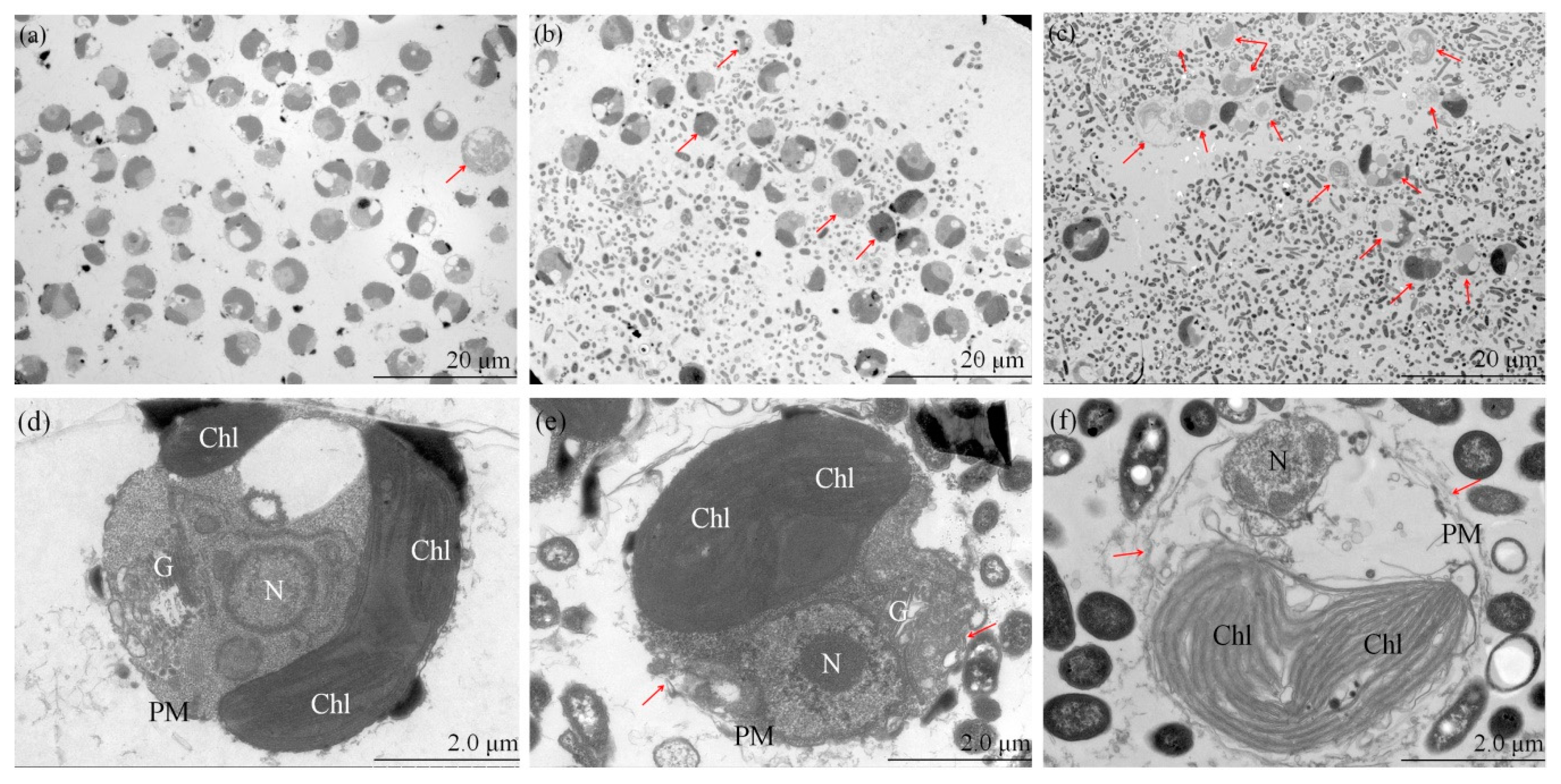
3.3. Ultrastructure of the Test Algae

The control samples exhibited almost no intracellular substances and most of the cells were healthy (Figure 4a). The cell membrane of the individual cell was distinct and the intracellular chloroplasts and nuclei did not exhibit a deformed or damaged structure, in addition the chloroplasts were tightly arranged together (Figure 4d). Additional intracellular substances were noted in the 1 mg/mL treatment group (Figure 4b). Some individual algal cell membranes were slightly ruptured, while some vacuolization areas could be found in the cytoplasm (Figure 4e). A high number of intracellular substances were made evident by TEM (Figure 4c), and the majority of the algal cells did not possess complete morphology. The intracellular organelles could not be distinguished, and the intracellular substances had outflowed, indicating necrosis of these cells (Figure 4f). The 1 mg/mL treatment group caused no or slight damage to part of the algal structure. However, the test algae were severely damaged by a 48-h treatment at 4 mg/mL, which was associated with significant inhibition.

3.4. The Main Compounds in C. rotundus Aqueous Extracts

The total ion chromatogram of the UPLC-HRMS obtained for the C. rotundus was satisfactory and the independent peaks could be identified distinctly. In terms of HRMS, the total ion current in response to the main component under the positive ion scanning mode was better than that of the negative ion mode. However, the response to some of the compounds was preferable in the negative ion mode; consequently, the structure was identified in both positive and negative modes.
A total of 17 major peaks were observed in the positive ion mode that corresponds to seven secondary metabolites (Table 2 and Figure 5a). A total of 21 major peaks were observed in the negative ion mode and 12 secondary metabolites were identified, of which quinic acid, 3-O-feruloylquinic acid, 3-O-trans-Coumaroylquinic acid, 4-O-trans-coumaroylquinic acid and 5-O-trans-coumaroylquinic acid exhibited unique chromatographic patterns (Table 2 and Figure 5c). 3-O-feruloylquinic acid was an ester formed between quinic acid and ferulic acid. 3-, 4-, and 5-O-trans-coumaroylquinic acid were an ester formed between quinic acid and p-coumaric acid, although the binding sites were different. These substances were classified as phenolic acids (No.1, 3, 4, 5, 6, 8, 9, 10, and 11), non-protein amino acids (No. 12), alkaloids (No. 13), coumarins (No. 15), and other compounds (No. 2, 7, and 14).

3.5. Quinic Acid Test for the Algal Growth Assessment

To better understand the algicidal traits of single phenolic acid, standard substances of phenolic acid with maximal content in the C. rotundus aqueous extracts were used. The concentration of the component in the extracts can be determined by the peak area, although no strong link was noted between the concentration and peak area. However, the concentration of the quinic acid could be compared among the peaks based on the peak area, since similar classes of compounds may possess similar chemical features and ionization potential [46]. The quinic acid exhibited the largest peak area (138266) which was prompted to be investigated (Table 2) and the algicidal activity of quinic acid was tested in the subsequent experiment. The results of the quinic acid test revealed that the IRqa of all the treatment groups shows no significant difference (p > 0.05) at the 24 and 48 h sampling time points, and the 0.05 mg/mL treatment group of quinic acid demonstrated a low IRqa in the following time. A significant difference (p < 0.05) was noted as compared to the results found in the other treatment groups at the 72 and 96 h sampling time points (Figure 6a). There was no significant difference (p > 0.05) within the chlorophyll-a content of all groups at 12 h time point, while the control grew over time in the following time and showed a significant difference (p < 0.05) compared to all treatment groups. The 0.05 mg/mL treatment group exhibited a higher chlorophyll-a content than other treatment groups at 72 and 96 h sampling periods and a significant difference (p < 0.05) was noted (Figure 6b). Finally, the median effective concentration of quinic acid at 96 h was 22 mg/L.

4. Discussion

4.1. Modes of Physiological Progression

Although observing the effect of plant extracts on the algal growth (in terms of inhibition rate) is the main purpose of this study, the mode of algal physiological progression can help understand the causes of this phenomenon, providing an insight into modifying the materials and extraction technologies for field implementation [47,48]. The algicidal substances used against HABs were involved in various physiological processes, including oxidative damage, interruption of the electron transfer chain of PSⅡ, cell division inhibition and DNA damage [49,50]. In the present study, the indexes, such as inhibition rate, chlorophyll-a content, photosynthetic efficiency and changes in the cell integrity and ultrastructure showed pronounced inhibition when exposed to ≥4 mg/mL C. rotundus aqueous extracts and the concentration of quinic acid was 0.027 mg/mL in 4 mg/mL C. rotundus aqueous extracts.
The PEA parameters reflect the photosynthetic efficiency, and Fv/Fm is one of the most widely characterized parameters. The Fv/Fm value is the intuitive parameter reflecting the potential maximum photosynthetic capacity in response to stress [51]. The present study indicated that the Fv/Fm values were normal with the exposure of low concentrations of aqueous extracts, but the growth of microalgae was slightly inhibited, while Fv/Fm values decreased significantly at high concentrations. To elucidate the action of PSII under the environmental stress, the chlorophyll-a fluorescence transient induction curves were drawn, which provided insights into the damage of the photosystem [52].
The OJIP curve is a highly efficient method of studying PSII without damaging the cells [53]. The O-J phase is related to the accumulation of the reduced electron acceptors(QA), the J-I phase is related to the complete closure of PSII RC or a QB-quenching mechanism, and the I-P phase is related to the plastoquinone pool reduction [54,55]. In the present study, the OJIP curve values decreased with the increased content of C. rotundus aqueous extracts. This phenomenon was essentially consistent with the previous reports, which showed that the photosynthetic efficiency of Scrippsiella trochoidea was inhibited by Gracilaria lemaneiformis [56]. Furthermore, the JIP-test parameters reflected the quantum efficiency of the reaction center (RC) through the trapped energy flux per RC (TR0/RC), heat dissipation per RC (DI0/RC), electron transport flux per RC (ET0/RC), and energy absorbed per RC (ABS/RC). Due to the exposure to a high concentration of C. rotundus aqueous extracts, the proportion of RC per unit area declined gradually, thereby increasing the absorption of light energy for the remaining active RC (manifested as the increase in ABS/RC). This feature suggested that high concentration might inactivate the RC or increase the functional PSII antenna size [57,58]. The decline in the RC increased the excitation energy, which reduced the QA, thereby indicating that the efficiency of the remaining active RC increased (manifested as the increase in TR0/RC). The reduction energy in the electron transport chain beyond the QA decreased, suggesting that the active RC captured less energy for electron transport (manifested as the decrease in ET0/RC); moreover, the heat dissipation increased (manifested as the increase in DI0/RC). Thus, the superfluous energy could be dissipated by non-photochemical quenching [59,60]. In addition, phenolic acids could hinder chlorophyll-a synthesis and reduce chlorophyll-a content [61,62]. Overall, the C. rotundus aqueous extracts interfered with photosynthesis efficiency by reducing its chlorophyll-a content and electron transfer efficiency.
Phenolic acids exhibit cell-permeability features due to their amphiphilic and lipophilic nature [63,64]. Wang et al. [65] and Zhang et al. [66] found that ferulic acid and p-coumaric acid disrupted the cell membrane integrity of Microcystis aeruginosa, respectively. In the present study, phenolic acids, the main components of C. rotundus aqueous extracts, penetrate the cells through passive diffusion, disrupt the cell membrane integrity and damaged the cell structure, leading to the efflux of intracellular material, including protein and nucleic acids. Oxidative damage induced by phenolic acids, such as vanillic acid, protocatechuic acid, ferulic acid and caffeic acid, is regarded as the main reason for the inhibitory effect of some microalgae [67,68]. Previous studies indicated that algicidal substances extracted from plants also involved relative gene expressions [69]. Shao et al. [70] found that the gene expressions of Microcystis aeruginosa were regulated and the antioxidant systems were damaged with the exposure of pyrogallol. Moreover, some phenolic acids can influence the phycosphere and interrupt the ecological relationship between microalgae and other microorganisms, thus influencing algal growth, decay and nutrient cycling [71].

4.2. Algicidal Potential of Phenolic Acids

The use of extracts from different plants to suppress HABs is considered as one of the most cost-effective and environment-friendly strategies. Numerous natural algicidal substances have been isolated and identified [20,25,69,72,73,74]. The majority of the algicidal substances are categorized as terpenoids, phenolics (flavonoids, tannins and phenolic acids) and nitrogen-containing secondary metabolites (alkaloids, cyanogenic glycosides and non-protein amino acids) [75], and most of the studies focused on terpenoids and phenolics [76]. Terpenoids are lipophilic and insoluble in water [64], among which monoterpenoids and sesquiterpenoids (constituents of volatile oils) are highly volatile with short retention time in the water [77], limiting the application of terpenoids in the treatment of HABs. Previous studies reported that flavonoids show strong algicidal activities [78]. Flavonoids are natural pigments (flavonols are colorless) [79,80] with low water solubility [81] and render difficultly in determining the effect of shading interference on algal growth. The algicidal effect of tannins was also explored [82]. Since tannins can form complexes with proteins, starches, and digestive enzymes [83], their usage for HABs should be limited.
Phenolic acids are widely distributed allelopathic chemicals in higher plants [84,85]; however, the inhibitory potential of phenolic acids on the growth of P. globosa has been rarely reported. Previous studies showed that the allelopathic potential of phenolic acids is the result of phenolic acids in the plants, and the allelopathic effect of single phenolic acid is not distinct [86,87]. The study by Nakai et al. [88] demonstrated that quinic acid, p-coumaric acid and ferulic acid (10 mg/L) had no inhibitory effect on M. aeruginosa, respectively. In the present study, no significant inhibitory effect was noted concerning the growth of three dinoflagellates, Scrippsiella trochoidea, Alexandrium tamarense and Karenia mikimotoi, following the exposure to quinic acid (concentration range of 0.1, 0.2, 0.3, 0.4 and 0.5 mg/mL) (data not shown). However, quinic acid exerted a specific inhibitory effect on the growth of P. globosa. This phenomenon could be attributed to the fact that the algicidal effect is species dependent. The concentration of quinic acid in the 4 mg/mL C. rotundus aqueous extracts was approximately the EC50–96h value, but the inhibition rate of the extracts was >90%, indicating that other components might take effect. To the best of our knowledge, 3-, 4-, and 5-O-trans-coumaroylquinic acids were first reported in the C. rotundus aqueous extracts. Therefore, the potential to inhibit HABs should be explored further.

4.3. Bioavailability of Phenolic Compounds

The ideal algicidal substances can be readily degraded in specific environments, can be easily gained in nature and, can exhibit potent inhibitory effect to target microalgae. Thus, the ideal algicidal substances exhibit limited or low toxicity to other microalgae of the phytoplankton communities, as well as animals [49,89]. The bioavailability of phenolic compounds can be realized through the utilization pattern of microalgae. In the complex aqueous environment, phenolic compounds can be mineralized into carbon dioxide or converted into a structurally similar molecule through biotransformation, and other degradative mechanisms [90,91]. Phenolic compounds are also potential alternative carbon sources required for the growth of microalgae [92,93]. The co-culture experiment by Escapa et al. [94] showed that Chlorella sorokiniana removes salicylic acid; additionally, the biomass of microalgae was stimulated. In the present study, monocyclic phenolic acid and phenolic acid ester were the main components of the C. rotundus aqueous extracts, which benefited to the bioavailability of microalgae due to the low molecular weight because the persistence of benzene series decrease with the decrease in molecular weight [95]. In addition, the use of C. rotundus aqueous extracts confronts the problem of dilution of the allelochemical molecules in the field, and thus degradation by microorganisms or even inefficiency of their bioactivity. However, excess amounts of extracts to water are not recommended because it might cause a risk of secondary pollution. Huang et al. [96] developed continuous-release beads that contain algicidal substances released continually during the bloom.
This study explored the algicidal mechanism of C. rotundus aqueous extracts and discussed the bioavailability of phenolic compounds, facilitating the extraction and utilization of secondary metabolites from marine plants.

5. Conclusions

The aqueous extracts of C. rotundus exhibited a significant inhibitory effect on the growth of P. globosa with increasing inhibition rate and decreasing chlorophyll-a content. The variables comprising photosynthetic efficiency and cell integrity were observed based on the parameters of the OJIP curve, JIP-test parameters and the images of the cell ultrastructure, respectively. UPLC-HRMS analysis revealed that phenolic acids could be major algicidal active components. The standard substance quinic acid was tested, and it was found that the algicidal effect of single phenolic acid was limited; the other components in the extracts might contribute to the algicidal effect. Furthermore, the impact of C. rotundus aqueous extracts on other species is unknown and should be evaluated with respect to the ecological risk before practical application in the natural environment. Since the use of C. rotundus aqueous extracts might confront the bioavailability of microbial species and the dilution in the field, affecting the algicidal efficiency, more techniques should be developed to prolong the retention time of the algicidal substances in water.

Author Contributions

In the manuscript, S.D. is the corresponding author. Y.L. tested the inhibitory effect of aqueous extracts and quinic acid, tested the photosynthetic efficiency, processed data and wrote the manuscript. Q.C. participated in the sample collection, pre-treatment and observation of ultrastructure of cells. T.G. analyzed the component of aqueous extracts. K.S. and D.S. reviewed the manuscript. S.D. designed the experiment and supervised the writing. J.Z. provided technical supports for the article modification. All authors have read and agreed to the published version of the manuscript.

Funding

This research was supported by the National Natural Science Foundation of China (No.41676099).

Conflicts of Interest

The authors declare no conflict of interest.

References

  1. Heisler, J.; Glibert, P.; Burkholder, J.; Anderson, D.; Cochlan, W.; Dennison, W.; Gobler, C.; Dortch, Q.; Heil, C.; Humphries, E.; et al. Eutrophication and Harmful Algal Blooms: A Scientific Consensus. Harmful Algae 2008, 8, 3–13. [Google Scholar] [CrossRef] [PubMed] [Green Version]
  2. Harrison, P.J.; Piontkovski, S.; Al-Hashmi, K. Understanding how physical-biological coupling influences harmful algal blooms, low oxygen and fish kills in the Sea of Oman and the Western Arabian Sea. Mar. Pollut. Bull. 2017, 114, 25–34. [Google Scholar] [CrossRef] [PubMed]
  3. Furuya, K.; Iwataki, M.; Lim, P.T.; Lu, S.; Leaw, C.-P.; Azanza, R.V.; Kim, H.-G.; Fukuyo, Y. Overview of harmful algal blooms in Asia. In Global Ecology and Oceanography of Harmful Algal Blooms; Springer: Berlin/Heidelberg, Germany, 2018; pp. 289–308. [Google Scholar]
  4. Schoemann, V.; Becquevort, S.; Stefels, J.; Rousseau, V.; Lancelot, C. Phaeocystis blooms in the global ocean and their controlling mechanisms: A review. J. Sea Res. 2005, 53, 43–66. [Google Scholar] [CrossRef]
  5. Qi, Y.; Chen, J.; Wang, Z.; Xu, N.; Wang, Y.; Shen, P.; Lu, S.; Hodgkiss, I.J. Some observations on harmful algal bloom (HAB) events along the coast of Guangdong, southern China in 1998. Hydrobiologia 2004, 512, 209–214. [Google Scholar] [CrossRef]
  6. Smith, W.O., Jr.; Liu, X.; Tang, K.W.; DeLizo, L.M.; Doan, N.H.; Nguyen, N.L.; Wang, X. Giantism and its role in the harmful algal bloom species Phaeocystis globosa. Deep Sea Res. Part II Top. Stud. Oceanogr. 2014, 101, 95–106. [Google Scholar] [CrossRef] [Green Version]
  7. Doan, H.; Nguyen, N.L.; Nguyen, C.; Ho, V.T.; Nguyen, M.A. Plankton assemblages during the late bloom of haptophyte algae in Binh Thuan province, Southern central Vietnam, in July 2002. Collect. Mar. Res. Work. 2003, 13, 105–118. [Google Scholar]
  8. Rousseau, V.; Lantoine, F.; Rodriguez, F.; LeGall, F.; Chretiennot-Dinet, M.J.; Lancelot, C. Characterization of Phaeocystis globosa (Prymnesiophyceae), the blooming species in the Southern North Sea. J. Sea Res. 2013, 76, 105–113. [Google Scholar] [CrossRef]
  9. Hai, D.-N.; Lam, N.-N.; Dippner, J.W. Development of Phaeocystis globosa blooms in the upwelling waters of the South Central coast of Viet Nam. J. Mar. Syst. 2010, 83, 253–261. [Google Scholar] [CrossRef]
  10. Mars Brisbin, M.; Mitarai, S. Differential gene expression supports a resource-intensive, defensive role for colony production in the bloom-forming haptophyte, Phaeocystis globosa. J. Eukaryot. Microbiol. 2019, 66, 788–801. [Google Scholar] [CrossRef] [Green Version]
  11. Rauch, M.; Denis, L.; Dauvin, J.-C. The effects of Phaeocystis globosa bloom on the dynamics of the mineralization processes in intertidal permeable sediment in the Eastern English Channel (Wimereux, France). Mar. Pollut. Bull. 2008, 56, 1284–1293. [Google Scholar] [CrossRef]
  12. Rousseau, V.; Chrétiennot-Dinet, M.-J.; Jacobsen, A.; Verity, P.; Whipple, S. The life cycle of Phaeocystis: State of knowledge and presumptive role in ecology. Biogeochemistry 2007, 83, 29–47. [Google Scholar] [CrossRef]
  13. Veldhuis, M.J.; Brussaard, C.P.; Noordeloos, A.A. Living in a Phaeocystis colony: A way to be a successful algal species. Harmful Algae 2005, 4, 841–858. [Google Scholar] [CrossRef]
  14. Rousseau, V.; Vaulot, D.; Casotti, R.; Cariou, V.; Lenz, J.; Gunkel, J.; Baumann, M. The life cycle of Phaeocystis (Prymnesiophycaea): Evidence and hypotheses. J. Mar. Syst. 1994, 5, 23–39. [Google Scholar] [CrossRef]
  15. Spilmont, N.; Denis, L.; Artigas, L.F.; Caloin, F.; Courcot, L.; Créach, A.; Desroy, N.; Gevaert, F.; Hacquebart, P.; Hubas, C. Impact of the Phaeocystis globosa spring bloom on the intertidal benthic compartment in the eastern English Channel: A synthesis. Mar. Pollut. Bull. 2009, 58, 55–63. [Google Scholar] [CrossRef] [Green Version]
  16. Blauw, A.; Los, F.; Huisman, J.; Peperzak, L. Nuisance foam events and Phaeocystis globosa blooms in Dutch coastal waters analyzed with fuzzy logic. J. Mar. Syst. 2010, 83, 115–126. [Google Scholar] [CrossRef]
  17. Seuront, L.; Vincent, D.; Mitchell, J.G. Biologically induced modification of seawater viscosity in the Eastern English Channel during a Phaeocystis globosa spring bloom. J. Mar. Syst. 2006, 61, 118–133. [Google Scholar] [CrossRef]
  18. Tian, F.; Zhou, J.Y.; Sun, Z.W.; Cai, Z.P.; Xu, N.; An, M.; Duan, S.S. Inhibitory effects of Chinese traditional herbs and herb-modified clays on the growth of harmful algae, Phaeocystis globosa and Prorocentrum donghaiense. Harmful Algae 2014, 37, 153–159. [Google Scholar] [CrossRef]
  19. Zhao, M.; Xiao, H.; Sun, D.; Duan, S. Investigation of the Inhibitory Effects of Mangrove Leaves and Analysis of Their Active Components on Phaeocystis globosa during Different Stages of Leaf Age. Int. J. Environ. Res. Public Health 2018, 15, 2434. [Google Scholar] [CrossRef] [Green Version]
  20. Xu, C.; Ge, Z.; Li, C.; Wan, F.; Xiao, X. Inhibition of harmful algae Phaeocystis globosa and Prorocentrum donghaiense by extracts of coastal invasive plant Spartina alterniflora. Sci. Total Environ. 2019, 696, 133930. [Google Scholar] [CrossRef]
  21. Brussaard, C.; Mari, X.; Van Bleijswijk, J.; Veldhuis, M. A mesocosm study of Phaeocystis globosa (Prymnesiophyceae) population dynamics: II. Significance for the microbial community. Harmful Algae 2005, 4, 875–893. [Google Scholar] [CrossRef]
  22. Sun, R.; Sun, P.F.; Zhang, J.H.; Esquivel-Elizondo, S.; Wu, Y.H. Microorganisms-based methods for harmful algal blooms control: A review. Bioresour. Technol. 2018, 248, 12–20. [Google Scholar] [CrossRef] [PubMed]
  23. Jancula, D.; Marsalek, B. Critical review of actually available chemical compounds for prevention and management of cyanobacterial blooms. Chemosphere 2011, 85, 1415–1422. [Google Scholar] [CrossRef] [PubMed]
  24. Gallardo-Rodríguez, J.J.; Astuya-Villalón, A.; Llanos-Rivera, A.; Avello-Fontalba, V.; Ulloa-Jofré, V. A critical review on control methods for harmful algal blooms. Rev. Aquac. 2018, 11, 661–684. [Google Scholar] [CrossRef]
  25. Zhu, J.; Xiao, H.; Chen, Q.; Zhao, M.; Sun, D.; Duan, S. Growth Inhibition of Phaeocystis Globosa Induced by Luteolin-7-O-glucuronide from Seagrass Enhalus acoroides. Int. J. Environ. Res. Public Health 2019, 16, 2615. [Google Scholar] [CrossRef] [PubMed] [Green Version]
  26. Patino, R.; Rashel, R.H.; Rubio, A.; Longing, S. Growth-suppressing and algicidal properties of an extract from Arundo donax, an invasive riparian plant, against Prymnesium parvum, an invasive harmful alga. Harmful Algae 2018, 71, 1–9. [Google Scholar] [CrossRef]
  27. Chen, S.; Zheng, T.; Ye, C.; Huannixi, W.; Yakefu, Z.; Meng, Y.; Peng, X.; Tian, Z.; Wang, J.; Ma, Y.; et al. Algicidal properties of extracts from Cinnamomum camphora fresh leaves and their main compounds. Ecotoxicol. Environ. Saf. 2018, 163, 594–603. [Google Scholar] [CrossRef]
  28. Hussain, I.; Singh, N.; Singh, A.; Singh, H. Allelopathic potential of sesame plant leachate against Cyperus rotundus L. Ann. Agrar. Sci. 2017, 15, 141–147. [Google Scholar] [CrossRef]
  29. Bryson, C.T.; Reddy, K.N.; Molin, W.T. Purple nutsedge (Cyperus rotundus) population dynamics in narrow row transgenic cotton (Gossypium hirsutum) and soybean (Glycine max) rotation. Weed Technol. 2003, 17, 805–810. [Google Scholar] [CrossRef]
  30. Dhima, K.; Vasilakoglou, I.; Stefanou, S.; Gatsis, T.; Paschalidis, K.; Aggelopoulos, S.; Eleftherohorinos, I. Differential competitive and allelopathic ability of Cyperus rotundus on Solanum lycopersicum, Solanum melongena and Capsicum annuum. Arch. Agron. Soil Sci. 2016, 62, 1250–1263. [Google Scholar] [CrossRef]
  31. Hu, Q.P.; Cao, X.M.; Hao, D.L.; Zhang, L.L. Chemical Composition, Antioxidant, DNA Damage Protective, Cytotoxic and Antibacterial Activities of Cyperus rotundus Rhizomes Essential Oil against Foodborne Pathogens. Sci. Rep. 2017, 7, 45231. [Google Scholar] [CrossRef] [Green Version]
  32. Peerzada, A.M.; Ali, H.H.; Naeem, M.; Latif, M.; Bukhari, A.H.; Tanveer, A. Cyperus rotundus L.: Traditional uses, phytochemistry, and pharmacological activities. J. Ethnopharmacol. 2015, 174, 540–560. [Google Scholar] [CrossRef] [PubMed]
  33. Lawal, O.A.; Oyedeji, A.O. Chemical composition of the essential oils of Cyperus rotundus L. from South Africa. Molecules 2009, 14, 2909–2917. [Google Scholar] [CrossRef] [PubMed]
  34. Kilani-Jaziri, S.; Bhouri, W.; Skandrani, I.; Limem, I.; Chekir-Ghedira, L.; Ghedira, K. Phytochemical, antimicrobial, antioxidant and antigenotoxic potentials of Cyperus rotundus extracts. S. Afr. J. Bot. 2011, 77, 767–776. [Google Scholar] [CrossRef] [Green Version]
  35. Li, L.; Lu, S.H.; Cen, J.Y. Spatio-temporal variations of Harmful algal blooms along the coast of Guangdong, Southern China during 1980–2016. J. Oceanol. Limnol. 2019, 37, 535–551. [Google Scholar] [CrossRef]
  36. Wang, S.F.; Tang, D.L.; He, F.L.; Fukuyo, Y.S.; Azanza, R.V. Occurrences of harmful algal blooms (HABs) associated with ocean environments in the South China Sea. Hydrobiologia 2008, 596, 79–93. [Google Scholar] [CrossRef]
  37. Hoshaw, R. Methods for microscopic algae. In Handbook of Phycological Methods: Culture Methods and Growth Measurements; Cambridge University Press: Cambridge, UK, 1973; pp. 53–68. [Google Scholar]
  38. Xu, N.; Huang, B.Z.; Hu, Z.X.; Tang, Y.Z.; Duan, S.S.; Zhang, C.W. Effects of temperature, salinity, and irradiance on the growth of harmful algal bloom species Phaeocystis globosa Scherffel (Prymnesiophyceae) isolated from the South China Sea. Chin. J. Oceanol. Limnol. 2017, 35, 557–565. [Google Scholar] [CrossRef]
  39. Guillard, R.R.; Ryther, J.H. Studies of marine planktonic diatoms. I. Cyclotella nana Hustedt, and Detonula confervacea (cleve) Gran. Can. J. Microbiol. 1962, 8, 229–239. [Google Scholar] [CrossRef]
  40. Sun, D.; He, N.; Chen, Q.; Duan, S. Effects of Lanthanum on the Photosystem II Energy Fluxes and Antioxidant System of Chlorella vulgaris and Phaeodactylum tricornutum. Int. J. Environ. Res. Public Health 2019, 16, 2242. [Google Scholar] [CrossRef] [Green Version]
  41. Strasser, R.J.; Tsimilli-Michael, M.; Srivastava, A. Analysis of the Chlorophyll a Fluorescence Transient. In Chlorophyll a Fluorescence: A Signature of Photosynthesis; Papageorgiou, G.C., Govindjee, G.C., Eds.; Springer: Dordrecht, The Netherlands, 2004; pp. 321–362. [Google Scholar]
  42. Strasser, R.J.; Tsimilli-Michael, M.; Qiang, S.; Goltsev, V. Simultaneous in vivo recording of prompt and delayed fluorescence and 820-nm reflection changes during drying and after rehydration of the resurrection plant Haberlea rhodopensis. Biochim. Biophys. Acta (BBA)-Bioenerg. 2010, 1797, 1313–1326. [Google Scholar] [CrossRef] [Green Version]
  43. Kula, M.; Kalaji, H.; Skoczowski, A. Culture density influence on the photosynthetic efficiency of microalgae growing under different spectral compositions of light. J. Photochem. Photobiol. B Biol. 2017, 167, 290–298. [Google Scholar] [CrossRef]
  44. Lin, S.J.; He, L.J.; Huang, P.S.; Han, B.P. Comparison and improvement on the extraction method for chlorophyll a in phytoplankton. Ecol. Sci. 2005, 24, 9–11. [Google Scholar]
  45. Zhang, J.; Li, R.; Zhang, X.; Bai, Y.; Cao, P.; Hua, P. Vehicular contribution of PAHs in size dependent road dust: A source apportionment by PCA-MLR, PMF, and Unmix receptor models. Sci. Total Environ. 2019, 649, 1314–1322. [Google Scholar] [CrossRef] [PubMed]
  46. St-Pierre, A.; Blondeau, D.; Lajeunesse, A.; Bley, J.; Bourdeau, N.; Desgagné-Penix, I. Phytochemical screening of quaking aspen (populus tremuloides) extracts by UPLC-QTOF-MS and evaluation of their antimicrobial activity. Molecules 2018, 23, 1739. [Google Scholar] [CrossRef] [PubMed] [Green Version]
  47. Joo, J.-H.; Kuang, Z.; Wang, P.; Park, B.S.; Patidar, S.K.; Han, M.-S. Ecological assessment of an algaecidal naphthoquinone derivate for the mitigation of Stephanodiscus within a mesocosm. Environ. Pollut. 2017, 229, 735–745. [Google Scholar] [CrossRef] [PubMed]
  48. Joo, J.-H.; Wang, P.; Park, B.S.; Byun, J.-H.; Choi, H.J.; Kim, S.H.; Han, M.-S. Improvement of cyanobacterial-killing biologically derived substances (BDSs) using an ecologically safe and cost-effective naphthoquinone derivative. Ecotoxicol. Environ. Saf. 2017, 141, 188–198. [Google Scholar] [CrossRef]
  49. Shao, J.H.; Li, R.H.; Lepo, J.E.; Gu, J.D. Potential for control of harmful cyanobacterial blooms using biologically derived substances: Problems and prospects. J. Environ. Manag. 2013, 125, 149–155. [Google Scholar] [CrossRef]
  50. Liu, H.; Huang, J.; Yang, S.; Li, J.; Zhou, L. Chemical Composition, Algicidal, Antimicrobial, and Antioxidant Activities of the Essential Oils of Taiwania flousiana Gaussen. Molecules 2020, 25, 967. [Google Scholar] [CrossRef] [Green Version]
  51. Huang, H.M.; Xiao, X.; Ghadouani, A.; Wu, J.P.; Nie, Z.Y.; Peng, C.; Xu, X.H.; Shi, J.Y. Effects of Natural Flavonoids on Photosynthetic Activity and Cell Integrity in Microcystis aeruginosa. Toxins 2015, 7, 66–80. [Google Scholar] [CrossRef] [Green Version]
  52. Zhu, J.Y.; Liu, B.Y.; Wang, J.; Gao, Y.N.; Wu, Z.B. Study on the mechanism of allelopathic influence on cyanobacteria and chlorophytes by submerged macrophyte (Myriophyllum spicatum) and its secretion. Aquat. Toxicol. 2010, 98, 196–203. [Google Scholar] [CrossRef]
  53. Strasser, R.J.; Srivastava, A.; Tsimilli-Michael, M. The fluorescence transient as a tool to characterize and screen photosynthetic samples. In Probing Photosynthesis: Mechanisms, Regulation and Adaptation; Routledge: London, UK, 2000; pp. 445–483. [Google Scholar]
  54. Gauthier, A.; Joly, D.; Boisvert, S.; Carpentier, R. Period-four Modulation of Photosystem II Primary Quinone Acceptor (QA) Reduction/Oxidation Kinetics in Thylakoid Membranes. Photochem. Photobiol. 2010, 86, 1064–1070. [Google Scholar] [CrossRef]
  55. Singh-Tomar, R.; Jajoo, A. Alteration in PS II heterogeneity under the influence of polycyclic aromatic hydrocarbon (fluoranthene) in wheat leaves (Triticum aestivum). Plant Sci. 2013, 209, 58–63. [Google Scholar] [PubMed]
  56. Ye, C.; Liao, H.; Yang, Y. Allelopathic inhibition of photosynthesis in the red tide-causing marine alga, Scrippsiella trochoidea (Pyrrophyta), by the dried macroalga, Gracilaria lemaneiformis (Rhodophyta). J. Sea Res. 2014, 90, 10–15. [Google Scholar]
  57. Gomes, M.T.G.; da Luz, A.C.; dos Santos, M.R.; Batitucci, M.d.C.P.; Silva, D.M.; Falqueto, A.R. Drought tolerance of passion fruit plants assessed by the OJIP chlorophyll a fluorescence transient. Sci. Hortic. 2012, 142, 49–56. [Google Scholar]
  58. Van Heerden, P.; Swanepoel, J.; Krüger, G. Modulation of photosynthesis by drought in two desert scrub species exhibiting C3-mode CO2 assimilation. Environ. Exp. Bot. 2007, 61, 124–136. [Google Scholar]
  59. Oukarroum, A. Change in photosystem II photochemistry during algal growth phases of Chlorella vulgaris and Scenedesmus obliquus. Curr. Microbiol. 2016, 72, 692–699. [Google Scholar] [PubMed]
  60. Li, X.; Cai, J.; Liu, F.; Zhou, Q.; Dai, T.; Cao, W.; Jiang, D. Wheat plants exposed to winter warming are more susceptible to low temperature stress in the spring. Plant Growth Regul. 2015, 77, 11–19. [Google Scholar]
  61. Yang, C.-M.; Lee, C.-N.; Chou, C.-H. Effects of three allelopathic phenolics on chlorophyll accumulation of rice (Oryza sativa) seedlings: I. Inhibition of supply-orientation. Bot. Bull. Acad. Sin. 2002, 43, 299–304. [Google Scholar]
  62. Jin, P.; Wang, H.; Huang, W.; Liu, W.; Fan, Y.; Miao, W. The allelopathic effect and safety evaluation of 3, 4-Dihydroxybenzalacetone on Microcystis aeruginosa. Pestic. Biochem. Physiol. 2018, 147, 145–152. [Google Scholar]
  63. Campos, F.; Couto, J.; Figueiredo, A.; Tóth, I.; Rangel, A.O.; Hogg, T. Cell membrane damage induced by phenolic acids on wine lactic acid bacteria. Int. J. Food Microbiol. 2009, 135, 144–151. [Google Scholar]
  64. Wagner, H.; Efferth, T. Introduction: Novel hybrid combinations containing synthetic or antibiotic drugs with plant-derived phenolic or terpenoid compounds. Phytomedicine 2017, 37, 1–3. [Google Scholar]
  65. Wang, R.; Hua, M.; Yu, Y.; Zhang, M.; Xian, Q.M.; Yin, D.Q. Evaluating the effects of allelochemical ferulic acid on Microcystis aeruginosa by pulse-amplitude-modulated (PAM) fluorometry and flow cytometry. Chemosphere 2016, 147, 264–271. [Google Scholar] [CrossRef] [PubMed]
  66. Zhang, T.-T.; Zheng, C.-Y.; Hu, W.; Xu, W.-W.; Wang, H.-F. The allelopathy and allelopathic mechanism of phenolic acids on toxic Microcystis aeruginosa. J. Appl. Phycol. 2010, 22, 71–77. [Google Scholar] [CrossRef]
  67. Gao, Y.-N.; Liu, B.-Y.; Xu, D.; Zhou, Q.-H.; Hu, C.-Y.; Ge, F.-J.; Zhang, L.-P.; Wu, Z.-B. Phenolic Compounds Exuded from Two Submerged Freshwater Macrophytes and Their Allelopathic Effects on Microcystis aeruginosa. Pol. J. Environ. Stud. 2011, 20, 1153–1159. [Google Scholar]
  68. Hua, Q.; Liu, Y.-G.; Yan, Z.-L.; Zeng, G.-M.; Liu, S.-B.; Wang, W.-J.; Tan, X.-F.; Deng, J.-Q.; Tang, X.; Wang, Q.-P. Allelopathic effect of the rice straw aqueous extract on the growth of Microcystis aeruginosa. Ecotoxicol. Environ. Saf. 2018, 148, 953–959. [Google Scholar] [CrossRef]
  69. Zhang, C.; Ling, F.; Yi, Y.L.; Zhang, H.Y.; Wang, G.X. Algicidal activity and potential mechanisms of ginkgolic acids isolated from Ginkgo biloba exocarp on Microcystis aeruginosa. J. Appl. Phycol. 2014, 26, 323–332. [Google Scholar] [CrossRef]
  70. Shao, J.; Wu, Z.; Yu, G.; Peng, X.; Li, R. Allelopathic mechanism of pyrogallol to Microcystis aeruginosa PCC7806 (Cyanobacteria): From views of gene expression and antioxidant system. Chemosphere 2009, 75, 924–928. [Google Scholar] [CrossRef]
  71. Chen, Q.; Zhu, B.; Sun, D.; Liu, W.; Sun, X.; Duan, S. The effect of protocatechuic acid on the phycosphere in harmful algal bloom species Scrippsiella trochoidea. Aquat. Toxicol. 2020, 227, 105591. [Google Scholar] [CrossRef]
  72. Ni, L.X.; Acharya, K.; Mao, X.Y.; Li, S.Y. Isolation and identification of an anti-algal compound from Artemisia annua and mechanisms of inhibitory effect on algae. Chemosphere 2012, 88, 1051–1057. [Google Scholar] [CrossRef]
  73. Zhang, C.; Yi, Y.-L.; Hao, K.; Liu, G.-L.; Wang, G.-X. Algicidal activity of Salvia miltiorrhiza Bung on Microcystis aeruginosa—Towards identification of algicidal substance and determination of inhibition mechanism. Chemosphere 2013, 93, 997–1004. [Google Scholar] [CrossRef]
  74. Sun, Y.-Y.; Xing, J.-Z.; Zhang, J.-S.; Zhou, W.-J.; Pu, Y.-F. Sesquiterpenoids with antialgal activity against the common red tide microalgae from marine macroalga Porphyra yezoensis. Environ. Sci. Pollut. Res. Int. 2018, 25, 7844–7859. [Google Scholar] [CrossRef]
  75. Bartwal, A.; Mall, R.; Lohani, P.; Guru, S.K.; Arora, S. Role of Secondary Metabolites and Brassinosteroids in Plant Defense Against Environmental Stresses. J. Plant Growth Regul. 2013, 32, 216–232. [Google Scholar] [CrossRef]
  76. Macías, F.A.; Galindo, J.L.; García-Díaz, M.D.; Galindo, J.C. Allelopathic agents from aquatic ecosystems: Potential biopesticides models. Phytochem. Rev. 2008, 7, 155–178. [Google Scholar] [CrossRef]
  77. Turek, C.; Stintzing, F.C. Stability of essential oils: A review. Compr. Rev. Food Sci. Food Saf. 2013, 12, 40–53. [Google Scholar] [CrossRef]
  78. Xiao, X.; Li, C.; Huang, H.; Lee, Y.P. Inhibition effect of natural flavonoids on red tide alga Phaeocystis globosa and its quantitative structure-activity relationship. Environ. Sci. Pollut. Res. Int. 2019, 26, 23763–23776. [Google Scholar] [CrossRef]
  79. Falcone Ferreyra, M.L.; Rius, S.; Casati, P. Flavonoids: Biosynthesis, biological functions, and biotechnological applications. Front. Plant Sci. 2012, 3, 222. [Google Scholar] [CrossRef] [PubMed] [Green Version]
  80. Koes, R.; Verweij, W.; Quattrocchio, F. Flavonoids: A colorful model for the regulation and evolution of biochemical pathways. Trends Plant Sci. 2005, 10, 236–242. [Google Scholar] [CrossRef]
  81. Tommasini, S.; Raneri, D.; Ficarra, R.; Calabrò, M.L.; Stancanelli, R.; Ficarra, P. Improvement in solubility and dissolution rate of flavonoids by complexation with β-cyclodextrin. J. Pharm. Biomed. Anal. 2004, 35, 379–387. [Google Scholar] [CrossRef]
  82. Laue, P.; Bährs, H.; Chakrabarti, S.; Steinberg, C.E. Natural xenobiotics to prevent cyanobacterial and algal growth in freshwater: Contrasting efficacy of tannic acid, gallic acid, and gramine. Chemosphere 2014, 104, 212–220. [Google Scholar] [CrossRef]
  83. Chung, K.-T.; Wei, C.-I.; Johnson, M.G. Are tannins a double-edged sword in biology and health? Trends Food Sci. Technol. 1998, 9, 168–175. [Google Scholar] [CrossRef]
  84. Brglez Mojzer, E.; Knez Hrnčič, M.; Škerget, M.; Knez, Ž.; Bren, U. Polyphenols: Extraction methods, antioxidative action, bioavailability and anticarcinogenic effects. Molecules 2016, 21, 901. [Google Scholar] [CrossRef]
  85. Khadem, S.; Marles, R.J. Monocyclic phenolic acids; hydroxy-and polyhydroxybenzoic acids: Occurrence and recent bioactivity studies. Molecules 2010, 15, 7985–8005. [Google Scholar] [CrossRef] [PubMed]
  86. Li, Z.-H.; Wang, Q.; Ruan, X.; Pan, C.-D.; Jiang, D.-A. Phenolics and plant allelopathy. Molecules 2010, 15, 8933–8952. [Google Scholar] [CrossRef] [PubMed] [Green Version]
  87. Olofsdotter, M.; Rebulanan, M.; Madrid, A.; Dali, W.; Navarez, D.; Olk, D.C. Why phenolic acids are unlikely primary allelochemicals in rice. J. Chem. Ecol. 2002, 28, 229–242. [Google Scholar] [CrossRef]
  88. Nakai, S.; Inoue, Y.; Hosomi, M. Algal growth inhibition effects and inducement modes by plant-producing phenols. Water Res. 2001, 35, 1855–1859. [Google Scholar] [CrossRef]
  89. Yu, X.-B.; Hao, K.; Ling, F.; Wang, G.-X. Aquatic environmental safety assessment and inhibition mechanism of chemicals for targeting Microcystis aeruginosa. Ecotoxicology 2014, 23, 1638–1647. [Google Scholar] [CrossRef] [PubMed]
  90. Lindner, A.V.; Pleissner, D. Utilization of phenolic compounds by microalgae. Algal Res. 2019, 42, 101602. [Google Scholar] [CrossRef]
  91. Surkatti, R.; Al-Zuhair, S. Microalgae cultivation for phenolic compounds removal. Environ. Sci. Pollut. Res. Int. 2018, 25, 33936–33956. [Google Scholar] [CrossRef]
  92. Ghasemi, Y.; Rasoul-Amini, S.; Fotooh-Abadi, E. The biotransformation, biodegradation, and bioremediation of organic compounds by microalgae 1. J. Phycol. 2011, 47, 969–980. [Google Scholar] [CrossRef]
  93. Maza-Márquez, P.; Martinez-Toledo, M.V.; Fenice, M.; Andrade, L.; Lasserrot, A.; Gonzalez-Lopez, J. Biotreatment of olive washing wastewater by a selected microalgal-bacterial consortium. Int. Biodeterior. Biodegrad. 2014, 88, 69–76. [Google Scholar] [CrossRef]
  94. Escapa, C.; Coimbra, R.; Paniagua, S.; García, A.; Otero, M. Paracetamol and salicylic acid removal from contaminated water by microalgae. J. Environ. Manag. 2017, 203, 799–806. [Google Scholar] [CrossRef]
  95. Haritash, A.; Kaushik, C. Biodegradation aspects of polycyclic aromatic hydrocarbons (PAHs): A review. J. Hazard. Mater. 2009, 169, 1–15. [Google Scholar] [CrossRef] [PubMed]
  96. Huang, H.; Xiao, X.; Lin, F.; Grossart, H.-P.; Nie, Z.; Sun, L.; Xu, C.; Shi, J. Continuous-release beads of natural allelochemicals for the long-term control of cyanobacterial growth: Preparation, release dynamics and inhibitory effects. Water Res. 2016, 95, 113–123. [Google Scholar] [CrossRef] [PubMed]
Figure 1. Inhibitory effect of C. rotundus aqueous extracts on P. globosa growth (a) and chlorophyll-a content (b). The data are presented as mean ± standard deviation (n = 4). Different letters indicated statistically significant differences among different groups at the same experimental time (p < 0.05).
Water 12 03256 g001
Figure 2. The chlorophyll-a fluorescence transient induction curves of P. globosa marked with OJIP phase (O: initial fluorescence value, J: fluorescence value in 2 ms, I: fluorescence value in 30 ms, P: maximum fluorescence value) at 0, 12, 24, 48, 72 and 96 h, respectively (af). The data are presented as mean values (n = 4).
Water 12 03256 g002
Figure 3. The bar graph of JIP-test parameters induced by C. rotundus aqueous extracts on photosynthetic efficiency (Fv/Fm) (a), performance index (PI) (b) and specific energy fluxes per active PSII reaction center (including ET0/RC, TR0/RC, DI0/RC and ABS/RC) (cf). The data are presented as mean ± standard deviation (n = 4). Different letters indicated statistically significant differences among different groups at the same experimental time (p < 0.05).
Water 12 03256 g003
Figure 4. The ultrastructure of P. globosa sampling at 48 h. (a,d) represented Control, (b,e) represented the 1 mg/mL treatment group, (c,f) represented the 4 mg/mL treatment group. Abbreviations: N: nuclei, Chl: chloroplast, PM: plasma membrane, G: Golgi apparatus, Red arrow: deformed or damaged structure or cell.
Water 12 03256 g004
Figure 5. Ultra-high-performance liquid chromatography-tandem time-of-flight mass spectrometer (UPLC-HRMS) profile of C. rotundus aqueous extracts. (a,c) represent the aqueous extracts in positive and negative ionization mode, respectively. (b,d) represent the modified seawater in positive and negative ionization mode, respectively.
Water 12 03256 g005
Figure 6. Inhibitory effect (a) and chlorophyll-a content (b) of different concentrations of quinic acid on the growth of P. globosa. The data are presented as mean ± standard deviation (n = 4). Different letters indicated statistically significant differences among different groups at the same experimental time (p < 0.05).
Water 12 03256 g006
Table 1. The selected parameters of fluorescence transients.
ParameterDefinition
Fv/FmMaximum photochemical efficiency of PSII
ABS/RCNumber of QA reducing reaction centers per PSII antenna chlorophyll
TR0/RCMaximum trapped exaction flux per PSII
ET0/RCElectron transport from QA to QB per PSII reaction center
DI0/RCHeat dissipation per PSII reaction center
PIPerformance index
Table 2. Identification of chemical constituents from C. rotundus aqueous extracts.
No.Rt
(min)
Main CompoundsFormula Exact Mass (m/z)Peak Area
[M+H]+[M−H]
10.62Quinic acid C7H12O6/191.0556138,266
22.192-hydroxy-2-(2-((4-hydroxy-3-(hydroxymethyl)but-2-en-1-yl)oxy)-2-oxoethyl)succinic acidC11H16O9/291.072724,200
33.063-O-trans-coumaroylquinic acid C16H16O8/337.091022,574
43.20Quinic acid derivative C13H24O9/323.132831,057
53.323-O-feruloylquinic acid C16H16O8/367.104511,735
63.544-O-trans-coumaroylquinic acid C16H16O8/337.091013,202
73.704-(1-carboxy-2-(3,4-dihydroxyphenyl)ethoxy)-2-hydroxy-4-oxobutanoic acidC13H14O9/313.054417,665
83.905-O-trans-coumaroylquinic acid C16H16O8/337.09102142
93.99trans-p-coumaric acid C9H8O3165.0543163.0398109,235
104.02cis-p- coumaric acid C9H8O3165.0543163.03982969
114.22Ferulic Acid C10H10O4195.0647193.048937,364
124.27N-acetyl-5-carboxytryptophan C14H14N2O5291.0976289.08082253
134.805,6,7,8-tetrahydroquinazoline-2,4(1H,3H)-dioneC8H10N2O3183.0777/77,655
146.362-(4-(methoxymethyl)phenyl)propan-2-olC11H16O2181.1224/47,605
156.92Toddanone C16H18O5291.1220/88,422
“/” indicates that no compound was identified.
Publisher’s Note: MDPI stays neutral with regard to jurisdictional claims in published maps and institutional affiliations.

