

Abstract
Composites of magnetically hard and soft phases are present in multiple and diverse applications, ranging from bulk permanent magnets in motors and generators to state-of-the-art recording media devices. The nature of the magnetic coupling between the hard and soft phases is of great technological relevance, as the macroscopic properties of the functional composite material ultimately depend on the atomic-scale interactions between phases. In this work, the hard/soft bilayer system SrFe12O19/Co has been studied based on photoemission electron microscopy combined with x-ray absorption and magnetic circular dichroism. Our experiments show that the magnetization of the hard magnetic oxide has a direction perpendicular to the layer plane, whereas the magnetization of the soft metallic overlayer remains in-plane. As a consequence, the magnetic domain patterns observed for the hard and soft phases are very different and completely uncorrelated to one another, indicating that no soft spins align with the hard phase by pure magnetodipolar arguments. The results are understood as the consequence of an absence of exchange-coupling between phases, in a scenario in which the shape anisotropy of the soft layer overcomes the Zeeman energy of the perpendicular magnetic field generated by the hard ferrite. Micromagnetic simulations of our system predict that low degrees of exchange-coupling effectively prevent substantial softening of the composite and lead to the alignment of soft and hard magnetic moments. A strategy thus emerges for the development of future hard-soft magnets, based on minimizing the degree of exchange-coupling while avoiding complete uncoupling.
Export citation and abstract BibTeX RIS
1. Introduction
The quest for permanent magnets with improved properties has been fuelled in recent years by limits in the supply of rare-earths, which are the building elements of the best magnets in the market [1, 2]. Hybrid systems composed of magnetically hard and soft phases hold promise for the development of not only novel permanent magnets, but future recording media devices as well [3–8]. By controlling the type of coupling at the interface and the size of the soft phase, the properties of the system as a whole can be tuned [9–12]. For instance, a decoupled soft layer on top of a hard film can drastically reduce the system's coercivity, which can be useful for writing in recording media devices [6]. At the other end of the spectrum of coupling regimes, rigid exchange-coupling, associated with soft-phase crystallite sizes below a certain threshold, leads to an increased magnetization at a low coercivity loss that is expected to enhance the performance of hard-soft permanent magnets [7, 13, 14]. This threshold is determined by the exchange-length of the hard phase, typically equal to twice the size of a domain wall.
However, a robust exchange-coupling between hard and soft phases has been reported to lead to a quite drastic decrease in coercivity as well [15–20], as a consequence of the fast propagation of unpinned domain walls generated at the soft phase. This has been debated as a reason for some of the disappointing results obtained in the development of novel hybrid permanent magnets [19–23]. It has even been observed in thin-film bilayers with soft layer thickness as low as 2 nm, leading to non-rigid spring-magnet behaviour instead of the expected rigid coupling [20]. In this framework, given that robust exchange-coupling may lead to a collapse of coercivity, the question exists as at what degree of exchange-coupling, remanence and coercivity (or their product) can be maximized. In addition, it is also relevant to ask under which conditions and to what extent soft spins may align with the magnetization of the hard by pure magnetodipolar interaction and/or in low exchange-coupling conditions. This could represent an alternative strategy to successfully increase the net magnetization without softening the system.
In an earlier work by our group, strontium ferrite (SrFe12O19, SFO)/Co thin-film bilayers were investigated [24]. In that system, the continuous SFO film under study was magnetized in-plane. We observed that there was no clear correlation between the magnetic domains of both layers, although the easy axes of the SFO and Co films were found to be parallel. Here, a complementary study is presented using, as the hard layer, SFO platelets (instead of a continuous thin-film) that are magnetized out-of-plane. Using photoemission electron microscopy (PEEM) combined with x-ray absorption (XAS) and magnetic circular dichroism (XMCD), we characterize the composition and magnetic domain structure of both layers of this hard/soft system. By studying the magnetization directions and the domain patterns observed experimentally, and by micro-magnetically modelling the system, the dominant contributions to the magnetic energy are brought forward and discussed.
2. Materials and methods
A hydrothermal method was used to synthesize SFO particles with platelet shape. Details of the synthesis can be found in reference [25], together with a thorough structural and electronic characterization of the platelets. Lateral sizes of the order of several micrometres and thicknesses of a few tens of nanometres are reported. The dry powder obtained was dispersed in ethanol and sonicated. Subsequently, a drop of the dispersion was deposited onto a Si (100) substrate, without removing the native Si oxide, and left to dry. The sample was then introduced in the ultra-high vacuum (UHV) chamber of a PEEM microscope and degassed at 100 °C for a few hours. All the deposited platelets observed in the microscope lie flat on their surface. After characterization of the SFO platelets by XAS and XMCD [25], a metallic overlayer was grown by molecular beam epitaxy inside the microscope chamber. A Co film 1.2 nm thick was grown on top of the sample surface [26].
XAS and XMCD experiments were carried out using the PEEM of the CIRCE beamline at the ALBA Synchrotron Light Facility [27]. Images are formed from the energy-filtered photoelectron distribution with an energy resolution of approximately 0.2 eV. The incoming photon beam hits the sample at an angle of 16° from the surface plane, while the azimuthal direction and the photon energy and polarization can be controlled at will. The azimuthal angle is defined as the angle, within the plane of the sample, with respect to the insertion position of the sample into the PEEM chamber. Hence, the initial insertion position of the sample is considered 0º. XMCD-PEEM images are extracted by subtracting pixel-by-pixel the XPEEM images obtained with opposite light helicities at an energy close to the maximum of the Fe and Co L3 absorption edges [28].
Micromagnetic simulations were performed with the MuMax3 software [29] in order to model the studied bilayer system, i.e. SFO/Co. The parameters used were, for SFO: exchange stiffness Aex = 6 · 10−12 J m−1, uniaxial magnetocrystalline anisotropy Ku = 3.6 · 105 J m−3( pointing out of the plane of the platelet), saturation magnetization Ms = 3.8 · 105 A m−1, thickness = 18 nm [25]. For Co: exchange stiffness Aex = 1.5 · 10−11 J m−1[ 30], saturation magnetization Ms = 1.4 · 106 A m−1[ 31], thickness = 1.2 nm. The magnetocrystalline anisotropy for Co was considered negligible, Ku = 0 J m−3, due to the fact that it is normally two orders of magnitude smaller than that of SFO and that the Co layer is expected to be polycrystalline being grown at room-temperature (RT), which averages the effective anisotropy of the film to a lower value [32]. The initial configuration was with the magnetization in the SFO platelet out of the plane, and a random magnetization of the Co layer. Each configuration was then relaxed using the 'relax' command in MuMax3, which minimizes first the energy and then the torque25. The cell size used is 4.2 × 4.2 × 1.2 nm, and the full simulation slab is 1.2 µm × 1.2 µm × 19.2 nm. The simulation was repeated for five different values of interlayer exchange-coupling strength (κ). This interlayer coupling κ is a term that multiplies the arithmetic mean of the exchange stiffness of the two layers to give the exchange stiffness between soft and hard layer.
3. Results and discussion
Figure 1 shows the characterization of an individual SFO platelet that lies flat on the Si (100) surface. In the XAS and XMCD spectra corresponding to the L3 absorption peak of Fe, we observe the shape and features expected for the Fe3+ cations in different octahedral environments that are hosted by the SFO structure [25, 33, 34].

Figure 1. (a) Fe L3-edge XAS spectrum of a SFO platelet. (b) Associated XMCD spectrum. (c) PEEM image at the L3 maximum. (d) and (e) Corresponding XMCD-PEEM images obtained at the energies indicated by the arrows.
Download figure:
Standard image High-resolution imageIn figure 1(c), a PEEM image of the isolated platelet is shown. Since it is collected at the L3 maximum, the SFO platelet appears bright over a dark background corresponding to the SiO2 native oxide layer. In the XMCD images in figures 1(d) and (e), magnetic contrast is observed at the platelet over the grey (i.e. no magnetic signal) of the background. The platelet appears either completely black or white, depending on the photon energy. This indicates that the SFO platelet is in a single-domain state, which is not surprising given that the size of the platelet is close to SFO single-domain threshold size of approximately 1 μm [35]. The contrast inversion at different energies is a consequence of the ferrimagnetic nature of SFO, with different Fe cation populations pointing antiparallel to each other, being probed at the different photon energies [25, 35].
After characterizing the platelets by XAS and XMCD [25], we proceeded to deposit 1.2 nm of Co on top of the surface, in-situ inside the microscope chamber. Figure 2 shows two series of XMCD-PEEM images obtained at three different azimuthal angles, at the Fe and Co L-edges respectively. As a consequence of the element specificity, the magnetic domain patterns of the Co layer and the underlying SFO platelets are separately revealed.

Figure 2. Upper line: XMCD-PEEM images for (a) 0°, (b) 60° and (c) 120° azimuthal angles obtained at the Fe L3-edge (708 eV). Lower line: XMCD-PEEM images for (e) 0°, (f) 60° and (g) 120° azimuthal angles obtained at the Co L3-edge (777.5 eV). On the right side, schemes of the magnetization of the platelet (d) and Co layer (h) with two azimuthal angles of incoming photons are sketched.
Download figure:
Standard image High-resolution imageIn the SFO platelet, the single-domain state is observed again, unaltered by Co deposition. In addition, dark contrast is reproduced at the three angles studied, which implies, given that the photons probe the sample at a 16° angle with respect to the surface plane, that SFO has its magnetization completely out-of-plane, as schematized in the drawing of figure 2(d). Perpendicular magnetization is the consequence of the magnetization being tightly bound to the c-axis of the SFO hexagonal structure, by means of its strong magnetocrystalline anisotropy [25] and is expected, as previous studies have shown that hydrothermally synthesized SrFe12O19 tends to form hexagonal platelets with the c-axis and thus the magnetization perpendicular to the platelet plane [36, 37]. In the Co layer, the magnetic domain pattern is completely different. First of all, several domains are observed across the platelet as opposed to SFO. As the azimuthal angle of the incident beam with respect to the sample is changed, the magnetic contrast reverses. For instance, the upper left side of the platelet shows a white domain at 0°, and the same domain appears black for 120°. As can be seen in the corresponding scheme, this indicates that the magnetization vector of the Co layer lies predominantly in the surface plane. Therefore, we can infer that the magnetic domains of the two layers appear to be uncorrelated, and their magnetization vectors orthogonal to each other.
Figure 3 shows the XAS and XMCD spectra of both the Co overlayer and the SFO platelet. Given the shape and features of the spectra, it is clear that the Co layer is metallic [38] while the typical Fe L-edge of SFO is observed again. These results are not surprising taking into account that the growth is performed in UHV and at RT and thus extremely mild Co oxidation and/or interlayer diffusion, if any, are expected.

Figure 3. XAS spectra and associated XMCD spectra of the SFO platelet covered by a Co overlayer at the (a) Co L2,3-edge and (b) Fe L2,3-edge.
Download figure:
Standard image High-resolution imageBased on a comparable work by Kang et al[ 39] on Fe/SFO bilayers and considering the growth was carried out at RT, one might expect the soft Co layer to be polycrystalline, although this was not experimentally verified in the present work.
It is important to remark that the exchange length Lex of SFO, which determines the soft phase size below which rigid coupling should occur, is approximately Lex = 21 nm [21], while the Co film in our bilayer is 1.2 nm thin. However, without thermal activation of diffusion/reaction at the interfaces, or structural match at the atomic scale [40, 41], exchange-coupling between hard and soft layers is not likely to occur, as chemical bonds and orbital overlap are required for the exchange interaction to be activated [42]. Instead, the magnetocrystalline anisotropy of each layer and magnetodipolar terms determine the domain configuration. A simplified quantitative estimation can be performed as follows: on the one hand, the SFO platelet creates a magnetic field HSFO = M(SFO), where M(SFO) is the SFO magnetization per unit volume. This field generates a Zeeman energy term on the Co layer that follows, across the whole volume of the sample, EZeeman = μ0 Ms(Co) HSFO[ 42]. As a scalar product, this energy term is minimized when the Co magnetization is aligned with the external SFO field, i.e. perpendicular to the film plane. On the other hand, the shape anisotropy energy of the Co film follows Eshape = ½ μ0 Ms 2 (Co) cos2 θ where θ is the angle between the magnetization and the normal to the thin film plane [43], and is thus minimized when the Co magnetization is in-plane. Thus, these two energy terms are minimized in opposite cases. Our observations suggest that shape anisotropy in the Co film dominates over the out-of-plane magnetic field imposed by the hard layer below and stabilizes in-plane magnetization. Given the equations above and that Ms(SFO) = 3.8 · 105 A m−1 and Ms(Co) = 1.4 · 106 A m−1[ 42], the energy terms have been calculated both for the out-of-plane and in-plane configurations. It is confirmed that minimizing shape anisotropy is energetically favourable. See Supplemental Information for details on the calculations (supporting information, available online at https://stacks.iop.org/JPhysD/54/054003/mmedia).
In order to further understand the balance between the different contributions to the magnetic energy and domain structure, micromagnetic simulations were performed. Figure 4 shows the spin configuration of a hexagonal SFO platelet with perpendicular magnetization in a single-domain state and a 1.2 nm Co film on top. Starting from a random initial configuration, the system is left to relax. The simulation was repeated for 5 different values of interlayer exchange-coupling strength.
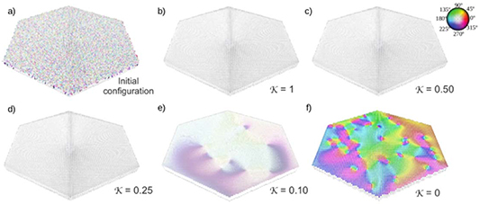
Figure 4. Micromagnetic simulation showing the configuration of spins of both an 18 nm SFO hexagonal platelet and a 1.2 nm Co film on top. White spins are pointing up in an out-of-plane configuration, while colour spins correspond to different in-plane directions following the colour scheme on the upper right corner. Images (a) to (f) correspond to the micromagnetic pattern of both layers with random initial configuration of spins and five different values of the exchange-coupling strengthκ, from completely coupled (κ = 1) to completely uncoupled (κ = 0).
Download figure:
Standard image High-resolution imageThe SFO spins appear white which means they follow an out-of-plane direction, pointing up in this case. The strength of exchange-coupling interaction κ is parametrized between 0 < κ < 1 [19], where 0 corresponds to complete absence of coupling and 1 to perfect coupling. For κ ≥ 0.25, the spins in the Co layer are perfectly aligned with those of SFO. For κ = 0.1, a mild colouring is detected in the Co spins, which indicates that they are slightly deviating from the out-of-plane direction and starting to follow their own multi-domain configuration. Finally, in the absence of exchange-coupling, the spins in the Co layer exhibit a completely in-plane multi-domain pattern. When comparing these simulated spin configurations with figure 2, and due to the resolution of our experimental images, it must be noted that a certain degree of coupling in selected small regions of the bilayer, that could lead to a small undetectable fraction of Co spins pointing out-of-plane, cannot be fully discarded.
The micromagnetic modelling results support the hypothesis that, for the material parameters at hand, shape anisotropy clearly overcomes the perpendicular magnetic field created by the hard layer and forces soft spins to lie within the plane. In addition, simulations strongly suggest that there is an absence of interlayer exchange-coupling, as they predict that even coupling values as low as κ = 0.1 should lead to a certain alignment of soft spins with the hard layer. We have followed up the study of the magneto-dipolar coupling in bilayer thin films by using hard layers with in plane magnetization, so that the competition with the strong influence of the soft layer shape anisotropy is avoided [24].
Figure 5 presents the simulated hysteresis loops for the system in figure 4. It reveals changes in the coercive field and the remanent magnetization of the bilayer system according to degree of coupling κ. As expected, the coercive field of the bilayer system decreases as the degree of exchange coupling is increased, because the soft layer acts as a source for domain walls and facilitates reversal. In the same way, the rise in κ involves a higher degree of alignment of the soft layer which increases remanence. The highest remanence value is achieved for a perfectly rigid coupling (κ = 1). Although this simplified simulated system may not quantitatively describe our system, the qualitative trends and underlying physical mechanisms are correctly predicted.

Figure 5. (a) Simulated hysteresis loops for the SrFe12O19/Co hard/soft system portrayed in figure 4 with various degrees of exchange coupling (κ).The magnetic field was applied perpendicular (001) to the bilayer sample.
Download figure:
Standard image High-resolution image
Our results bring forward the fact that close interphase contact and a soft phase size clearly below the threshold for rigid coupling is not sufficient to achieve exchange-coupling. Orbital overlap at the interface is required. In fact, studies carried out by Fullerton [7] and Sabet [23] for different hard/soft bilayer systems infer the need for controlled crystallinity, epitaxy and rearrangement of the atoms between both phases in order to tailor the degree of exchange coupling at the interphase.
Based on these results and the existing literature [19], it is concluded that in order to successfully fabricate a hard-soft composite where magnetization is enhanced at low coercivity penalty, a low degree of exchange-coupling is desirable since it may be sufficient to align the soft spins with the hard phase without facilitating massive domain wall propagation. In that sense, the composite bilayers presented here, and associated systems with low degrees of coupling, present the advantage of a larger coercivity when compared to fully exchange-coupled systems [16, 17, 44, 45]. However, a complete absence of coupling must be avoided, as it leads to no soft spin alignment and therefore no increase in remanence. Similar conclusions were extracted in our works on CoFe2O4/FeCo powder composites [19, 20], which brings forward this strategy as potentially extendable to other metal-oxide hard-soft magnetic systems in either powder or thin-film form. Approaches to tune the degree of exchange-coupling in this and similar systems will be the focus of future work. In this sense, the graded-interface concept introduced by Jiang et al[ 46] for spring magnets could be of great interest.
4. Conclusions
We have studied the correlation between the magnetic domains in a bilayer system composed of magnetically hard strontium hexaferrite platelets and a soft metallic Co film. The isolated SFO platelets, adhered onto the native oxide of a Si (100) surface, present an out-of-plane magnetization. The growth of the metallic overlayer is performed in UHV and at RT, which leads to an absence of exchange-coupling between both layers. As a consequence of this lack of coupling and the shape anisotropy of the Co layer, the magnetic domains in the soft layer appear completely uncorrelated with that of the underlying hexaferrite. Soft magnetization lies in the plane and is thus orthogonal to the magnetization of the hard SFO platelet. Our results are interpreted as a consequence of the shape anisotropy of the metallic layer dominating over the external perpendicular field created by the hexaferrite. We conclude that a low, non-zero, degree of exchange-coupling is required in order to align, at least partially, the soft-layer moments with the hard phase and at the same time prevent domain wall propagation activated in rigid coupling conditions. The strategy of implementing a low degree of exchange-coupling between magnetically hard and soft interfaces as a means to develop improved permanent magnets is further supported by our results.
Acknowledgments
This work is supported by the Spanish Ministerio de Economía y Competitividad (MINECO) through Projects no. MAT2017-86450-C4-1-R, RTI2018-095303-B-C51, RTI2018-095303-B-C53, RTI2018-095303-A-C52, and FIS2017-82415-R and by the European Comission through Project H2020 no. 720853 (AMPHIBIAN). The work is funded as well by the Regional Government of Madrid through Project S2018/NMT-4321 (NANOMAGCOST). C.G.-M. acknowledges financial support from Spanish Ministerio de Ciencia e Innovación (MICINN) through the 'Juan de la Cierva' Program (FJC2018-035532-I). Spanish Ministerio de Economía y Competitividad (MINECO).
Author contributions
The manuscript was written through contributions of all authors. All authors have given approval to the final version of the manuscript.