
Abstract
Purpose
To develop and validate a deep learning and thresholding-based model for automatic kidney stone detection and scoring according to S.T.O.N.E. nephrolithometry.
Procedures
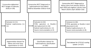
Abdominal noncontrast computed tomography (NCCT) images were retrospectively archived from February 2018 to April 2019 for three parts: a segmentation dataset (n = 167), a hydronephrosis classification dataset (n = 282), and test dataset (n = 117). The model consisted of four steps. First, the 3D U-Nets for kidney and renal sinus segmentation were developed. Second, the deep 3D dual-path networks for hydronephrosis grading were developed. Third, the thresholding methods were used to detect and segment stones in the renal sinus region. The stone size, CT attenuation, and tract length were calculated from the segmented stone region. Fourth, the stone’s location was determined. The stone detection performance was estimated with sensitivity and positive predictive value (PPV). The hydronephrosis grading and stone size, tract length, number of involved calyces, and essence grading were estimated with the area under the curve (AUC) method and linear-weighted κ statistics, respectively.
Results
The stone detection algorithm reached a sensitivity of 95.9 % (236/246) and a PPV of 98.7 % (236/239). The hydronephrosis classification algorithm achieved an AUC of 0.97. The scoring model results showed good agreement with radiologist results for the stone size, tract length, number of involved calyces, and essence grading (κ = 0.95, 95 % confidence interval [CI]: 0.92, 0.98; κ = 0.97, 95 % CI: 0.95, 1.00; κ = 0.95, 95 % CI: 0.92, 0.98; and κ = 0.97, 95 % CI: 0.94, 1.00), respectively.
Conclusions
The scoring model was constructed that can automatically detect and score stones in NCCT images.


Abbreviations
- 3D:
-
Three dimensional
- CT:
-
Computed tomography
- NCCT:
-
Noncontrast computed tomography
- PCNL:
-
Percutaneous nephrolithotomy
- ROC:
-
Receiver operating characteristic curve
- AUC:
-
Area under the curve
- PPV:
-
Positive predictive value
- CI:
-
Confidence interval
References
Park S (2007) Medical management of urinary stone disease. Expert Opin Pharmacother 8:1117–1125
Scales CD, Tasian GE, Schwaderer AL, Goldfarb DS, Star RA, Kirkali Z (2016) Urinary stone disease: advancing knowledge, patient care, and population health. Clin J Am Soc Nephrol 11:1305–1312
Allison SJ (2014) Stones: ultrasonography and computed tomography: performance in detection of kidney stones. Nat Rev Nephrol 10:611
Turk C, Petrik A, Sarica K et al (2016) EAU guidelines on interventional treatment for urolithiasis. Eur Urol 69:475–482
De S, Autorino R, Kim FJ et al (2016) Corrigendum re: “Percutaneous nephrolithotomy versus retrograde intrarenal surgery: a systematic review and meta-analysis” [Eur Urol 2015;67:125-37]. Eur Urol 69:e85
Okhunov Z, Friedlander JI, George AK, Duty BD, Moreira DM, Srinivasan AK, Hillelsohn J, Smith AD, Okeke Z (2013) S.T.O.N.E. nephrolithometry: novel surgical classification system for kidney calculi. Urology 81:1154–1159
Noureldin YA, Elkoushy MA, Andonian S (2015) External validation of the S.T.O.N.E. nephrolithometry scoring system. Can Urol Assoc J 9:190–195
Kambadakone AR, Eisner BH, Catalano OA, Sahani DV (2010) New and evolving concepts in the imaging and management of urolithiasis: urologists’ perspective. Radiographics 30:603–623
Okhunov Z, Helmy M, Perez-Lansac A, Menhadji A, Bucur P, Kolla SB, Cho JS, Osann K, Lusch A, Landman J (2013) Interobserver reliability and reproducibility of s.T.o.N.e. nephrolithometry for renal calculi. J Endourol 27:1303–1306
Chartrand G, Cheng PM, Vorontsov E, Drozdzal M, Turcotte S, Pal CJ, Kadoury S, Tang A (2017) Deep learning: a primer for radiologists. Radiographics 37:2113–2131
Demehri S, Kalra MK, Rybicki FJ, Steigner ML, Lang MJ, Houseman EA, Curhan GC, Silverman SG (2011) Quantification of urinary stone volume: attenuation threshold-based CT method-a technical note. Radiology 258:915–922
Duan X, Wang J, Qu M, Leng S, Liu Y, Krambeck A, McCollough C (2012) Kidney stone volume estimation from computerized tomography images using a model based method of correcting for the point spread function. J Urol 188:989–995
Hoffmann U, Kwait DC, Handwerker J, Chan R, Lamuraglia G, Brady TJ (2003) Vascular calcification in ex vivo carotid specimens: precision and accuracy of measurements with multi-detector row CT. Radiology 229:375–381
Jacobs C, van Rikxoort EM, Scholten ET, de Jong PA, Prokop M, Schaefer-Prokop C, van Ginneken B (2015) Solid, part-solid, or non-solid?: classification of pulmonary nodules in low-dose chest computed tomography by a computer-aided diagnosis system. Investig Radiol 50:168–173
Fernbach SK, Maizels M, Conway JJ (1993) Ultrasound grading of hydronephrosis-introduction to the system used by the society-for-fetal-urology. Pediatr Radiol 23:478–480
Maizels M, Keays M, Snyder H, Leonard M (2008) Reliability assessment of Society for Fetal Urology ultrasound grading system for hydronephrosis-discussion. J Urol 180:1683–1683
Eisner BH, Kambadakone A, Monga M, Anderson JK, Thoreson AA, Lee H, Dretler SP, Sahani DV (2009) Computerized tomography magnified bone windows are superior to standard soft tissue windows for accurate measurement of stone size: an in vitro and clinical study. J Urol 181:1710–1715
Yushkevich PA, Piven J, Hazlett HC, Smith RG, Ho S, Gee JC, Gerig G (2006) User-guided 3D active contour segmentation of anatomical structures: significantly improved efficiency and reliability. Neuroimage 31:1116–1128
Ward AD, Hamarneh G, Ashry R, Schweitzer ME (2007) 3D shape analysis of the supraspinatus muscle: a clinical study of the relationship between shape and pathology. Acad Radiol 14:1229–1241
Çiçek Ö, Abdulkadir A, Lienkamp SS, Brox T, Ronneberger O (2016) 3D U-Net: learning dense volumetric segmentation from sparse annotation. Springer International Publishing, Cham, pp. 424–432
Zhu WT, Liu CC, Fan W, Xie XH (2018) DeepLung: deep 3D dual path nets for automated pulmonary nodule detection and classification. 2018 Ieee Winter Conference on Applications of Computer Vision (Wacv 2018) 2018-:673-681
Ziemba JB, Li P, Gurnani R, Kawamoto S, Fishman EK, Fung G, Ludwig WW, Stoianovici D, Matlaga BR (2018) A user-friendly application to automate CT renal stone measurement. J Endourol 32:685–691
Jendeberg J, Geijer H, Alshamari M, Liden M (2018) Prediction of spontaneous ureteral stone passage: automated 3D-measurements perform equal to radiologists, and linear measurements equal to volumetric. Eur Radiol 28:2474–2483
Selby MG, Vrtiska TJ, Krambeck AE, McCollough CH, Elsherbiny HE, Bergstralh EJ, Lieske JC, Rule AD (2015) Quantification of asymptomatic kidney stone burden by computed tomography for predicting future symptomatic stone events. Urology 85:45–50
Oliveira B, Torres HR, Queiros S, Morais P, Vilaca JL (2018) Segmentation of kidney and renal collecting system on 3D computed tomography images 2018 IEEE 6th International Conference on Serious Games and Applications for Health (SeGAH),
Papalia R, Abreu ALD, Panebianco V et al (2015) Novel kidney segmentation system to describe tumour location for nephron-sparing surgery. World J Urol 33:865–871
Langkvist M, Jendeberg J, Thunberg P, Loutfi A, Liden M (2018) Computer aided detection of ureteral stones in thin slice computed tomography volumes using convolutional neural networks. Comput Biol Med 97:153–160
Author information
Authors and Affiliations
Corresponding author
Ethics declarations
Conflict of Interest
The authors declare that they have no conflict of interest.
Ethical Approval
All procedures performed in studies involving human participants were in accordance with the ethical standards of the institutional research committee and with the 1964 Helsinki declaration and its later amendments. For this type of study, formal consent is not required.
Additional information
Publisher’s Note
Springer Nature remains neutral with regard to jurisdictional claims in published maps and institutional affiliations.
Electronic Supplementary Material
ESM 1
(DOCX 732 kb)
Rights and permissions
About this article

Cite this article
Cui, Y., Sun, Z., Ma, S. et al. Automatic Detection and Scoring of Kidney Stones on Noncontrast CT Images Using S.T.O.N.E. Nephrolithometry: Combined Deep Learning and Thresholding Methods. Mol Imaging Biol 23, 436–445 (2021). https://doi.org/10.1007/s11307-020-01554-0
Received:
Revised:
Accepted:
Published:
Issue Date:
DOI: https://doi.org/10.1007/s11307-020-01554-0