Article

The Duckweed, Lemna minor Modulates Heavy Metal-Induced Oxidative Stress in the Nile Tilapia, Oreochromis niloticus

1
Water Pollution Research Department, Centre of Excellence for Advanced Sciences (CEAS), National Research Centre, Dokki, Giza 12622, Egypt
2
Department of Cell Biology, Centre of Excellence for Advanced Sciences (CEAS), National Research Centre, Dokki, Giza 12622, Egypt
3
Aquaculture Research Institute, Department of Animal and Veterinary Science, University of Idaho, Moscow, ID 83844, USA
4
Comparative Endocrinology Lab, Department of Biology, University of Naples Federico II, 80126 Naples, Italy
5
Laboratory of Zebrafish Developmental Genomics, International Institute of Molecular and Cell Biology, 02-109 Warsaw, Poland
6
Marine Science Department, Canal Suez University, Ismailia 41522, Egypt
7
Environmental Sciences and Industrial Development Department, Faculty of Postgraduate Studies for Advanced Sciences (PSAS), Beni-Suef University, Beni-Suef 62511, Egypt
8
Interdepartmental Research Centre for Environment, University of Naples Federico II, 80134 Naples, Italy
*
Authors to whom correspondence should be addressed.
Authors contributed equally.
Water 2020, 12(11), 2983; https://doi.org/10.3390/w12112983
Submission received: 18 September 2020 / Revised: 18 October 2020 / Accepted: 21 October 2020 / Published: 24 October 2020

Abstract

:
A two-fold integrated research study was conducted; firstly, to understand the effects of copper (Cu) and zinc (Zn) on the growth and oxidative stress in Nile tilapia, Oreochromis niloticus; secondly, to study the beneficial effects of the duckweed Lemna minor L. as a heavy metal remover in wastewater. Experiments were conducted in mesocosms with and without duckweed. Tilapia fingerlings were exposed to Cu (0.004 and 0.02 mg L−1) and Zn (0.5 and 1.5 mg L−1) and fish fed for four weeks. We evaluated the fish growth performance, the hepatic DNA structure using comet assay, the expression of antioxidative genes (superoxide dismutase, SOD; catalase, CAT; glutathione peroxidase, GPx and glutathione-S-transferase, GST) and GPx and GST enzymatic activity. The results showed that Zn exhibited more pronounced toxic effects than Cu. A low dose of Cu did not influence the growth whereas higher doses of Cu and Zn significantly reduced the growth rate of tilapia compared to the control, but the addition of duckweed prevented weight loss. Furthermore, in the presence of a high dose of Cu and Zn, DNA damage decreased, antioxidant gene expressions and enzymatic activities increased. In conclusion, the results suggest that duckweed and Nile tilapia can be suitable candidates in metal remediation wastewater assessment programs.

Graphical Abstract

1. Introduction

Among the major health concerns worldwide is the massive release of toxic compounds into the natural environment including soil and water [1,2]. Many of these compounds are defined as metals and are dangerous even at minimal concentrations and which may be cytotoxic, carcinogenic and mutagenic in nature [3,4]. They occur in the environment from natural and anthropogenic sources [5,6]. Dietary contamination by these chemical elements gives rise to numerous adverse effects on human and animal physiology [1,2,7,8]. These compounds may seriously affect cellular processes [7]. Their toxicity involves the generation of reactive oxygen and nitrogen species, which disturb redox systems [9], and antioxidants [10,11,12]. An overexpression of free radical production or a downregulation of radicals-scavenging activity alters cellular functions through the direct modification of biomolecules and by the alteration of signaling pathways [7].
The most effective antioxidative physiological defense systems are comprised of enzymes such as superoxide dismutase (SOD), catalase (CAT), glutathione peroxidase (GPx) and glutathione-S-transferase (GST), which are known as biomarkers of oxidative stress [9,13]. The SOD converts superoxides (O2−) generated in peroxisomes and mitochondria to hydrogen peroxide detoxified by the CAT enzyme. The SOD and CAT systems provide the first mechanism for combating oxygen toxicity. The GPx catalyzes the reduction of hydrogen peroxide and lipid peroxides; GST acts as a catalytic agent in the biotransformation process by the conjugation of metabolites as xenobiotic metabolites. Antioxidant enzymes have been shown to have different responses and significantly lower activities in the polluted sites [14].
Thus, to make the environment safer and healthier for humans with regards to food consumption, and to ensure adequate fish growth performance, contaminated waters and lands need to be decontaminated to lower levels of heavy metals and trace compounds [15,16,17]. Several techniques are currently used to remove heavy metals. Most of them, in particular physico-chemical methods, become ineffective when heavy metal concentrations are under 100 mg L−1 [18]. In fact, metal salts are present in water in a dissolved form and cannot be separated using physical approaches [19]. The introduction of aquatic phytoremediation plant species and adsorbents should be performed in land management plans in order to reduce risks due to their contamination [20]. Therefore, plants represent an alternative remediation approach which has escalated in recent decades [21,22].
The eco-friendly macrophyte Lemna minor (family Lemnaceae, genera Lemna), commonly known as duckweed, is present worldwide [23], and is used as a standard ecotoxicological test species [24]. As macrophytes are more sensitive than equivalent indicators which lack a vascular system, they are more environmentally protective, thus confirming this plant as an acceptable species for toxic metal remediation [25,26]. Lemna minor is also used for the elimination of organic matter, nutrients such as phosphorus and nitrogen, soluble salts, as well as the reduction of fecal coliform densities and suspended solids [27,28]. It is also well known to be able to accumulate Cu and Zn from contaminated wastewater [29,30,31,32]. Specifically, Lemna minor can accumulate a wide range of pollutants in its root tissue [32,33], and is able to keep the hyperaccumulated metal out of the cytoplasm as nontoxic or less toxic complexes and to sequester the complexes’ metal ions in the vacuoles by chelation and such mechanistic evidence provides the best example of bioremediation programs [5,26,34].
Fish, as aquatic organisms, are subject to a vast array of water pollutants and as such, may serve as indicators for contamination assessment. Therefore, a series of biomarkers, including oxidative stress biomarkers, can be successfully applied for the detection of biological impacts and for environmental quality assessment [35]. These biomarkers provide a clear and useful link between pollution exposure, tissue contamination, and early adverse effects in organisms [9,36,37,38].
The Nile tilapia, Oreochromis niloticus, is a domesticated fish species extensively used in environmental studies because it is easily handled and maintained in the laboratory; it readily adapts to confinement; it is susceptible to various pollutants; it has economic importance but at same time it is very invasive. Moreover, the Nile tilapia and its primary tissue for detoxification, the liver, has been widely used for the toxicity evaluation of several contaminants in aquatic ecosystems [39,40].
In our toxicological studies, we utilized zinc and copper which are essential trace minerals for teleost fish and all vertebrates, present in all organs, tissues, and fluids. These metals have structural and catalytic functions and also play a regulatory role in multiple metalloenzymes as a specific cofactor and catalyst. Their toxicity is often linked to the physiological processes’ disruption. Zn, in fact, is one of the most important essential trace elements involved in animal growth and the most widely used metal cofactor in many enzymes. Cu acts as a catalyst in many enzyme systems, mainly for cytochrome oxidase and the electron carrier plastocyanin and is actively taken up by liver mitochondria via an energy-dependent system [35,41,42]. Nonetheless, it is also known that under normal conditions, these elements are essential micronutrients.
The chief aim of this research was to detect the low and high concentrations of zinc and copper effects on hepatic antioxidative biomarkers in tilapia and to examine the efficacy of the duckweed Lemna minor for their bioremediation in the environment.

2. Materials and Methods

2.1. Fish and Mesocosm

Tilapia fish, Oreochromis niloticus (n = 810, monosex type of body weight 36 ± 3.2 g), were transferred from the National Research Centre farm in Nubaria, Egypt. The tilapia fish were treated with lidocaine, CHNO (5 mg L−1), during the transportation, for stress reduction. Over an approximate two-hour transport period, the fish were transferred to an outdoor experimental system (mesocosms) at the laboratory at National Research Centre in a fiberglass container (1 m3 water capacity) supplied with battery-powered aerators for oxygen supply. The fish underwent 40 days of acclimatization in a 40 L glass mesocosm under natural light (45 × 60 × 30 cm, N = 9 mesocosms with ten fish each), un-chlorinated, well aerated and tap water (27.2 ± 1.8 °C and pH 7–8, dissolved oxygen 7–8 mg L−1). A pelleted diet (32% protein ration, 6.1% crude lipid, 4.5% pure crude fiber, and total energy 4080 Kcal/Kg, Zoo-Control Co., Giza, Egypt) was provided daily at a rate of 3% of fish body weight and the water was removed daily. This experiment followed the Egyptian ethical guidance for animal research of the Institutional Animal Care and Use Committee (IACUC), 2013.

2.2. Experimental Design

The fish were distributed into nine experimental groups and were exposed to water with copper and zinc for four weeks, as follows: the first fish group was exposed to regular, uncontaminated water as a control. The second and third groups were exposed to water contaminated with low and high doses of copper sulfate of 0.004 mg L−1 (CuL) and 0.02 (CuH) mg L−1 respectively. The fourth and fifth groups were exposed to water with the same doses of copper as in the previous groups plus one layer of duckweed, Lemna minor, covering the water surface. The sixth and seventh groups were contaminated with low (ZnL) and high (ZnH) doses of zinc acetate of 0.5 and 1.5 mg L−1, respectively. The eighth and ninth groups were exposed to water with the same concentrations of zinc as in the previous groups, plus one layer of duckweed covering the water surface.
Copper was added in the form of copper sulfate pentahydrate, CuSO4·5H2O at 25% Cu, and zinc from the zinc acetate dehydrate, Zn(CH3COO)2·2H2O at ≥98% Zn. Cu and Zn ions were determined by inductively coupled plasma mass spectrometry in each mesocosm. The applied doses of copper and zinc were based on the permissible concentrations in natural water [43] and the estimated levels in polluted areas in Egypt [44]. The amount of around 2000 g of Lemna minor was used in each mesocosm with duckness treatment. At the end of the four weeks, the fish were euthanized in 2-phenoxyethanol and dissected. Growth performance was evaluated in the mesocosms treated and untreated with metals, and with and without duckweed.
Each liver (1 ± 0.3 g weight for the control and treated animals) was divided into 4 aliquots: two of 50 mg and two of 250 mg. One of the two aliquots of 50 mg was used directly to perform a comet assay, the other to study the gene expression by qRT-PCR. Both aliquots of 250 g were used to constitute a pool of 500 mg with one other liver aliquot of tilapia of the same experimental group to perform the biochemical measurement in triplicate. Aliquots (n = 8 for each experimental group) and pools (n = 8 for each experimental group) and the additional tissue were stored at −80 °C until the analyses.

2.3. Growth Performance

Growth performance was measured as follows: weight gain (g) = W2 − W1; where W2 is the final weight after the experimental periods (four weeks), and W1 is the initial weight.

2.4. Analysis of DNA

The Comet Assay

Comet assay followed the protocol established by Blasiak et al. (2004) [45]. Images from 100 randomly selected cells (fifty counts on each duplicate slide) were analyzed for each sample by DNA damage analysis software (Comet Score, Tri Tek Corp., Sumerduck, VA, USA). In each comet class were calculated the mean score and standard deviation. Different classes were distinguished as follows: class 0 (no visible tail), class 1 (low fluorescence, round head and low damage—tail length no more than 30 µm), class 2 (equally brightly fluorescent for head and tail, medium damage tail length between 30 and 50 µm), and class 3 (bright and head small and weakly fluorescent and high damage—tail length between 50 and 70 µm). Comets with a completely disintegrated head and only visible tails were considered apoptotic and were not included in the analysis.

2.5. Gene Expression Analysis

2.5.1. RNA Extraction

RNA was extracted from each tilapia liver tissue (n = 8 of each treatment group) using TRIzol Reagent (Invitrogen, Darmstadt, Germany). Then, 1 mL of TRIzol reagent buffer was used to homogenize 50 mg of liver at room temperature for 15 min. Subsequently, 0.2 mL of chloroform was added. The samples were vortexed for 15 s, incubated for 3 min and then centrifuged at 4 °C at 12,000× g. for 15 min. The upper aqueous layer was transferred to a fresh tube and mixed to 0.5 mL isopropyl alcohol for RNA precipitation. Samples were first incubated at 30 °C for 10 min and then centrifuged at 4 °C, 12,000× g for 10 min. The RNA pellet obtained was washed with 1 mL of 75% ethanol, centrifuged at 4 °C, 7500× g for 5 min, air-dried for 10 min, dissolved in 100 µL of diethylpyrocarbonate (DEPC)-treated water and stored at −80 °C.

2.5.2. Reverse Transcription (RT) Reaction

The RNA from the tilapia liver was transcribed in 20 µL of cDNA using RevertAidTM First Strand cDNA Synthesis Kit (MBI Fermentas, St. Leon-Roth, Germany). The RNA (5 µg) was mixed to 50 UM- MuLV reverse transcriptase, 20 U ribonuclease inhibitor (50 kDa recombinant enzyme to inhibit RNase activity), 50 µM oligo-dT primer, 10 mM of each dNTP, 50 mM MgCl2 and 5× reverse transcription (RT) buffer. The RT thermal reaction program was 25 °C for 10 min followed by 1 h at 42 °C with a final heating at 99 °C for 5 min. The final reaction was cooled in ice and then used for quantitative real time-polymerase chain reaction (qRT-PCR).

2.5.3. Quantitative Real Time-Polymerase Chain Reaction (qRT-PCR)

Reaction mixtures (35 μL) of qRT-PCR consisted of 5 μL of cDNA template, 5 μL 0.2 μM of each primer, 12.5 μL of 1× SYBR® Premix Ex Taq TM (TaKaRa, Biotech. Co. Ltd., Dalian, China) and 7.5 μL dH2O was used. The PCR was performed as follows: 95.0 °C for 3 min, then 28 cycles of 95 °C, 1 min; 60 °C, 1 min; 72 °C, 1 min then, 71 cycles at 60 °C and then changed every 10 s at about 0.5 °C until reaching 95 °C. By the end of each qRT-PCR, a melting curve analysis was carried out at 95 °C to check the the quality of primers used in the reaction [46]. All reactions were performed using the Step One Real-Time PCR system (Applied Biosystems, Forster City, CA, USA), and each run contained distilled water as a control. The expression level of the following antioxidant enzyme genes was quantified in the liver tissues of tilapia fish: SOD, CAT, GPx and GST. The primers were designed using Primer3 software (http://bioinfo.ut.ee/primer3/) Table 1. The quantitative values of the RT-PCR were normalized using housekeeping genes β-actin [47].

2.6. Biochemical Measurements

2.6.1. Glutathione-S-Transferase (GST) Activity

GST activity was estimated in tilapia liver pools (n = 4 of each treatment group) according to methods described by Habig et al. (1974) [48]. The GST was evaluated with a spectrophotometer for 5 min at 25 °C due to the conjugation of reduced glutathione with 1- chloro-2,4-dinitrobenzene (CDNB) at 1 mM final concentration, 1 mM 1-chloro-2,4-dinitrobenzene, and 100 mM potassium phosphate buffer (pH 6.5) considering the blank values. A Bradford protein assay was used to determine the protein concentration, using bovine serum albumin (Sigma, St. Louis, MO, USA) as standard. GST activity was expressed as µM/min/mg protein.

2.6.2. Glutathione Peroxidase (GPx) Activity

GPx activity was measured in tilapia liver pools (n = 4 of each treatment group) group according to methods described by Mannervik (1985) [49]. The enzymatic reaction was estimated using the consecutive glutathione reductase reaction, the oxidation of NADPH (nicotinamide adenine dinucleotide phosphate oxidase) and the substrate t-butyl hydroperoxide. A Bradford protein assay was used to determine the protein concentration, using bovine serum albumin (Sigma) as a standard. In accordance with Flohé and Gunzler (1984) [50], a unit of GPx activity was defined as the amount of GPx needed to reduce the initial glutathione concentration. The GPx activity was expressed as µM/min/mg protein.

2.7. Statistical Analysis

One-way ANOVA, and when appropriate, the Scheffé post-hoc test, were used to analyze multiple group data. Data are shown as the mean ± standard error of the mean (SEM). The level of statistical significance was set at p < 0.05.

3. Results

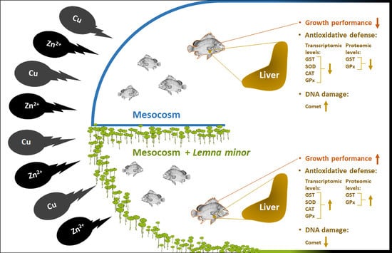
3.1. Effect of Duckweed on Growth Performance

The results for the fish weight gain reported in Table 2 show that the tilapia fish exposed to a low dose of Cu did not have a significantly reduced final body weight compared to the control fish.
However, a high dose of Cu resulted in significantly reduced final body weights of tilapia compared to the control fish. Likewise, low and high doses of Zn reduced significantly the final body weight of tilapia compared with the control fish.

3.2. Effect of Duckweed against Heavy Metals Induced DNA Damage

The results for the percentage of DNA-damaged cells reported in Table 3 revealed that the fish exposed to Zn exhibited rates of DNA damage more significant than those exposed to Cu compared to the control group. Furthermore, the fish exposed to a low dose of Cu and Zn revealed relatively similar rates of DNA damage compared to those in control fish. However, the high dose of Cu and Zn induced higher frequencies of DNA damage with percentages of 17.4% and 19.6% for Cu and Zn, respectively, compared to the control group. Results for the percentage of DNA damaged cells assessed in Oreochromis niloticus liver indicated less damage when Lemna minor was added with respect to the treatment with both metals. Specifically, DNA damage reduction was 1.6% for CuL concentration and 6.2% for CuH concentration and 2.0% for ZnL and 7.2% for ZnH concentration.

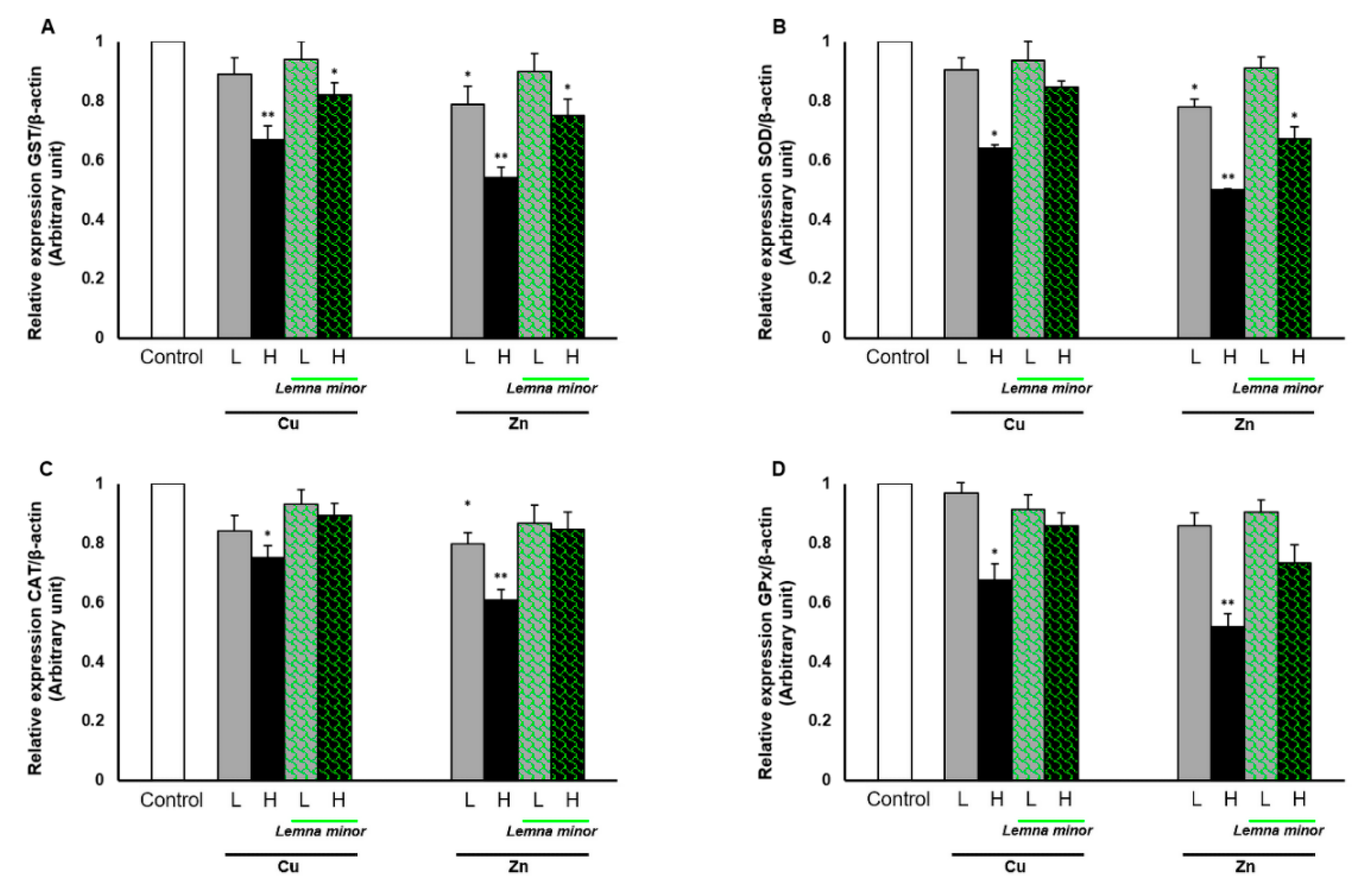
3.3. Effect of Duckweed on Antioxidants Gene Expression

The quantitative expression of antioxidant enzyme-related genes including glutathione-s-transferase (GST), catalase (CAT), superoxide dismutase (SOD) and glutathione peroxidase (GPx) genes in the liver tissues of Nile tilapia is summarized in Figure 1A–C. GST, SOD, CAT and GPx genes were significantly downregulated in the liver tissues of tilapia exposed to a high dose of Zn (1.5 mg L−1) and Cu (0.02 mg L−1) compared to the control group. In particular, even at a low Zn dose (0.02 mg L−1), the GST, SOD and CAT (p < 0.01) were affected in comparison to control fish. Interestingly, the SOD, CAT and GPx expression, which were reduced with high doses of Cu, were not affected in the presence of Lemna minor. While for the low Zn treatment, a decreased expression of SOD and CAT was not observed for the same treatment with the Lemna minor addition. Surprisingly, the reduced CAT expression (p < 0.01) observed at high Zn exposure remained at control levels in experiments where Lemna minor was added.

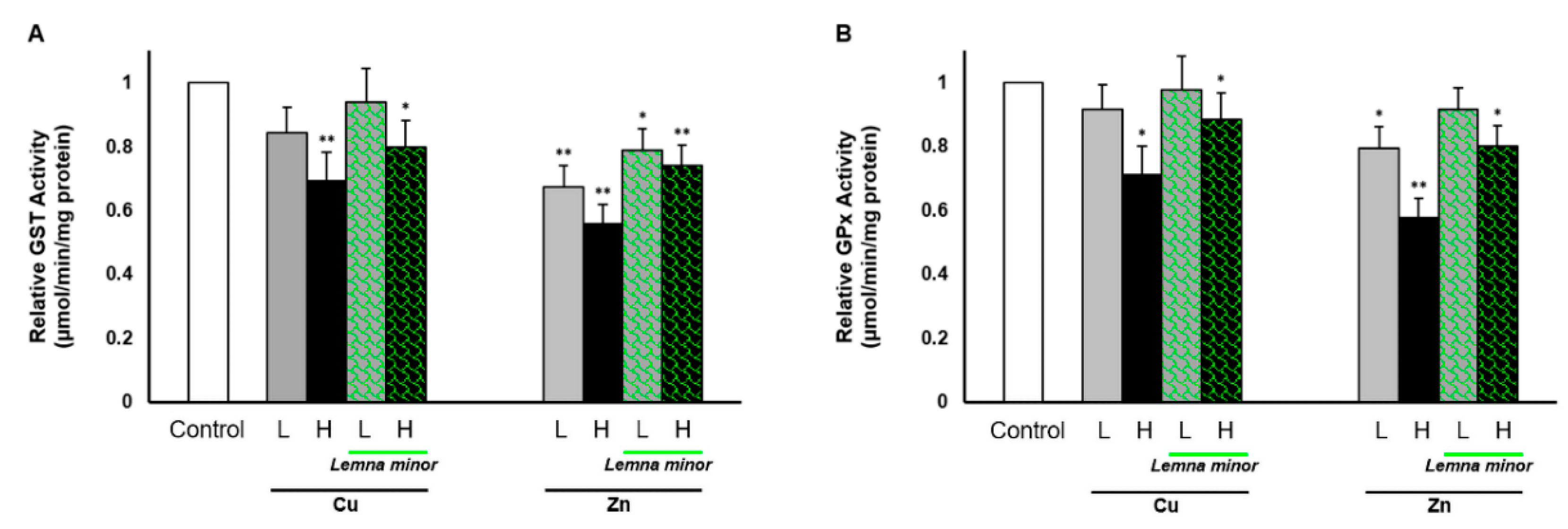
3.4. Effect of Duckweed on the GST and GPx Activities

Results show damaged liver cells of Nile tilapia Oreochromis niloticus exposed to different concentrations (low, L and high, H) of heavy metals, Cu and Zn alone or combined with duckweed, Lemna minor. The applied doses of Cu and Zn were chosen based on the estimated levels in polluted areas in Egyptian river water in the last [43,44] and recent assessment [51].
Biochemical measurements were performed to examine the hepatic GST and GPx activities in Oreochromis niloticus (Figure 2). The results show that a high dose of Cu (0.02 mg L−1) and Zn (1.5 mg L−1) induced significantly lower activity levels of GST and GPx. In particular, for both enzymes, Zn doses induced the lowest activity levels of the enzymes even at low concentrations (0.05 mg L−1). Moreover, the significant decrease in GPx activity subjected to low Zn concentration (0.5 mg L−1) was not affected in the presence of Lemna minor (Figure 1B).

4. Discussion

Heavy metal accumulation in fish has the potential to induce toxicological effects [1,2] and cause oxidative damage to tissues determining cell function loss [11,12]. As ionic Cu and Zn inhibit a number of enzymes, it follows that the basis for their toxicity may be due to their diminished activity. In particular, it is well known that the liver, among all the tissues, is the site of multiple oxidative reactions and maximal free radical generation [52,53,54].
Thus, to reduce the excessive free radical production and their effects, in the present study, the Nile tilapia chosen were male because their blood volume is higher than that of female and as a consequence there is less toxic concentration of metals in the plasma, since they were exposed for a period of four weeks to copper or zinc in the presence of duckweed Lemna minor.
The concentrations used in the mesocosm water (Cu: 0.004 mg L−1 and 0.02 mg L−1; Zn: 0.05 mg L−1 and 1.5 mg L−1) match those estimated in polluted areas in Egypt [44]. However, at the proteomic level, after high Cu exposure and both low and high Zn exposure, the magnitude of hepatic activity of GST and GPx decreased, as has been reported in the liver of Nile tilapia [13,40]. Such changes in the antioxidative capacity in Nile tilapia could be attributed to the metal ion of Cu and Zn concentration and duration of exposure [13,40].
Cu acts as a cofactor for a wide range of metal-binding enzymes, fluctuating between the oxidized and reduced copper forms. These forms, which have a high affinity for protein sites, act as potential ligands that lead to the displacement of essential metal ions from their active sites [55]. Furthermore, their excess leads to their involvement in the overexpression of free radicals able to damage DNA, lipids and proteins [56].
Zn is well known for its role as a cofactor for SOD, and it protects biological structures from the damage caused by free radicals. However, at high levels, Zn can also cause osmoregulatory disturbances in aquatic organisms, and may also cause cytotoxic effects in the presence of hydrogen peroxide [57]. In fact, a significant correlation between GST, GPx and Zn, as well as Cu levels supports our results (Figure 1). Zinc exhibited more toxic effects than Cu in fish in terms of liver cell damage which led to the reduced weight gain of fish in the Zn exposed group compared to the Cu exposed group. Our results concur with other studies which reported that although Zn may be present at allowable normal levels, it can be toxic at both conventional and at permissible high-level standards [57,58].
This is the first study wherein we have studied the impacts of duckweed (Lemna minor) on the hepatic oxidative/redox status of Nile tilapia in the presence of heavy metals in a mesocosm [59].
Our present results are in agreement with a previous study in which fish show liver SOD inhibition when exposed to 5 mg L−1 [60]. It has also been shown that copper oxide nanoparticles suppressed the activity levels of GPx and GST and also inhibited levels of GSH and resulted in increased oxidative stress in the digestive gland of the freshwater snail [61]. Moreover, the treatment of Nile tilapia with 1 and 2 mg L−1 ZnONPs resulted in the suppression of antioxidant activity. ZnONPs also decreased the gene expression of SOD and CAT in the liver and gills of Nile tilapia [62].
At transcriptional levels, the SOD, CAT, GPx and GST gene expression patterns have been validated as biomarkers of exposure to oxidative stress-inducing chemical pollutants and also to abiotic factors such as hyperthermia [12].
In our study, exposure to Cu and Zn caused the greatest reduction in SOD, CAT and GPx and GST transcription and an increase in DNA damage. However, Zn may have the more deleterious effect by notably decreasing enzymatic activity even at low concentrations (Figure 2). These results are in accordance with other studies on antioxidative mRNA expression, in which the hepatopancreas, gills and kidney were shown to be downregulated by the exposure to Cd, Cu and Zn [13,63,64]. In contrast, much research has shown an increase in hepatic gene expression in relation to toxic metals exposure [65,66,67]. Thus, it has been suggested that the expression of antioxidant biomarkers can be enhanced or reduced depending on many factors as the chemical stress intensity and duration, as well as the investigated species sensitivity [12,41,65]. These studies of antioxidative expressions at transcriptional and translational levels can answer fundamental questions linked to the xenobiotic type, exposure times, data on seasonal time of sampling, and the sex and sexual maturity of fish [39].
Our results on fish growth performance and DNA structure together with our analysis of genes expression and biochemical measurements highlight the potential use of Lemna minor for reducing oxidative stress and enhancing the capacity for heavy metal tolerance in Nile tilapia. This is important because when the antioxidative capacity is lowered, protection against cell damage is also impacted due to a reduction in the scavenging ability for free radicals leading to increased oxidative stress.
Our DNA damage analysis provides high concentrations of metals, either individually or in combination [68,69,70], which induced both sub-lethal and lethal effects in fish. The parameters most markedly affected include: tissue genotoxicity, immunity suppression, endocrine disruption, enzyme and vitamin degradation and morphological alteration in cells [5,71,72].
Interestingly, the expression levels of all examined genes were significantly increased, and the rate of DNA damage decreased in the fish treated with duckweed Lemna minor, highlighting the inhibition of the deleterious effect posed by Cu and Zn exposure in water. Thus, the consistency between the change of enzyme activities and gene mRNA abundance exposed to toxic substances underscores how activities of antioxidant enzymes could be regulated. This strengthens our data showing that the decrease in antioxidant activity reflects the reduction in the gene expression, and the addition of duckweed Lemna minor prevents the alteration of enzymatic activity and gene expression previously diminished by metal exposure [65].
Finally, it was demonstrated that Lemna minor prevented the decrease in final body weight of fish exposed to low doses of Cu and Zn compared to control fish. This result confirms the duckweed Lemna minor as a successful treatment for preventing the deteriorating effects of water-borne metals, copper and zinc, on the growth performance and health of Nile tilapia. These effects resulting from a series of events as well as cell surface biosorption/precipitation of metals, the exclusion of metal chelates into the extracellular space and enzymatic redox reaction through the conversion of metal ions into a non-toxic or less toxic state, afforded protection and nourishment [15,16,34].

5. Conclusions

In summary, Lemna minor is a potential remediator for the protection of one of the most important aquaculture species in Egypt and worldwide, the Nile tilapia Oreochromis niloticus. This remediation may be achieved by reducing oxidative stress and enhancing the heavy metal tolerance of these fish. In this regard, tilapia can be introduced as an in vivo model through the utilization of liver antioxidants as biomarkers for remediation screening in the countries with scarce or absent native species, whereas in countries rich in biodiversity, their use must be very restricted as this species is very invasive.
Understanding the relationships between stressors, stress responses, and the recovery process contribute to the effective management and restoration of aquatic ecosystems.

Author Contributions

Conceptualization, F.K.A.-G. and G.G.; methodology, F.K.A.-G., S.M.B., W.K.B.K., G.G.; software, S.M.B., T.A.T.; validation, F.K.A.-G., S.M.B., V.K., C.P. and G.G.; formal analysis and investigation, S.M.B., T.A.T., H.F.N.; resources, F.K.A.-G., W.K.B.K., H.F.N. and G.G.; data curation, W.K.B.K., C.P., V.K., S.I.; writing—original draft preparation, F.K.A.-G. and G.G.; writing—review and editing, F.K.A.-G. and G.G.; visualization, all; supervision, all; project administration, F.K.A.-G., W.K.B.K., H.F.N. and G.G.; funding acquisition, F.K.A.-G., W.K.B.K., H.F.N. and G.G. All authors have read and agreed to the published version of the manuscript.

Funding

This research received no external funding.

Acknowledgments

This work was performed within the framework of the Memorandum of Understanding between the National Research Centre of Giza (Egypt) and Federico II University. The authors acknowledge Italian and Egyptian students in mobility for the logistic technical support, the International Office of Federico II University for the publication fee and Emidio Sivieri, Biomedical Engineer at Children’s Hospital of Philadelphia, Philadelphia (USA), for the English revision.

Conflicts of Interest

The authors declare no conflict of interest.

References

  1. Guerriero, G.; Parisi, C.; Abdel-Gawad, F.K.; Hentati, O.; D’Errico, G. Seasonal and pharmaceutical-induced changes in selenoprotein glutathione peroxidase 4 activity in the reproductive dynamics of the soil biosentinel Podarcis sicula (Chordata: Reptilia). Mol. Reprod. Dev. 2019, 86, 1378–1387. [Google Scholar] [CrossRef] [PubMed]
  2. Lecomte, S.; Habauzit, D.; Charlier, T.; Pakdel, F. Emerging estrogenic pollutants in the aquatic environment and breast cancer. Genes 2017, 8, 229. [Google Scholar] [CrossRef] [PubMed] [Green Version]
  3. Fasulo, S.; Guerriero, G.; Cappello, S.; Colasanti, M.; Schettino, T.; Leonzio, C.; Mancini, G.; Gornati, R. The “SYSTEMS BIOLOGY” in the study of xenobiotic effects on marine organisms for evaluation of the environmental health status: Biotechnological applications for potential recovery strategies. Rev. Environ. Sci. Bio/Technol. 2015, 14, 339–345. [Google Scholar] [CrossRef]
  4. Diaconu, M.; Pavel, L.V.; Hlihor, R.-M.; Rosca, M.; Fertu, D.I.; Lenz, M.; Corvini, P.X.; Gavrilescu, M. Characterization of heavy metal toxicity in some plants and microorganisms—A preliminary approach for environmental bioremediation. New Biotechnol. 2020, 56, 130–139. [Google Scholar] [CrossRef] [PubMed]
  5. Conte, B.; Sorbo, S.; Piscopo, M.; Rabbito, D.; De Ruberto, F.; Guerriero, G.; Basile, A. Antioxidant activity and ultrastructural alterations in the biosensor Lemna minor l. exposed in bags in sarno river (South Italy). Fresenius Environ. Bull. 2017, 26, 225–236. [Google Scholar]
  6. Sabiha, J.; Mehmood, T.; Chaudhry, M.M.; Tufail, M.; Irfan, N. Heavy metal pollution from phosphate rock used for the production of fertilizer in Pakistan. Microchem. J. 2009, 91, 94–99. [Google Scholar] [CrossRef]
  7. Jan, A.T.; Azam, M.; Siddiqui, K.; Ali, A.; Choi, I.; Haq, Q.M. Heavy metals and human health: Mechanistic insight into toxicity and counter defense system of antioxidants. Int. J. Mol. Sci. 2015, 16, 29592–29630. [Google Scholar] [CrossRef] [Green Version]
  8. Strungaru, S.-A.; Nicoara, M.; Teodosiu, C.; Baltag, E.; Ciobanu, C.; Plavan, G. Patterns of toxic metals bioaccumulation in a cross-border freshwater reservoir. Chemosphere 2018, 207, 192–202. [Google Scholar] [CrossRef]
  9. Abdel-Gawad, F.K.; Nassar, H.F.; Bassem, S.M.; Guerriero, G.; Khalil, W.K.B. Effect of polycyclic aromatic hydrocarbons (PAHs) on modulate genes encoding stress-related proteins and antioxidant enzymes in different marine fish species of Red Sea Water. World Appl. Sci. J. 2014, 32, 2337–2346. [Google Scholar] [CrossRef]
  10. Arena, C.; Vitale, L.; Bianchi, A.R.; Mistretta, C.; Vitale, E.; Parisi, C.; Guerriero, G.; Magliulo, V.; De Maio, A. The Ageing Process Affects the Antioxidant Defences and the Poly (ADPribosyl) ation Activity in Cistus Incanus L. Leaves. Antioxidants 2019, 8, 528. [Google Scholar] [CrossRef] [Green Version]
  11. Guerriero, G.; Trocchia, S.; Abdel-Gawad, F.K.; Ciarcia, G. Roles of reactive oxygen species in the spermatogenesis regulation. Front. Endocrinol. 2014, 5, 56. [Google Scholar] [CrossRef] [PubMed] [Green Version]
  12. Parisi, C.; Guerriero, G. Antioxidative Defense and Fertility Rate in the Assessment of Reprotoxicity Risk Posed by Global Warming. Antioxidants 2019, 8, 622. [Google Scholar] [CrossRef] [PubMed] [Green Version]
  13. Mohamed, A.A.-R.; El-Houseiny, W.; Abd Elhakeem, E.-M.; Ebraheim, L.L.; Ahmed, A.I.; Abd El-Hakim, Y.M. Effect of hexavalent chromium exposure on the liver and kidney tissues related to the expression of CYP450 and GST genes of Oreochromis niloticus fish: Role of curcumin supplemented diet. Ecotoxicol. Environ. Saf. 2020, 188, 109890. [Google Scholar] [CrossRef]
  14. Nofal, M.I. Effects of heavy metal pollution on nile tilapia in manzala farm: Oxidative stress biomarkers and histopathological findings. Int. J. Fish Aquat. Stud. 2019, 7, 315–328. [Google Scholar]
  15. Abdel-Tawwab, M.; El-Sayed, G.O.; Shady, S.H. Growth, biochemical variables, and zinc bioaccumulation in Nile tilapia, Oreochromis niloticus (L.) as affected by water-born zinc toxicity and exposure period. Int. Aquat. Res. 2016, 8, 197–206. [Google Scholar] [CrossRef] [Green Version]
  16. Ali, A.; Al-Ogaily, S.M.; Al-Asgah, N.A.; Gropp, J. Effect of sublethal concentrations of copper on the growth performance of Oreochromis niloticus. J. Appl. Ichthyol. 2003, 19, 183–188. [Google Scholar] [CrossRef]
  17. Plavan, G.; Jitar, O.; Teodosiu, C.; Nicoara, M.; Micu, D.; Strungaru, S.-A. Toxic metals in tissues of fishes from the Black Sea and associated human health risk exposure. Environ. Sci. Pollut. Res. 2017, 24, 7776–7787. [Google Scholar] [CrossRef]
  18. Ahluwalia, S.S.; Goyal, D. Microbial and plant derived biomass for removal of heavy metals from wastewater. Bioresour. Technol. 2007, 98, 2243–2257. [Google Scholar] [CrossRef]
  19. Hussein, H.; Farag, S.; Moawad, H. Isolation and characterization of Pseudomonas resistant to heavy metals contaminants. Arab J. Biotechnol. 2003, 7, 13–22. [Google Scholar]
  20. Kumar, V.; Parihar, R.D.; Sharma, A.; Bakshi, P.; Sidhu, G.P.S.; Bali, A.S.; Karaouzas, I.; Bhardwaj, R.; Thukral, A.K.; Gyasi-Agyei, Y. Global evaluation of heavy metal content in surface water bodies: A meta-analysis using heavy metal pollution indices and multivariate statistical analyses. Chemosphere 2019, 236, 124364. [Google Scholar] [CrossRef]
  21. Suhag, A.; Gupta, R.; Tiwari, A. Biosorptive removal of heavy metals from waste water using duckweed. Int. J. Biomed. Adv. Res. 2011, 2, 281–290. [Google Scholar] [CrossRef] [Green Version]
  22. Danner, R.I.; Mankasingh, U.; Anamthawat-Jonsson, K.; Thorarinsdottir, R.I. Designing aquaponic production systems towards integration into greenhouse farming. Water 2019, 11, 2123. [Google Scholar] [CrossRef] [Green Version]
  23. Zimmo, O.R.; Van Der Steen, N.P.; Gijzen, H.J. Effect of organic surface load on process performance of pilot-scale algae and duckweed-based waste stabilization ponds. J. Environ. Eng. 2005, 131, 587–594. [Google Scholar] [CrossRef]
  24. Organization for Economic Cooperation and Development. 221: Lemna sp. growth inhibition test. In OECD Guidelines for the Testing of Chemicals; Organization for Economic Cooperation and Development: Paris, France, 2006. [Google Scholar]
  25. Liu, C.; Gu, W.; Dai, Z.; Li, J.; Jiang, H.; Zhang, Q. Boron accumulation by Lemna minor L. under salt stress. Sci. Rep. 2018, 8, 1–6. [Google Scholar] [CrossRef] [Green Version]
  26. Beheary, M.; M Sheta, B.; Hussein, M.; Nawareg, M.; A El-Matary, F.; Hyder, A. Environmental Remediation of Tilapia Aquaculture Wastewater Using Ceratophyllum demersum and Lemna minor. Egypt. J. Aquat. Biol. Fish. 2019, 23, 379–396. [Google Scholar] [CrossRef] [Green Version]
  27. Nassar, H.; Shaban, M.; Bassem, S.; Abdel-Gawad, F. Utilization of duckweed (DW) in nutrient removal from agricultural waste water and producing alternative economic animal fodder. Der Pharma Chem. 2015, 7, 280–285. [Google Scholar]
  28. Nieder, W.C.; Barnaba, E.; Findlay, S.E.; Hoskins, S.; Holochuck, N.; Blair, E.A. Distribution and abundance of submerged aquatic vegetation and Trapa natans in the Hudson River estuary. J. Coast. Res. 2004, 150–161. [Google Scholar] [CrossRef]
  29. Dirilgen, N.; Inel, Y. Effects of zinc and copper on growth and metal accumulation in duckweed, Lemna minor. Bull. Environ. Contam. Toxicol. 1994, 53. [Google Scholar] [CrossRef]
  30. Zayed, A.; Gowthaman, S.; Terry, N. Phytoaccumulation of trace elements by wetland plants: I. Duckweed. J. Environ. Qual. 1998, 27, 715–721. [Google Scholar] [CrossRef]
  31. Megateli, S.; Semsari, S.; Couderchet, M. Toxicity and removal of heavy metals (cadmium, copper, and zinc) by Lemna gibba. Ecotoxicol. Environ. Saf. 2009, 72, 1774–1780. [Google Scholar] [CrossRef]
  32. Kastratović, V.; Jaćimović, Ž.; DJurović, D.; Bigović, M.; Krivokapić, S. Lemna minor L.: As bioindicator of heavy metal pollution in Skadar lake: Montenegro. Kragujev. J. Sci. 2015, 123–134. [Google Scholar] [CrossRef] [Green Version]
  33. Appenroth, K.-J.; Krech, K.; Keresztes, Á.; Fischer, W.; Koloczek, H. Effects of nickel on the chloroplasts of the duckweeds Spirodela polyrhiza and Lemna minor and their possible use in biomonitoring and phytoremediation. Chemosphere 2010, 78, 216–223. [Google Scholar] [CrossRef] [PubMed]
  34. Ma, Y.; Oliveira, R.S.; Freitas, H.; Zhang, C. Biochemical and Molecular Mechanisms of Plant-Microbe-Metal Interactions: Relevance for Phytoremediation. Front. Plant Sci. 2016, 7. [Google Scholar] [CrossRef] [PubMed]
  35. Guerriero, G.; Ciarcia, G. Stress biomarkers and reproduction in fish. Fish Endocrinol. 2006, 2, 665–692. [Google Scholar] [CrossRef] [Green Version]
  36. Kroon, F.; Streten, C.; Harries, S. A protocol for identifying suitable biomarkers to assess fish health: A systematic review. PLoS ONE 2017, 12, e0174762. [Google Scholar] [CrossRef] [PubMed]
  37. Abdel-Gawad, F.K.; Guerriero, G.; Khalil, W.K.; Abbas, H.H. Evaluation of oxidative stress, genotoxicity and gene expression alterations as oil pollution markers in Solea vulgaris, from Suez canal. Quantum Matter 2016, 5, 291–296. [Google Scholar] [CrossRef]
  38. Jitar, O.; Teodosiu, C.; Nicoara, M.; Plavan, G. Study of heavy metal pollution and bioaccumulation in the Black Sea living environment. Environ. Eng. Manag. J. 2013, 12, 271–276. [Google Scholar]
  39. Wang, J.; Xiao, J.; Zhang, J.; Chen, H.; Li, D.; Li, L.; Cao, J.; Xie, L.; Luo, Y. Effects of dietary Cu and Zn on the accumulation, oxidative stress and the expressions of immune-related genes in the livers of Nile tilapia (Oreochromis niloticus). Fish Shellfish Immunol. 2020. [Google Scholar] [CrossRef]
  40. Abd-Allah, M.M.; Ramadan, A.A.; Said, N.M.; Ibrahim, I.H.; Abdel-karim, E.A. Effects of Cadmium Chloride and Glyphosate on Antioxidants as Biochemical Biomarkers in Nile Tilapia. J. Aquac. Res. Dev. 2019, 10, 2. [Google Scholar] [CrossRef]
  41. Guerriero, G.; Di Finizio, A.; Ciarcia, G. Oxidative Defenses in the Sea Bass, Dicentrarchus labrax. In Oxygen Transport to Tissue XXIV; Dunn, J.F., Swartz, H.M., Eds.; Springer: Boston, MA, USA, 2003; pp. 681–688. ISBN 978-1-4613-4912-9. [Google Scholar]
  42. Yuan, S.-S.; Lv, Z.-M.; Zhu, A.-Y.; Zheng, J.-L.; Wu, C.-W. Negative effect of chronic cadmium exposure on growth, histology, ultrastructure, antioxidant and innate immune responses in the liver of zebrafish: Preventive role of blue light emitting diodes. Ecotoxicol. Environ. Saf. 2017, 139, 18–26. [Google Scholar] [CrossRef]
  43. Mohamed, A.; El Safty, M. Current situation of water pollution and its effect on aquatic life in Egypt. Egypt. J. Occup. Med. 2013, 37, 95–115. [Google Scholar] [CrossRef]
  44. Alawy, A.E.; El-Tras, W.F.; El Raiy, H.R. Impact of industrial wastewater on water and fish quality of Nile River in Kafr El-Zayat, Egypt. Benha Vet. Med. J. 2015, 28, 78–87. [Google Scholar] [CrossRef] [Green Version]
  45. Blasiak, J.; Arabski, M.; Krupa, R.; Wozniak, K.; Zadrozny, M.; Kasznicki, J.; Zurawska, M.; Drzewoski, J. DNA damage and repair in type 2 diabetes mellitus. Mutat. Res./Fundam. Mol. Mech. Mutagenesis 2004, 554, 297–304. [Google Scholar] [CrossRef] [PubMed]
  46. Kumar, V.; Khalil, W.K.B.; Weiler, U.; Becker, K. Influences of incorporating detoxified Jatropha curcas kernel meal in common carp (Cyprinus carpio L.) diet on the expression of growth hormone-and insulin-like growth factor-1-encoding genes. J. Anim. Physiol. Anim. Nutr. 2013, 97, 97–108. [Google Scholar] [CrossRef]
  47. AbdEl-Rahim, W.M.; Khalil, W.K.; Eshak, M.G. Evaluation of the gene expression changes in Nile tilapia (Oreochromis niloticus) as affected by the bio-removal of toxic textile dyes from aqueous solution in small-scale bioreactor. Environmentalist 2010, 30, 242–253. [Google Scholar] [CrossRef]
  48. Habig, W.H.; Pabst, M.J.; Jakoby, W.B. Glutathione S-transferases the first enzymatic step in mercapturic acid formation. J. Biol. Chem. 1974, 249, 7130–7139. [Google Scholar]
  49. Mannervik, B. Glutathione peroxidase. In Methods in Enzymology; Academic Press: New York, NY, USA, 1985; pp. 490–495. ISSN 0076-6879. [Google Scholar]
  50. Flohé, L.; Günzler, W.A. [12] Assays of glutathione peroxidase. In Methods in Enzymology; Academic Press: New York, NY, USA, 1984; pp. 114–120. ISSN 0076-6879. [Google Scholar]
  51. Shalaby, B.; Samy, Y.M.; Mashaly, A.O.; El Hefnawy, M.A.A. Comparative Geochemical Assessment of Heavy Metal Pollutants among the Mediterranean Deltaic Lakes Sediments (Edku, Burullus and Manzala), Egypt. Egypt. J. Chem. 2017, 60, 361–378. [Google Scholar] [CrossRef] [Green Version]
  52. Heath, A.G. Behavior and nervous system function. In Water Pollution and Fish Physiology; CRC Press: Boca Raton, FL, USA, 1987; pp. 181–196. [Google Scholar]
  53. Atli, G.; Alptekin, Ö.; Tükel, S.; Canli, M. Response of catalase activity to Ag+, Cd2+, Cr6+, Cu2+ and Zn2+ in five tissues of freshwater fish Oreochromis niloticus. Comp. Biochem. Physiol. Part C Toxicol. Pharmacol. 2006, 143, 218–224. [Google Scholar] [CrossRef] [PubMed]
  54. Saglam, D.; Atli, G.; Dogan, Z.; Baysoy, E.; Gurler, C.; Eroglu, A.; Canli, M. Response of the antioxidant system of freshwater fish (Oreochromis niloticus) exposed to metals (Cd, Cu) in differing hardness. Turk. J. Fish. Aquat. Sci. 2014, 14, 43–52. [Google Scholar] [CrossRef]
  55. Rae, T.D.; Schmidt, P.J.; Pufahl, R.A.; Culotta, V.C.; O’Halloran, T.V. Undetectable intracellular free copper: The requirement of a copper chaperone for superoxide dismutase. Science 1999, 284, 805–808. [Google Scholar] [CrossRef] [PubMed] [Green Version]
  56. Halliwell, B.; Gutteridge, J.M.C. Role of free radicals and catalytic metal ions in human disease: An overview. In Oxygen Radicals in Biological Systems Part B: Oxygen Radicals and Antioxidants; Methods in Enzymology; Academic Press: New York, NY, USA, 1990; pp. 1–85. [Google Scholar]
  57. McGeer, J.C.; Szebedinszky, C.; McDonald, D.G.; Wood, C.M. Effects of chronic sublethal exposure to waterborne Cu, Cd or Zn in rainbow trout. 1: Iono-regulatory disturbance and metabolic costs. Aquat. Toxicol. 2000, 50, 231–243. [Google Scholar] [CrossRef]
  58. Trevisan, R.; Flesch, S.; Mattos, J.J.; Milani, M.R.; Bainy, A.C.D.; Dafre, A.L. Zinc causes acute impairment of glutathione metabolism followed by coordinated antioxidant defenses amplification in gills of brown mussels Perna perna. Comp. Biochem. Physiol. Part C Toxicol. Pharmacol. 2014, 159, 22–30. [Google Scholar] [CrossRef] [Green Version]
  59. Omar, W.A.; Mikhail, W.Z.; Abdo, H.M.; Abou El Defan, T.A.; Poraas, M.M. Ecological risk assessment of metal pollution along greater Cairo sector of the river Nile, Egypt, using nile tilapia, Oreochromis niloticus, as Bioindicator. J. Toxicol. 2015, 2015. [Google Scholar] [CrossRef] [Green Version]
  60. Xiong, D.; Fang, T.; Yu, L.; Sima, X.; Zhu, W. Effects of nano-scale TiO2, ZnO and their bulk counterparts on zebrafish: Acute toxicity, oxidative stress and oxidative damage. Sci. Total Environ. 2011, 409, 1444–1452. [Google Scholar] [CrossRef] [PubMed]
  61. Ali, D.; Ali, H. Susceptibility of the freshwater pulmonate snail Lymnea luteola L. to copper oxide nanoparticle. Toxicol. Environ. Chem. 2015, 97, 576–587. [Google Scholar] [CrossRef]
  62. Alkaladi, A.; Afifi, M.; Mosleh, Y.; AbuZinada, O. Histopathological effects of zinc oxide nanoparticles on the liver and gills of Oreochromis niloticus, protective effect of vitamins C and E. Microbiol 2014, 8, 4549–4558. [Google Scholar]
  63. Hansen, B.H.; Rømma, S.; Garmo, Ø.A.; Pedersen, S.A.; Olsvik, P.A.; Andersen, R.A. Induction and activity of oxidative stress-related proteins during waterborne Cd/Zn-exposure in brown trout (Salmo trutta). Chemosphere 2007, 67, 2241–2249. [Google Scholar] [CrossRef] [PubMed]
  64. Kim, K.-Y.; Lee, S.Y.; Cho, Y.S.; Bang, I.C.; Kim, K.H.; Kim, D.S.; Nam, Y.K. Molecular characterization and mRNA expression during metal exposure and thermal stress of copper/zinc- and manganese-superoxide dismutases in disk abalone, Haliotis discus discus. Fish Shellfish Immunol. 2007, 23, 1043–1059. [Google Scholar] [CrossRef] [PubMed]
  65. Craig, P.M.; Wood, C.M.; McClelland, G.B. Oxidative stress response and gene expression with acute copper exposure in zebrafish (Danio rerio). Am. J. Physiol. Regul. Integr. Comp. Physiol. 2007, 293, R1882–R1892. [Google Scholar] [CrossRef] [Green Version]
  66. Varela-Valencia, R.; Gómez-Ortiz, N.; Oskam, G.; de Coss, R.; Rubio-Piña, J.; del Río-García, M.; Albores-Medina, A.; Zapata-Perez, O. The effect of titanium dioxide nanoparticles on antioxidant gene expression in tilapia (Oreochromis niloticus). J. Nanoparticle Res. 2014, 16, 2369. [Google Scholar] [CrossRef]
  67. El-Sayed, Y.S.; El-Gazzar, A.M.; El-Nahas, A.F.; Ashry, K.M. Vitamin C modulates cadmium-induced hepatic antioxidants’ gene transcripts and toxicopathic changes in Nile tilapia, Oreochromis niloticus. Environ. Sci. Pollut. Res. 2016, 23, 1664–1670. [Google Scholar] [CrossRef] [PubMed]
  68. Obiakor, M.O.; Okonkwo, J.C.; Ezeonyejiaku, C.D.; Ezenwelu, C.O. Genotoxicology: Single and joint action of copper and zinc to Synodontis clarias and Tilapia nilotica. J. Appl. Sci. Environ. Manag. 2010, 14. [Google Scholar] [CrossRef] [Green Version]
  69. Shahzad, K.; Khan, M.N.; Jabeen, F.; Kosour, N.; Chaudhry, A.S.; Sohail, M.; Ahmad, N. Toxicity of zinc oxide nanoparticles (ZnO-NPs) in tilapia (Oreochromis mossambicus): Tissue accumulation, oxidative stress, histopathology and genotoxicity. Int. J. Environ. Sci. Technol. 2019, 16, 1973–1984. [Google Scholar] [CrossRef]
  70. Shahzad, K.; Khan, M.N.; Jabeen, F.; Kosour, N.; Chaudhry, A.S.; Sohail, M. Evaluating toxicity of copper (II) oxide nanoparticles (CuO-NPs) through waterborne exposure to tilapia (Oreochromis mossambicus) by tissue accumulation, oxidative stress, histopathology, and genotoxicity. Environ. Sci. Pollut. Res. 2018, 25, 15943–15953. [Google Scholar] [CrossRef] [PubMed] [Green Version]
  71. De Maio, A.; Trocchia, S.; Guerriero, G. The amphibian Pelophylax bergeri (Günther, 1986) testis poly (ADP-ribose) polymerases: Relationship to endocrine disruptors during spermatogenesis. Ital. J. Zool. 2014, 81, 256–263. [Google Scholar] [CrossRef] [Green Version]
  72. Scalici, M.; Traversetti, L.; Spani, F.; Malafoglia, V.; Colamartino, M.; Persichini, T.; Cappello, S.; Mancini, G.; Guerriero, G.; Colasanti, M. Shell fluctuating asymmetry in the sea-dwelling benthic bivalve Mytilus galloprovincialis (Lamarck, 1819) as morphological markers to detect environmental chemical contamination. Ecotoxicology 2017, 26, 396–404. [Google Scholar] [CrossRef]
Figure 1. RTqPCR expression analysis of liver antioxidant enzyme genes (A: GST; B: CAT; C: SOD; D: GPx) of Nile tilapia, Oreochromis niloticus. The relative expression indicated in arbitrary units defines the expression change in comparison to that of the reference housekeeping β-actina rRNA gene in samples exposed to metals, Cu and Zn in mesocosm with or without duckweed, Lemna minor (Cu L: 0.004 and H: 0.02 mg L−1); (Zn, L: 0.5 and H: 1.5 mg L−1) with respect to samples without treatment used as control. * p < 0.05 and ** p < 0.01 for the treated groups compared with the control group.
Water 12 02983 g001
Figure 2. Activity levels of GST (A) and GPx (B) in the pools of the the liver tissues of Nile tilapia, Oreochromis niloticus exposed to metals (Cu and Zn) in the mesocosm with or without the duckweed, Lemna minor. Cu = copper exposure (L: 0.004 and H: 0.02 mg L−1), Zn = zinc exposure (L: 0.5 and H: 1.5 mg L−1). Data are presented as the mean ± SEM. * p < 0.05 and ** p < 0.01 for the treated groups compared with the control group.
Water 12 02983 g002
Table 1. Primer sequences for the Nile tilapia Oreochromis niloticus genes encoding antioxidant enzymes.
GeneForward Primer Sequence (5′-3′)Reverse Primer Sequence (5′-3′)
SODGGTGCCCTGGAGCCCTAATGCGAAGTCTTCCACTGTC
CATTCCTGAATGAGGAGGAGCGAATCTTAGATGAGGCGGTGATG
GPxCCAAGAGAACTGCAAGAACGACAGGACACGTCATTCCTACAC
GSTTAATGGGAGAGGGAAGATGGCTCTGCGATGTAATTCAGGA
β-actinCAATGAGAGGTTCCGTTGCAGGATTCCATACCAAGGAAGG
Superoxide dismutase, SOD; catalase, CAT; glutathione peroxidase, GPx and glutathione-S-transferase, GST.
Table 2. Growth performance of Nile tilapia, Oreochromis niloticus exposed to metals: low and high dose Cu (CuL, CuH), and low and high dose Zn (ZnL, ZnH) in mesocosm with or without the duckweed, Lemna minor.
TreatmentInitial Weight (g) Final Weight (g)
Control36.2 ± 2.499.3 ± 3.2 a
CuL37.1 ± 3.288.1 ± 4.1 ab
CuH35.4 ± 1.977.6 ± 2.9 bc
CuL + L. minor38.2 ± 2.793.2 ± 4.8 ab
CuH + L. minor36.4 ± 1.684.4 ± 5.2 b
ZnL36.2 ± 1.581.5 ± 3.7 b
ZnH37.5 ± 2.271.2 ± 2.4 c
ZnL + L. minor38.2 ± 3.389.1 ± 3.8 ab
ZnH + L. minor36.6 ± 2.180.3 ± 3.1 b
Data are presented as the mean ± SEM. a,b,c Mean values within tissue with unlike superscript letters were significantly different (p < 0.05, Scheffé Test).
Table 3. Total comets, class of comet and % DNA damaged liver cells in Nile tilapia, Oreochromis niloticus exposed to metals: low and high dose Cu (CuL, CuH), and low and high dose Zn (ZnL, ZnH) in mesocosm with or without the duckweed, Lemna minor, using the comet assay.
TreatmentTotal CometsComet ClassDNA % Damaged Cells
0123
Control33467221106.6 ± 1.1 c
CuL464541716139.2 ± 1.6 bc
CuH8741326322917.4 ± 2.4 a
CuL + L. minor384621215117.6 ± 1.2 c
CuH + L. minor5644416211911.2 ± 1.6 b
ZnL494511815169.8 ± 1.5 bc
ZnH9840228373319.6 ± 2.2 a
ZnL + L. minor39461211177.8 ± 1.3 c
ZnH + L. minor6243818222112.4 ± 1.8 b
Data are presented as the mean ± SEM. a,b,c Mean values within tissue with unlike superscript letters were significantly different (p < 0.05, Scheffé Test) (n = 5).
Publisher’s Note: MDPI stays neutral with regard to jurisdictional claims in published maps and institutional affiliations.

