Article

The Novel Quantitative Assay for Measuring the Antibiofilm Activity of Volatile Compounds (AntiBioVol)

1
Department of Pharmaceutical Microbiology and Parasitology, Faculty of Pharmacy Wrocław Medical University, 50-556 Wrocław, Poland
2
Department of Microbiology and Biotechnology, Faculty of Biotechnology and Animal Husbandry, West Pomeranian University of Technology, 70-311 Szczecin, Poland
3
Faculty of Medicine, Lazarski University, 02-662 Warszawa, Poland
4
Bioimaging Laboratory, Łukasiewicz Research Network—PORT Polish Center for Technology Development, 54-066 Wrocław, Poland
5
Department of Environment, Hygiene and Animal Welfare, Faculty of Biology and Animal, Science, Wrocław University of Environmental and Life Sciences, 51-630 Wrocław, Poland
6
Laboratory of Microbiology, Institute of Genetics and Microbiology, University of Wrocław, 51-148 Wrocław, Poland
7
Laboratory of Microbiology, Łukasiewicz Research Network—PORT Polish Center for Technology Development, 54-066 Wrocław, Poland
*
Author to whom correspondence should be addressed.
Appl. Sci. 2020, 10(20), 7343; https://doi.org/10.3390/app10207343
Submission received: 31 July 2020 / Revised: 14 October 2020 / Accepted: 14 October 2020 / Published: 20 October 2020
(This article belongs to the Special Issue Biofilm and Applications in Medicine and Industry)

Abstract

:
Herein, we present a new test, dubbed AntiBioVol, to be used for the quantitative evaluation of antibiofilm activity of volatile compounds in vitro. AntiBioVol is performed in two 24-well plates using a basic microbiological laboratory equipment. To demonstrate AntiBioVol usability, we have scrutinized the activity of volatilized eucalyptus, tea tree, thyme essential oils, and ethanol (used for method suitability testing) against biofilms of Staphylococcus aureus, Pseudomonas aeruginosa, and Candida albicans. We have also compared AntiBioVol with the standard disc volatilization method, placing a special stress on evaluating the impact of various technical parameters on the outcomes of the latter method. The obtained results indicate that AntiBioVol allows analyzing the antibiofilm activity of volatile compounds in a high number of repeats and provides semi-quantitative or quantitative results of high repeatability. In comparison to disc volatilization, AntiBioVol is a more space- and cost-effective method that allows analyzing various types of microbial aggregates. Moreover, we have indicated that the possible reasons for the discrepancies in the results obtained by means of the standard disc volatilization method may be related to various parameters of the testing dishes used (height, volume, diameter) and to various volumes of the agar medium applied. In turn, the application of a 24-well plate and a strictly defined AntiBioVol protocol provide a higher control of experimental conditions. Therefore, the application of AntiBioVol may enable an optimization of and introduction of volatile compounds to the fight against infective biofilms.

1. Introduction

Biofilm is a diversified and adaptive community of microbial cells that displays a high tolerance to conventional antimicrobials (antibiotics, antiseptics, disinfectants) due to the presence of a protective extracellular matrix, diversification of metabolism within specific biofilm layers, and coordinated reactivity to stimuli [1]. High tolerance to stressors and antibiotic resistance mechanisms displayed by microbial cells within the biofilm make the structure highly persistent and are the reasons why science has recognized the biofilm’s central role in the pathogenesis of infective diseases [2,3,4]. Such opportunistic pathogens as Staphylococcus aureus, Pseudomonas aeruginosa, and Candida albicans are particularly frequently associated with serious biofilm-related infections and strong antibiotic resistance [5]. The health complications resulting from infections caused by the above-mentioned pathogens are of severe and life-threatening nature. Moreover, they are also a significant economic burden for healthcare systems [6].
Therefore, the introduction of novel antibiofilm countermeasures is one of the most pressing needs of contemporary medicine [7]. The use of such antimicrobial substances of plant origin as essential oils (EOs) in biofilm eradication is considered a promising direction to follow [8]. Thanks to their high lipophilicity, EOs bind to and break the integrity of microbial cell walls and membrane structures, which results in cell lysis through a mechanism resembling the one displayed by antiseptics [9]. Moreover, individual EOs are characterized by low toxicity, a broad spectrum of effectiveness, biodegradability, as well as anti-inflammatory and immune-stimulating properties [10]. The above-mentioned features also predispose EOs to be used either in monotherapy or as adjuvant substances for antibiotics [11]. We have chosen the essential oils of confirmed biological activity, including eucalyptus oil (Eucalyptus globulus Labill.), which exhibits antibacterial, antifungal, analgesic, and anti-inflammatory properties and has also been widely used in pharmaceutical, food, and cosmetics products [12]; thyme oil (Thymus vulgaris L.), which is known for its anti-inflammatory and antibacterial properties [13], and tea tree oil (Melaleuca alternifolia Cheel.), which is employed largely for its antimicrobial properties and incorporated as an active ingredient in many topical formulations used to treat cutaneous infections [14].
Both liquid and volatile fractions of EOs may display antimicrobial (and antibiofilm) activity. However, while the testing methods for EOs liquid fractions rely on well-defined EUCAST (European Committee for Antimicrobial Susceptibility Testing) and pharmacopeial recommendations [15,16], the methodology of testing EOs’ volatile activity against microbes is highly diversified. These differences concern not only the experimental setting itself but also the various types of microbial aggregates scrutinized (lawn, biofilm) [17,18,19]. Moreover, also, the EOs are highly intra-species differentiated (with regard to the composition and concentration of antimicrobial compounds) caused by geographic and location factors, seasonal effects, and genetic factors, which determine the so-called chemotypes commonly found in EO-bearing plant species [20]. The above may explain why various scientific studies of the activity of the same type of EO against the same microorganism obtained using a single testing method produce varying data [21,22]. Such a lack of standardization impedes the introduction of EOs to the ensemble of clinical, anti-infective measures. Since there is an urgent need for new “anti-biofilm” compounds, the development of an in vitro test for screening the antibiofilm activity of volatile fractions of EOs is of high importance.
The main aim of the current study was to improve the consistency of EO analysis and to design a sensitive, cost-effective, and easy-to-perform test of antibiofilm activity of volatile compounds (later abbreviated as AntiBioVol). We also wanted to evaluate the AntiBioVol usability to test the aforementioned EOs’ activity against biofilms formed by S. aureus, P. aeruginosa, and C. albicans. AntiBioVol was also compared with the standard disc volatilization method.

2. Materials and Methods

2.1. Microorganisms and Essential Oils

  • For experimental purposes, the following reference strains from the American Type Culture Collection (ATCC) were applied: S. aureus 6538, P. aeruginosa 15442, and C. albicans 10321.
  • The EOs chosen for the experiment purposes were as follows:
    -
    Eucalyptus oil (Eucalyptus globulus Labill.), (PharmaTech, Poland), later referred to as E-EO;
    -
    Thyme oil (Thymus vulgaris L.), (Etja, Poland), later referred to as T-EO;
    -
    Tea tree oil (Melaleuca alternifolia Cheel.), (PharmaTech, Poland), later referred to as TT-EO.
    -
    The above-mentioned EOs were chosen for experimental purposes because of their confirmed antimicrobial activity. This fact allows comparing the results presented in this work with the results of other research teams.
  • Gas Chromatography-Mass Spectrometry Analysis of the Tested EOs Composition
TT-EOs, T-EOs, and E-EOs were diluted with hexane (JTB, Great Britain), vortexed, and immediately analyzed. The analysis was performed using the Agilent 7890B GC system coupled with the 7000GC/TQ system connected to PAL RSI85 autosampler (Agilent Technologies, Palo Alto, CA, USA). The column used was HP-5 MS; 30 m × 0.25 mm × 0.25 µm (J & W, Agilent Technologies, Palo Alto, CA, USA) with helium as a carrier gas at a total flow of 1 mL/min. Chromatographic conditions were as follows: split injection at a ratio of 100:1, the injector was set at 250 °C, the oven temperature program was 50 °C held for 1 min, then 4 °C/min up to 130 °C, 10 °C/min to 280 °C and then isothermal for 2 min. The MS detector operated in the electronic impact ionization mode at 70 eV. The transfer line, source, and quadrupole temperatures were set at 320, 230, and 150 °C, respectively. Masses were registered in the range from 30 to 400 m/z. The peaks were identified in the MassHunter Workstation Software Version B.08.00 coupled with the NIST17 mass spectra library and accomplished by a comparison with linear retention indexes. The relative abundance of each EO constituent was expressed as a percentage content based on the peak area normalization. All analyses were performed in triplicate.

2.2. Antibiofilm Activity of Volatile Fraction Test (AntiBioVol) Test Performance

The general principles of the AntiBioVol test are presented in Figure 1, while a photographic presentation of the AntiBioVol test performance is presented in Figure S2.

2.2.1. Preparation of Agar Plugs

First, 2.5 mL of 2% Brain Heart Infusion (BHI, Graso, Poland) agar was poured into the wells of a 24-well plate (BioFil, Warsaw, Poland). The plate was left in a sterile chamber until the agar has solidified (this plate is further referred to as Plate A, where “A” stands for “Agar”). Next, the agar plugs were cut out using a cork-borer (ChemLand, Warsaw, Poland) with a diameter of 8 mm. Subsequently, the plugs were cut evenly crosswise into two discs. One disc was placed in a well of a new 24-well plate (later referred to as Plate B, where “B” stands for “Biofilm”) and another disc was placed back to the agar-containing well of Plate A (Figure 1, Part I). The latter plate was sealed with an adhesive tape and kept refrigerated until further analysis.

2.2.2. Biofilm Formation on Agar Disc in Plate B

Using a densitometer (Densitomat II, BioMerieux, Poland), the analyzed strain’s suspension of 0.5 McFarland (MF) density in Tryptic Soya Broth (TSB, Graso Biotech, Poland) medium was obtained and subsequently diluted (1500x for bacteria and 14x for fungi) to obtain approximately 105 Colony Forming Units (CFU)/mL. In the next step, 2 mL of the strain’s suspension was added to Plate B wells containing the agar disc. Plate B was incubated for 24 h at 37 °C. After incubation, the discs were taken gently out using tweezers (Conbest, Warsaw, Poland) so as not to disrupt the agar surface and the biofilm structure on it. Next, the discs were rinsed twice with 0.9% saline (Stanlab, Wroclaw, Poland) to remove non-adhered microorganisms. The discs were subsequently placed in the agar wells of Plate A on top of the sterile discs to fill the hole of the well (Figure 1, Part II).

2.2.3. Exposure of Biofilms to EOs’ Volatile Fractions

First, 0.5 mL of the tested EO was added to a well of a fresh 24-well plate, which is later referred to as Plate C (EO-Containing plate). Then, Plate A with biofilm-containing agar discs was put upside down on Plate C in such a manner that the EO-containing well was placed directly under the biofilm-containing well. Next, the rims of both plates were taped around using an adhesive tape (Diall, Poland) and incubated for 24 h at 37 °C (Figure 1, Part II).

Control Settings

To check whether the applied experimental setting itself had no potentially impeding effect on the microorganisms’ growth, 0.9% saline was applied instead of EOs. Thus, the aforementioned setting served as a control of the microorganism’s growth (positive control, latter abbreviated as “C+”). To confirm the method’s suitability, 96% ethanol (Stanlab, Wroclaw, Poland), whose volatile form has a well-recognized antimicrobial activity, was applied. The above-mentioned control samples were performed in separate plates. Each experimental and control setting was tested in six replicates.

2.2.4. Assessment of Bacterial Biofilm Survival after Exposure to EOs

Following the exposure described in Section 2.2.3 of Materials and Methods, bacterial biofilm-containing discs were transferred to fresh wells of a 24-well plate. Next, 2 mL of 0.1% tetrazolium chloride solution (2,3,5-triphenyl-2H-tetrazolium chloride, TTC) (PanReac AppliChem, Darmstadt, Germany) was introduced gently in order to not de-attach the formed biofilm from the disc surface. The plates were incubated for 1.5 h at 37 °C. TTC turns into red formazan in the presence of metabolically active microorganisms. Next, the TTC-containing medium was removed, and 2 mL of 96% ethanol was introduced to the wells to extract red formazan crystals out of the biofilm-forming cells. Subsequently, the plates were incubated for 30 min at room temperature in a microplate shaker (Schuttler MTS-4, IKA, Germany) at a speed of 300 rpm/min. After this time, 200 μL of formazan-containing solution was transferred to the wells of a 96-well plate (Biofil, Warsaw. Poland). Absorbance was measured at 490 nm using a MultiScan Go spectrophotometer (Thermo Fischer Scientific, Waltham, MA, USA). Then, the values of absorbance measured for formazan in the samples of EO-treated biofilms were compared to the corresponding values obtained for biofilms incubated with 0.9% saline (Figure 1, Part III). In order to confirm the linear relationship between the values from TTC assay and the number of CFU, standard calibration curves were performed. A calibration curve for absorbance measurements at 490 nm versus the number of CFU has been established before the experiment (Figure S3).

2.2.4.1. Assessment of Fungal Biofilm Survival after Exposure to EOs

Since the use of tetrazolium salt-based assays in studies of Candida biofilm has significant limitations [23], we used this fact as an opportunity to present the possibility of direct quantitative assessment within the AntiBioVol setting. Following the exposure described in Section 2.2.3 of Materials and Methods, fungal biofilm-containing discs were transferred to 1 mL of 0.5% mild detergent saponin solution (Merck, KenilWorth, NJ, Germany) and subjected to vigorous vortex-shaking for 1 min to de-attach the biofilm-forming cells from the agar surface. Next, serial dilutions of the obtained suspension were cultured on the Sabouraud agar plates (Graso, Poland) and incubated for 24 h at 37 °C. The CFU number was counted on the next day.

2.2.5. Application of the AntiBioVol Test for Various Concentrations of EOs in Single vs. Separate Test Plates

The aim of this experimental setting was to investigate whether a liquid EO introduced to a single 24-well plate (to which other concentrations of the same type of liquid EO were also introduced) may get across, after volatilization, to another test well. We hypothesized that if such phenomenon occurs, we would observe a higher biofilm eradication in the well in which a lower EO concentration was applied in comparison to the setting in which only one EO concentration in a single test plate was used. To check it, two concentrations of TT-EO (25%, 50%) v/v diluted in polyethylene glycol (Pol-Aura, Zabrze, Poland) and an un-diluted TT-EO (100% v/v) were applied in a single AntiBioVol setting against S. aureus biofilm.
Then, 25%, 50%, 100% TT-EO concentrations were introduced to a 24-well plate in such a manner that the 6 wells containing 100% TT-EO were placed between the 6 wells containing the 25% concentration and between the 6 wells containing the 50% concentration of the TT-EO [Figure S3]. Additionally, the same experiments were performed in separate AntiBioVol settings (single plate/single EO concentration). The remaining steps of biofilm exposure and further quantification were performed according to the procedures presented in Section 2.2.3, Section 2.2.4, and Section 2.2.4.1 of Materials and Methods. The additional set of controls applied in this experiment included the use of EOs’ solvent—50% and 75% (v/v) of polyethylene glycol (PEG); this control set was performed in a separate plate for S. aureus as the microorganism tested.

2.3. Additional Control Experiments

This part of the manuscript is deliberately presented at the end of this section because the techniques presented below were not part of the standard AntiBioVol test, and they were performed to provide additional data confirming the correctness of the AntiBioVol setting.

2.3.1. Assessment of Antimicrobial Activity of Liquid and Volatile EO Fractions Using Standard Methods

To compare AntiBioVol with the methods previously developed for the assessment of antimicrobial activity of liquid EO fractions, we used the disc diffusion method [24], while to assess the activity of volatile fractions, we applied the inverted Petri plate method (also referred to as the Disc Volatilization Method [25]).
This part of the experiment was performed as a proof of concept for S. aureus biofilm vs. T-EO, because the activity of this oil was the highest against this particular pathogen, and it could be hypothesized that positive outcomes (with regard to the halo zone) might also be obtained.
In the case of the disc diffusion method, microbial suspension at 0.5 MF density was cultured on a Muller–Hinton (BioCorp, Warsaw, Poland) agar plate (diameter of 9 cm, height of 1.4 cm). Next, standard paper discs (6 mm in diameter, thickness of 0.5 mm) saturated with EOs were placed on the agar plates (one disc per plate). Then, the plates were incubated for 24 h at 37 °C. After incubation, microbial growth inhibition zones were measured (in mm) using a ruler (Leniar, Krakow, Poland).
In the inverted Petri plate method, paper discs saturated with EOs were placed on the inside of Petri-dish lids (one disc per plate). Next, the lids were placed on the base of the dish containing microbial-seeded agar. Subsequently, the whole dishes (lids and agar-containing bases) were incubated in such a manner that the lids were lying on the incubator shelf. Such a setting prevented EO-saturated discs and EO droplets from falling down on the microbial lawn formed on the agar. Inhibition of growth (if occurred) was visible as zones of reduced microbial growth and was measured using a ruler. Moreover, in order to evaluate the correlation between the parameters relating to Petri dish sizes and the antimicrobial activity of the EOs applied, this version of the experiment was performed on Petri dishes 15, 9, and 6 cm in diameter; plate height was equal to 2.5 cm (for plates 15 cm in diameter) and 1.4 cm (for plates 6 and 9 cm in diameter); poured agar height was 1.5, 0.5, and 0.3 cm resulting in agar volume equal to 100, 20, and 10 mL, respectively. When the test involved dishes 9 cm in diameter specifically, the range of agar volume was 30, 20, and 10 mm.

2.3.2. Scanning Electron Microscopy Analysis of the Strains’ Biofilm-Forming Ability in the Applied In Vitro Setting

The aim of this procedure was to confirm the microorganisms’ ability to form biofilm on the agar surface. Fresh, 24-h liquid cultures of S. aureus, P. aeruginosa, and C. albicans strains were diluted to 105 CFU/mL. Next, 2 mL of such a suspension was introduced to the wells of a 24-well plate containing agar discs, which was prepared as described in Section 2.2.1 of Materials and Methods of this manuscript. The plates with the discs and microbial suspensions were incubated for 24 h at 37 °C. Subsequently, the discs were carefully taken out and rinsed twice with 0.9% saline to remove non-adhered microorganisms. Next, the agar plugs were fixed by immersion in 2% glutarate (ChemPur, Piekary, Poland) for 4 h at 4 °C. After incubation, the samples were rinsed three times (for 2 min) with distilled water to remove the fixative. Subsequently, 10 min dehydration for each of the following ethanol concentrations (10%, 25%, 50%, 70%, 80%, 90%) was performed; the last dehydration lasted for 15 min and was performed in 100% EtOH. After dehydration, the ethanol was removed and the samples were dried at 37 °C. Then, the biofilm-containing agar discs were sputtered with Au:Pd mixture using a sputter device (Quorum International, Fort Worth, TX, USA) and examined using a scanning electron microscope (Auriga 60, ZEISS, Germany).

2.3.3. Evaluation of Minimal Inhibitory and Minimal Biofilm Eradication Concentrations of Liquid EOs

MIC assessment was prepared in 96-well titration microplates. The wells of the plate were filled with 100 μL of TSB medium. Next, 100 μL of undiluted EO was added to the first of the wells and mixed with the medium. Subsequently, geometric dilutions of the EOs in TSB were performed. Next, 100 μL of the bacterial suspension (105 CFU/mL) was introduced to each well containing different concentrations of the EOs. The plate, wrapped with adhesive tape, was incubated for 24 h at 37 °C in a microplate shaker. The culture with only the medium added served as a positive control of the microorganisms’ growth, while the well containing only the medium served as the sterility control of the experiment. After incubation, 5 μL of TTC was added to each well and incubated for 5 h at 37 °C. The EO concentration in the first colorless well, neighboring the red well, was taken as the MIC value.
The minimum biofilm eradication concentration was estimated in a manner similar to the MIC analysis. Briefly, 100 μL of the microbial suspension (105 CFU/mL) was introduced to each well and incubated for 24 h at 37 °C without shaking. Next, the whole medium (containing non-adhered microorganisms) was removed, leaving only biofilm-forming organisms attached to the bottom of the 96-well plate. Subsequently, geometric dilutions of EOs in the medium were applied to the wells and left for another 24 h at 37 °C. The following procedures containing TTC introduction and subsequent analysis were performed similarly to the ones described for MIC assessment. In the case of C. albicans, due to the aforementioned limitations of tetrazolium salt-based assays [23], a different check of cell viability was performed. Namely, fungus-containing suspensions were removed from the 96-well plates and spotted on stable Sabouraud agar plates (dedicated for fungi growth and cultivation) and incubated for 24 h. The presence of living colonies on the agar indicated the lack of viable cells, while the absence of colonies in the place where spotting was performed confirmed the survival of Candida cells.

3. Statistical Analysis
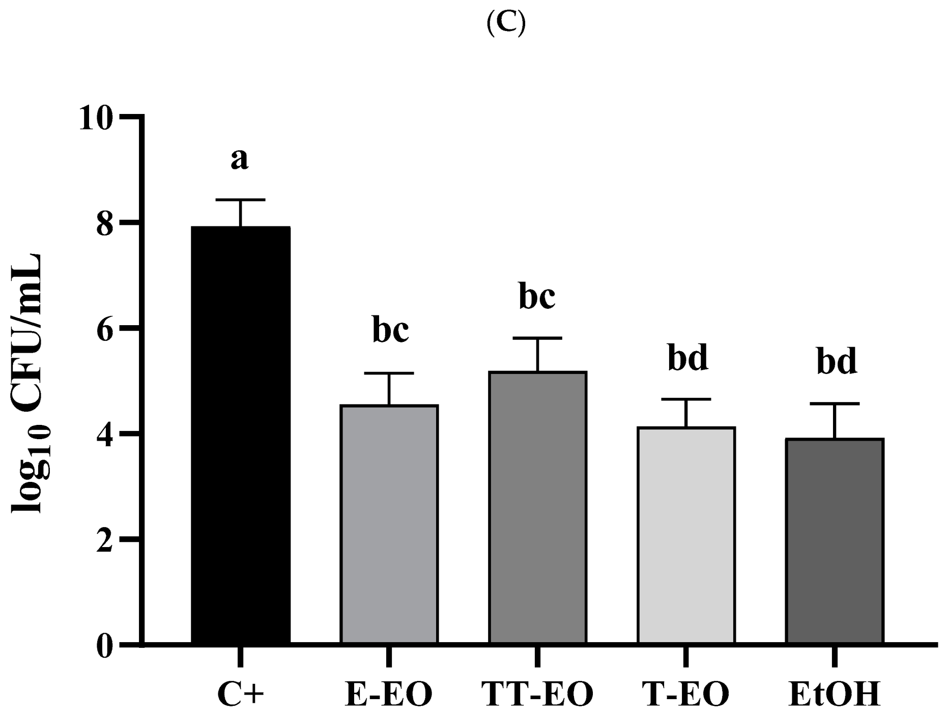
Calculations were performed using the GraphPad Prism (version 7) software. Normality distribution was calculated by means of D’Agostino–Pearson omnibus test. Since all values were non-normally distributed, the Kruskal–Wallis test with post-hoc Tukey’s analysis was applied. The results of statistical analyses were considered significant if they produced p-values < 0.05.

4. Results

In the first line of the investigation, we have scrutinized the E-, T-, and TT-EOs with regard to their content of antimicrobial substances. The GC-MS analysis confirmed the presence of specific active compounds within E-EO (including 1,8-cineole and γ—terpinene), T-EO (including thymol and p-cymene), and TT-EO (including γ—terpinene and terpinen-4-ol). The full list of active compounds identified is presented in Figure S1 and Table S1. Having proven that the tested EOs contain antimicrobial substances, we have analyzed the antimicrobial and antibiofilm activity of EOs’ liquid fractions against microorganisms in planktonic and biofilm forms using the microdilution method. The rationale behind this preliminary research was the fact that the crucial EOs’ components (such as thymol) display activity in both liquid and volatile fractions. Thus, the confirmation of antimicrobial activity of the liquid fraction would be a strong assumption for the potential activity of the volatile fraction. The results presented in Table S2 indicate a higher activity of all the tested liquid EOs against the applied microorganisms in planktonic than in biofilm form. Next, we analyzed the ability of S. aureus, C. albicans, and P. aeruginosa strains to form biofilm (Figure 2) on the agar surface. A confirmation of the presence of a settled, multi-cellular biofilm formed on agar was a prerequisite condition for the performance of the core part of this research, namely AntiBioVol test for the E-, T-, and TT-EOs.
The results of the performed AntiBioVol test suitability with the use of ethanol (which is a substance of known volatile antimicrobial activity) showed the correctness of the applied experimental setting—the number of biofilm-forming cells remaining after exposure to ethanol was lower than in the positive control of biofilm growth, regardless of the microorganism applied (Figure 3A–C). With regard to EOs, the results presented in Figure 3A–C show that none of the applied volatile fractions of EOs were able to completely eradicate bacterial or yeast biofilm, although the number of biofilm-forming cells after the exposure to all types of EOs was lower than in non-treated control settings. The volatile T-EO acted more efficiently against S. aureus biofilm than TT- and E-EO, while TT- and T-EO was stronger against P. aeruginosa biofilm than E-EO.
Next, we have measured the activity of three concentrations of TT-EO against S. aureus biofilm within a single measurement plate [Figure 4]. The comparison of TT-EO’s antibiofilm activity in a single vs. separate settings revealed a lack of statistical significance between them (K–W test p < 0.05, with post-hoc Tukey’s analysis) indicating no transmission of TT-EO volatile fractions between various plate wells within a single test plate. Similarly, no transmission of TT-EO solvent (PEG) was detected in the control setting (Figure S5).
Finally, we have performed the conventional assessment of EOs’ volatile fraction’s antimicrobial activity referred to as the inverted Petri plate method to check whether such initial technical parameters as plate diameter, plate height, poured agar volume, and agar height translate into an outcome such as the size of the microbial growth’s inhibition zone (Table 1 and Table 2). Moreover, the reader can find a comparison of the inverted Petri dish method and the conventional disc diffusion method (which, being referred to as the aromatogram [26], is also applied for EOs’ volatile fraction activity measurement) in Figures S6 and S7.
As can be seen in Table 1, the differences between agar height obtained in Petri dishes of 15, 9, and 6 cm translate into differences in air volume between the agar surface and the lid of the dish. In the case of dishes 15 cm in diameter vs. dishes 9 and 6 cm in diameter, the aforementioned parameter differed by 75% and 83%, respectively. In turn, the difference in air volume between the 9 and 6 cm diameter dish was 31%. It translated into differences in the concentrations of volatile compound in experimental settings and finally into ambiguous results of the microbial growth inhibition zone (from 0 cm observed if a 15 cm diameter dish was applied vs. a 4 cm inhibition zone observed when a 6 cm diameter dish was applied).
When Petri dishes were of the same diameter and height but various agar volumes were poured (the difference between them was 10 mL, see Table 2), a relatively small difference in agar height was observed (2 mm). Nevertheless, it translated into 19–36% of difference in air volume between the plates and differences in inhibition zone outcomes from 0 cm (lack of volatile compound’s antimicrobial activity) to 2.7 cm (detection of antimicrobial activity).

5. Discussion

The aim of this work was to develop a reliable and easy-to-perform test for the evaluation of antibiofilm activity of volatile fractions of antimicrobial compounds. We have chosen EOs as an example because, due to their plant-derived origin resulting in a variability of microbiological outcomes, EOs present a higher challenge than compounds obtained in a strictly defined process of chemical synthesis [20]. Another reason behind choosing EOs were promising reports on their antimicrobial and antibiofilm activity [9,27]. To stay in line with methodological requirements, we have also scrutinized ethanol fumes of known antimicrobial activity (usability control). Moreover, to allow other research teams to replicate our experiment, we have confirmed the presence of antimicrobial substances in the analyzed EOs and presented the results in Figure S1 and Table S1.
While the methodology for liquid EOs’ antimicrobial activity is practically based on the protocol for antibiotic-susceptibility testing provided by EUCAST [15], the assessment of EOs’ volatile fraction activity against settled forms of microbial aggregates is performed by means of diverse approaches, which are fraught with certain disadvantages [28,29,30,31]. Therefore, a methodology is needed allowing the performance of a high number of technical repeats within a single screening test, providing results in the form of a parametric ratio of microbial cells’ eradication (and not in the form of the size of the halo zone, as is the case for the inverted Petri dish method, see Section 2.2.4.1 of Materials and Methods). Such methodology should provide standardized test conditions and the ability to analyze the impact of volatile compounds against (among others) microbial biofilm, which is basically missing in the standard test methods.
The prerequisite condition for AntiBioVol performance was to obtain a robust biofilm in in vitro conditions. As can be seen in Figure 2, the Gram-positive bacteria, Gram-negative bacteria, and the fungus formed multicellular biofilms on the agar surface. A confirmation of this fact allowed us to perform the analyses presented in Figure 3 and Figure 4. It should be emphasized that methods simpler than Scanning Electron Microscopy can also be used to visualize biofilm presence, including dyeing with tetrazolium salts or quantitative culturing. These methods are sufficient for the above purpose, as we have indicated in our previous research [31,32,33]. Although we have performed an analysis of EOs’ activity specifically against biofilms, the AntiBioVol methodology (thanks to using agar discs) also enables testing such surface-attached cellular aggregates as microbial lawns or even single agar colonies, unlike standard Petri dish-based methods, where only planktonic cells seeded on agar are used.
The results presented in Figure 3 indicate that the application of AntiBioVol translates into low standard errors of mean (expressed as box whiskers in Figure 3) in outcomes of technical repeats. The number of S. aureus biofilm-forming cells of the growth control differed maximally by 27.6% between six biofilm samples used to produce the average value presented in Figure 3A. The difference in outcomes measured for four out of these six samples did not exceed 8% (please refer to the corresponding raw data of the experiment in Tables S3 and S4). One of the reasons behind these low deviations in outcomes is the fact that the AntiBioVol test is performed not in Petri dishes (as in standard methods) but in 24-well plates. This simple change allows performing 24 assays and obtaining 24 outcomes using only one test plate, while a similar assessment performed by means of the inverted Petri dish method requires 24 vessels. The high number of easy-to-achieve technical repeats gained owing to the AntiBioVol test correlates positively with the quality of data obtained and processed during post-laboratory statistical calculations. This is of paramount importance with regard to biofilm studies, where high deviations in outcomes is a common phenomenon hindering the obtaining of statistically significant results [34].
The above-mentioned reduction of experimental setting size observed for AntiBioVol vs. various Petri dish-based methods translates also into significant space savings in the microbiological incubator [Figure 5] and into materials’ cost savings.
One of the key challenges of in vitro biofilm testing is its low reproducibility of results due to aggressive pipette-based washing. It frequently causes a random de-attachment of large amounts of biofilm [35]. Therefore, in our test, we put special stress on the careful performance of the procedures concerning agar discs rinsing, washing, and transferring (please refer to Materials and Method Section 2.2.2 and Section 2.2.4). Moreover, we have deliberately chosen agar discs in the AntiBioVol test as a surface for biofilm growth. While a majority of other in vitro biofilm-oriented tests apply various types of polished polystyrene/polypropylene inserts or plugs for this purpose [36], we hypothesized that a porous agar surface used in the AntiBioVol test may be more appropriate for enabling the cells’ adhesion. The correlation between higher biofilm adherence and surface porosity has already been demonstrated by other research teams [37]. Indeed, as shown in Figure 6, the microorganisms used agar pores to anchor to the surface.
Another issue related to the testing of volatile compounds activity against microorganisms is the possibility of obtaining biased results due to the natural tendency of fumes to spread in the air and to get across from one experimental setting to another. To prevent the passing of volatile EOs from the plates to the microbiological incubator, we have wrapped the two AntiBioVol plates up tightly (using an adhesive tape) (A and C) (Figure S2, II). After 24 h of AntiBioVol test incubation, no EO scent was organoleptically detected in the incubator. The aforementioned wrapping resulted in another beneficial effect, i.e., the relevant wells of plate A and C were placed directly one over another (Figure 1, Part III). We hypothesized that such a setup would prevent EO volatile fractions placed in a specific well from reaching another well and from cross-reacting with other biofilm-covered agar discs. To test this hypothesis, we have introduced three different concentrations (25%, 50%, and 100%) of TT-EO to a single AntiBioVol setting and checked whether the obtained results of biofilm reduction differed from the results obtained in settings where each of the aforementioned TT-EO concentrations was tested in separate plates (for TT-EO concentration distribution, please refer to Figure S4). Moreover, we have confirmed that EO solvent does not affect the microorganisms’ viability [Figure S5]. As shown in Figure 4, the differences in these two set-ups with regard to the level of biofilm reduction were statistically insignificant (K–W test, p > 0.05). Therefore, it can be concluded that AntiBioVol allows testing various concentrations of volatile compounds within a single experimental setting. It should be pointed out that the issue of a possible interference of various volatile compounds in a single experimental set-up, resulting in a potentially biased outcome, is often neglected in other in vitro tests. For example, in a test referred to as the aromatogram, not only various concentrations, but also various types of EOs are analyzed in a single Petri dish [38,39]. Although the aromatogram is designed basically to assess the antimicrobial activity of liquid fractions of EOs, one should be aware that during incubation lasting for 24 h at 37 °C, also volatile fractions of EOs will be released and will interact with the microbial cells. Thus, the observed outcome of the aromatogram (expressed as the halo zone) would be an effect of a combined activity of liquid EO fractions and mixed-up plethora of volatile compounds. Therefore, a general principle with regard to standard Petri dish-based tests should be to apply a single type of EO, in a single concentration, per one Petri dish. This statement significantly supports the considerations concerning the advantage of AntiBioVol vs. Petri dish-based tests in the aforementioned context of space savings in the microbiological incubator.
As already mentioned above, the variety of Petri dish-based assays on antimicrobial testing translates into inconsistent results reported in the literature. For example, Tyagi et al. indicated that E-EO vapors are effective against P. aeruginosa [21], while Kloucek et al. [22] reported that the volatile E-EO had no activity against this pathogen. The standard disc volatilization method, and the modification of this method, where one Petri dish was divided into four parts, were applied by the first and the second team, respectively. Chao et al. [40] observed a reduction in the viability of P. aeruginosa cells following exposure to TT-EO, while the study performed by Lopez et al. showed no effect of volatile TT-EO against P. aeruginosa [28]. Among other methods, both teams applied disc-diffusion methods. These opposite outcomes concerning EOs activity may not only be a result of variances in EOs’ composition and various testing methods; they may be a consequence of differences in such seemingly trivial technical parameters as Petri dish diameter and the height of agar poured. These parameters correlate with the volume of air between the agar surface and the lid, translating into the concentration of volatilized compound inside the dish, and finally into the obtained size of the microbial growth inhibition zone. It should be noted that commercially available Petri dishes of the same diameter often vary in height (from 14 through 16 to 20 mm [41,42,43], most typically), and this difference has a substantial impact on the air volume within them and on the final concentration of the applied volatilized compound.
To investigate the relationship between the above parameters and the obtained halo zone size, we have performed a disc volatilization method using Petri dishes of various diameters, agar volumes, and heights (Table 1) or of the same diameter but of various agar volumes (Table 2). As can be seen in Table 1, along with the decrease in air volume, the final outcome (halo zone) increased. It is worthwhile to note that the outcomes obtained for dishes of 9 vs. 15 cm were opposite (confirmation of antimicrobial activity vs. lack of antimicrobial activity, respectively). Moreover, a similar discrepancy of results was observed if Petri dishes of the same diameter but of various agar volumes were scrutinized (Table 2). Agar heights differed from each other by 2 mm only, which may easily be overlooked during the test performance. In conclusion, such technicalities as dish height (or dish diameter) contributing to the final parameter (halo zone) are frequently not reported in the methods sections of manuscripts [44,45,46], and we believe it may be one of the reasons for the observed result discrepancies. The above-presented consideration explicitly indicates the need to standardize the test, which the AntiBioVol setting offers. The differences in the diameter to height ratio (which serves to calculate air volume) in the wells of the 24-well plate used in the AntiBioVol test are too faint to have a substantial impact on the outcome, and hence at least one important factor of variability is eliminated. Moreover, we have explicitly indicated the volume of agar needed to be poured to the well (2.5 mL, please refer to Section 2.2.1 of Materials and Methods).
Although the AntiBioVol test provides many advantages as compared to standard Petri dish tests, we are aware of certain limitations of our methodology. First of all, AntiBioVol is not designed to analyze anaerobic microorganisms, which are a frequent etiological factor of oral, bone, and chronic wound infections [47]. An adaptation of the AntiBioVol methodology for the analysis of anaerobic biofilm would require an implementation of expensive anaerobic chambers and nucleic acid sequencing methods, significantly increasing the time and cost of the procedure. Secondly, the biofilms cultured in laboratory conditions, as described in Section 2.2.2 of Materials and Methods, do not fully resemble the ones infecting a patient’s body in terms of cell number, matrix composition, and dimensional structures [48]. Therefore, one should be careful with translating the results obtained by means of AntiBioVol into clinical conclusions. Thirdly, the basic AntiBioVol setting relies on a TTC-based assay whose sensitivity differs depending on the specific microbial strain applied (and its ability to reduce tetrazolium salts). However, as we have shown in Figure S3, although the TTC test is able to detect from 10 to up 1010 microbial cells, the performance of additional tests to evaluate a strain’s ability to metabolize TTC may be required, especially for strains able to switch their metabolism from aerobic to anaerobic. Another limitation of our study, strictly related to the previous one, is the fact that microorganisms form various types of biofilm (as regards cell number and yield of the exopolymeric matrix) in various types of culturing media [49]. This fact makes us lean toward a conclusion that a more labor-consuming version of AntiBioVol (in which quantitative culturing (QC) is performed, please refer to Figure 3C) would be of higher usability than a quick, TTC-based version of AntiBioVol (please refer to Figure 3A,B). In such case, a plausible solution would be to use the TTC-based version of AntiBioVol for the rapid screening of a high number of strains and, subsequently, the QC-based version of AntiBioVol for further analyses of selected strains. Thus, the AntiBioVol methodology and study is of preliminary character and should be investigated further.
Despite the above-mentioned limitations, the AntiBioVol test set provides a powerful tool for a consisted, rapid analysis of already used or newly designed volatile antimicrobials of known or suspected anti-biofilm activity. Bearing in mind the fact that biofilm often exists in body sites hardly reached by liquid or stable medicinal products, the application of EOs and the data provided by AntiBioVol on their efficacy may be of importance for not only basic but also clinical studies on the eradication of biofilm formed in the pathogenesis of infections.

6. Conclusions

In this paper, the protocol for a test we dubbed AntiBioVol is provided. Furthermore, the antibiofilm activity of volatilized EOs against pathogenic biofilm was scrutinized in vitro using the AntiBioVol methodology. This method allowed a high number of repeats to be performed in a space- and cost-efficient manner. AntiBioVol has been compared to the standard Petri dish-based test. Not only do the results obtained by AntiBioVol display low standard deviations but also the use of resources is lower in comparison to standard tests. No expensive/advanced equipment is required for AntiBioVol performance. The application of AntiBioVol facilitates and improves the assessment of the effectiveness of antibiofilm volatile compounds.

Supplementary Materials

The following are available online at https://www.mdpi.com/2076-3417/10/20/7343/s1, Figure S1: GC-MS chromatograms of main EO ingredients. E-EO—eucalyptus oil; TT-EO—tea tree oil; T-EO—thyme oil. Results are presented as percentage content based on the peak area normalization. Figure S2: Photographic presentation of AntiBioVol test performance. Figure S3: Calibration curves for absorbance measurements at 490 nm (TTC assay) versus number of log10 colony-forming units (CFU)/mL [A] S. aureus, [B] P. aeruginosa. Figure S4: Distribution of 3 TT-EO’s concentrations in a single test plate. TT-EO—tea tree oil; 25%, 50%, 100%: applied concentrations [v/v] of TT-EO. Figure S5: Lack of PEG antimicrobial activity against S. aureus in the AntiBioVol experimental setting. C+—positive control; 50%, 25% PEG—concentrations (v/v) of polyethylene glycol. No significant difference (p < 0.05, K–W test, followed by Tukey’s analysis) between control and PEGs was detected with relation to S. aureus viability. Figure S6: Antibacterial activity of thyme essential oil liquid against S. aureus determined by the disc diffusion method (A) and inverted Petri plate method (B). Figure S7: Antibacterial activity of thyme essential oil liquid against P. areuginosa determined by the disc diffusion method (A) and inverted Petri plate method (B). Table S1: Composition of main ingredients of tested EOs. E-EO—eucalyptus essential oil; TT-EO—tea tree essential oil; T-EO—thyme essential oil; RI—retention index; RT—retention time. Table S2: Minimal Inhibitory Concentration (MIC) and Minimum Biofilm Eradication Concentration (MBEC) of eucalyptus, thyme, tea-tree EOs (E-EO, T-EO, and TT-EO, respectively) and ethanol (EtOH). Table S3: Antibiofilm activity of volatile fractions of EOs and ethanol against S. aureus, P. aeruginosa, and C. albicans biofilm. Table S4: Comparison of antibiofilm activity of three concentrations of TT-EO against S. aureus biofilm.

Author Contributions

Conceptualization, M.B. (Malwina Brożyna) and A.J.; methodology, M.B. (Malwina Brożyna), A.J., K.F., A.Ż.; validation, A.J., K.F.; formal analysis, M.B. (Marzenna Bartoszewicz); investigation, M.B. (Malwina Brożyna), A.J., K.F., A.Z., D.G., K.D., M.O.-W., B.D., P.M.; resources, K.F., M.B. (Malwina Brożyna), M.B. (Malwina Brożyna); data curation, M.B. (Malwina Brożyna); writing—original draft preparation, M.B. (Malwina Brożyna), A.J.; writing—review and editing, K.F., A.J., A.Ż., M.B. (Malwina Brożyna); visualization, M.B. (Malwina Brożyna), A.Ż.; supervision, K.F., A.J.; project administration, A.J., K.F.; funding acquisition, K.F., A.J., M.B. (Marzenna Bartoszewicz). All authors have read and agreed to the published version of the manuscript.

Funding

This study was supported by the National Science Center (Grant No. 2017/27/B/NZ6/02103) and STMD 230.20.126 funding from Wroclaw Medical University.

Conflicts of Interest

The authors declare no conflict of interest.

References

  1. Muhammad, M.H.; Idris, A.L.; Fan, X.; Guo, Y.; Yu, Y.; Jin, X.; Qiu, J.; Guan, X.; Huang, T. Beyond Risk: Bacterial Biofilms and Their Regulating Approaches. Front. Microbiol. 2020, 11, 1–20. [Google Scholar] [CrossRef] [PubMed]
  2. Römling, U.; Kjelleberg, S.; Normark, S.; Nyman, L.; Uhlin, B.E.; Åkerlund, B. Microbial Biofilm Formation: A Need to Act. J. Intern. Med. 2014, 276, 98–110. [Google Scholar] [CrossRef]
  3. Rabin, N.; Zheng, Y.; Opoku-Temeng, C.; Du, Y.; Bonsu, E.; Sintim, H.O. Biofilm Formation Mechanisms and Targets for Developing Antibiofilm Agents. Future Med. Chem. 2015, 7, 493–512. [Google Scholar] [CrossRef] [PubMed]
  4. Ventola, C.L. The Antibiotic Resistance Crisis. Pharm. Ther. 2015, 40, 277–283. [Google Scholar] [CrossRef] [Green Version]
  5. European Committee on Antimicrobial Susceptibility Testing. Clinical Breakpoints—Breakpoints and Guidance. Available online: https://eucast.org/clinical_breakpoints/ (accessed on 29 July 2020).
  6. Dewasthale, S.; Mani, I.; Vasdev, K. Microbial Biofilm: Current Challenges in Health Care Industry. J. Appl. Biotechnol. Bioeng. 2018, 5, 156–160. [Google Scholar] [CrossRef] [Green Version]
  7. Verderosa, A.D.; Totsika, M.; Fairfull-Smith, K.E. Bacterial Biofilm Eradication Agents: A Current Review. Front. Chem. 2019, 7, 1–17. [Google Scholar] [CrossRef] [Green Version]
  8. Jafri, H.; Ansari, F.A.; Ahmad, I. Prospects of Essential Oils in Controlling Pathogenic Biofilm. In New Look to Phytomedicine; Khan, M.S.A., Ahmad, I., Chattopadhyay, D., Eds.; Elsevier Inc.: New York, NY, USA, 2019; pp. 203–236. ISBN 978-0-12-814619-4. [Google Scholar]
  9. Chouhan, S.; Sharma, K.; Guleria, S. Antimicrobial Activity of Some Essential Oils—Present Status and Future Perspectives. Medicines 2017, 4, 58. [Google Scholar] [CrossRef] [Green Version]
  10. Peterfalvi, A.; Miko, E.; Nagy, T.; Reger, B.; Simon, D.; Miseta, A.; Czéh, B.; Szereday, L. Much More than a Pleasant Scent: A Review on Essential Oils Supporting the Immune System. Molecules 2019, 24, 4530. [Google Scholar] [CrossRef] [Green Version]
  11. Yap, P.S.X.; Yiap, B.C.; Ping, H.C.; Lim, S.H.E. Essential Oils, A New Horizon in Combating Bacterial Antibiotic Resistance. Open Microbiol. J. 2014, 8, 6–14. [Google Scholar] [CrossRef]
  12. Mulyaningsih, S.; Sporer, F.; Reichling, J.; Wink, M. Antibacterial activity of essential oils from Eucalyptus and of selected components against multidrug-resistant bacterial pathogens. Pharm. Biol. 2011, 49, 893–899. [Google Scholar] [CrossRef]
  13. De Oliveira, J.; de Jesus Viegas, J.; Martins, A.; Carvalho, C.; Soares, C.; Camargo, S.; Jorge, A.; de Oliveira, L. Thymus vulgaris L. extract has antimicrobial and anti-inflammatory effects in the absence of cytotoxicity and genotoxicity. Arch. Oral Biol. 2017, 82, 271–279. [Google Scholar] [CrossRef] [PubMed] [Green Version]
  14. Carson, D.; Hammer, K.; Riley, V. Melaleuca alternifolia (Tea Tree) Oil: A Review of Antimicrobial and Other Medicinal Properties. Clin. Microbiol. Rev. 2006, 19, 50–62. [Google Scholar] [CrossRef] [PubMed] [Green Version]
  15. European Committee on Antimicrobial Susceptibility Testing. EUCAST Reading Guide for Broth Microdilution. Available online: https://www.eucast.org/fileadmin/src/media/PDFs/EUCAST_files/Disk_test_documents/2020_manuals/Reading_guide_BMD_v_2.0_2020.pdf (accessed on 29 July 2020).
  16. Moser, C.L.; Meyer, B.K. Comparison of Compendial Antimicrobial Effectiveness Tests: A Review. Aaps Pharmscitech 2011, 12, 222–226. [Google Scholar] [CrossRef] [PubMed] [Green Version]
  17. Balouiri, M.; Sadiki, M.; Ibnsouda, S.K. Methods for In Vitro Evaluating Antimicrobial Activity: A Review. J. Pharm. Anal. 2016, 6, 71–79. [Google Scholar] [CrossRef] [Green Version]
  18. Yala, J.-F.; Mabika, R.M.; Camara, B.; Tuo, S.; Souza, A.; Lepengue, A.N.; Koné, D.; M’batchi, B. Assessment of the Antibacterial Activity of Four Essential Oils and the Biobactericide Neco. Int. J. Phytomed. 2017, 9, 443–450. [Google Scholar] [CrossRef]
  19. Budzyńska, A.; Sadowska, B.; Paszkiewicz, M.; Rózalska, B. Assessment of essential oils activity used in liquid or volatile phase against biofilm cultures of Staphylococcus aureus and Candida albicans. The methodological study. Med. Doswiadczalna Mikrobiol. 2011, 63, 327–331. [Google Scholar]
  20. Sadgrove, N.; Nicolas, J. A Contemporary Introduction to Essential Oils: Chemistry, Bioactivity and Prospects for Australian Agriculture. Agriculture 2015, 5, 48–102. [Google Scholar] [CrossRef] [Green Version]
  21. Tyagi, A.K.; Malik, A. Antimicrobial Potential and Chemical Composition of Eucalyptus Globulus Oil in Liquid and Vapour Phase against Food Spoilage Microorganisms. Food Chem. 2011, 126, 228–235. [Google Scholar] [CrossRef]
  22. Kloucek, P.; Smid, J.; Frankova, A.; Kokoska, L.; Valterova, I.; Pavela, R. Fast Screening Method for Assessment of Antimicrobial Activity of Essential Oils in Vapor Phase. Food Res. Int. 2012, 47, 161–165. [Google Scholar] [CrossRef]
  23. Kuhn, D.M.; Balkis, M.; Chandra, J.; Mukherjee, P.K.; Ghannoum, M.A. Uses and Limitations of the XTT Assay in Studies Of. J. Clin. Microbiol. 2003, 41, 506–508. [Google Scholar] [CrossRef] [Green Version]
  24. Clinical and Laboratory Standards Institute. Performance Standards for Antimicrobial Disc Susceptibility Tests; Approved Standard M2-A9. Available online: https://clsi.org/media/1631/m02a12_sample.pdf (accessed on 29 July 2020).
  25. Bueno, J. Models of Evaluation of Antimicrobial Activity of Essential Oils in Vapour Phase: A Promising Use in Healthcare Decontamination. Nat. Volatiles Essent. Oils 2015, 2, 16–29. [Google Scholar]
  26. Bismarck, D.; Dusold, A.; Heusinger, A.; Müller, E. Antifungal In Vitro Activity of Essential Oils against Clinical Isolates of Malassezia Pachydermatis from Canine Ears: A Report from a Practice Laboratory. Complement. Med. Res. 2020, 27, 143–154. [Google Scholar] [CrossRef] [PubMed]
  27. Zhang, D.; Gan, R.Y.; Zhang, J.R.; Farha, A.K.; Li, H.-B.; Zhu, F.; Wang, X.H.; Corke, H. Antivirulence Properties and Related Mechanisms of Spice Essential Oils: A Comprehensive Review. Compr. Rev. Food Sci. Food Saf. 2020, 19, 1018–1055. [Google Scholar] [CrossRef] [Green Version]
  28. López, P.; Sánchez, C.; Batlle, R.; Nerín, C. Vapor-Phase Activities of Cinnamon, Thyme, and Oregano Essential Oils and Key Constituents against Foodborne Microorganisms. J. Agric. Food Chem. 2007, 55, 4348–4356. [Google Scholar] [CrossRef]
  29. Ashraf, S.A.; Al-Shammari, E.; Hussain, T.; Tajuddin, S.; Panda, B.P. In-Vitro Antimicrobial Activity and Identification of Bioactive Components Using GC–MS of Commercially Available Essential Oils in Saudi Arabia. J. Food Sci. Technol. 2017, 54, 3948–3958. [Google Scholar] [CrossRef]
  30. Amatiste, S.; Sagrafoli, D.; Giacinti, G.; Rosa, G.; Carfora, V.; Marri, N.; Tammaro, A.; Bovi, E.; Rosati, R. Antimicrobial Activity of Essential Oils against Staphylococcus Aureus in Fresh Sheep Cheese. Ital. J. Food Saf. 2014, 3, 1696. [Google Scholar] [CrossRef] [Green Version]
  31. Junka, A.; Szymczyk, P.; Ziółkowski, G.; Karuga-Kuzniewska, E.; Smutnicka, D.; Bil-Lula, I.; Bartoszewicz, M.; Mahabady, S.; Sedghizadeh, P.P. Bad to the Bone: On In Vitro and Ex Vivo Microbial Biofilm Ability to Directly Destroy Colonized Bone Surfaces without Participation of Host Immunity or Osteoclastogenesis. PLoS ONE 2017, 12, e0169565. [Google Scholar] [CrossRef]
  32. Junka, A.F.; Żywicka, A.; Szymczyk, P.; Dziadas, M.; Bartoszewicz, M.; Fijałkowski, K.A.D.A.M. Test (Antibiofilm Dressing’s Activity Measurement)—Simple Method for Evaluating Anti-Biofilm Activity of Drug-Saturated Dressings against Wound Pathogens. J. Microbiol. Methods 2017, 143, 6–12. [Google Scholar] [CrossRef]
  33. Junka, A.; Bartoszewicz, M.; Smutnicka, D.; Secewicz, A.; Szymczyk, P. Efficacy of Antiseptics Containing Povidone-Iodine, Octenidine Dihydrochloride and Ethacridine Lactate against Biofilm Formed by Pseudomonas Aeruginosa and Staphylococcus Aureus Measured with the Novel Biofilm-Oriented Antiseptics Test. Int. Wound J. 2014, 11, 730–734. [Google Scholar] [CrossRef]
  34. Kragh, K.N.; Alhede, M.; Kvich, L.; Bjarnsholt, T. Into the Well—A Close Look at the Complex Structures of a Microtiter Biofilm and the Crystal Violet Assay. Biofilm 2019, 1, 100006. [Google Scholar] [CrossRef]
  35. Tasse, J.; Cara, A.; Saglio, M.; Villet, R.; Laurent, F. A Steam-Based Method to Investigate Biofilm. Sci. Rep. 2018, 8, 13040. [Google Scholar] [CrossRef] [PubMed]
  36. Azeredo, J.; Azevedo, N.F.; Briandet, R.; Cerca, N.; Coenye, T.; Costa, A.R.; Desvaux, M.; di Bonaventura, G.; Hébraud, M.; Jaglic, Z.; et al. Critical Review on Biofilm Methods. Crit. Rev. Microbiol. 2017, 43, 313–351. [Google Scholar] [CrossRef] [PubMed] [Green Version]
  37. Bordeleau, E.; Mazinani, S.A.; Nguyen, D.; Betancourt, F.; Yan, H. Abrasive Treatment of Microtiter Plates Improves the Reproducibility of Bacterial Biofilm Assays. RSC Adv. 2018, 8, 32434–32439. [Google Scholar] [CrossRef] [Green Version]
  38. Djelloul, R.; Mokrani, K.; Hacini, N. Study of the Antibacterial Activity of the Extract from the Essential Oil of Eucalyptus Globulus and Rosmarinus Officinalis on Three Bacterial Strains. Int. J. Appl. Environ. Sci. 2017, 12, 47–56. [Google Scholar]
  39. Institute of Microecology. Aromatogram: Essential Oils against Infections. Available online: https://www.mikrooek.de/en/laboratory-diagnostics/for-the-medical-community/extras/aromatogram/ (accessed on 29 July 2020).
  40. Chao, S.C.; Young, D.G.; Oberg, C.J. Screening for inhibitory activity of essential oils on selected bacteria, fungi and viruses. J. Essent. Oil Res. 2000, 12, 639–649. [Google Scholar] [CrossRef]
  41. Katalog Gama Group A.S. Available online: https://www.gama.cz/en/diameter-of-petri-dishes-90-mm (accessed on 29 July 2020).
  42. Merk. BRAND® Disposable Petri Dish with Lid. Available online: https://www.sigmaaldrich.com/catalog/product/aldrich/br452005?lang=pl&region=PL&cm_sp=Insite-_-caContent_prodMerch_cooccuranceModel-_-prodMerch10-5 (accessed on 29 July 2020).
  43. Amazon. Simport D210-7 Polystyrene Petri Dish. Available online: https://www.amazon.com/Simport-D210-7-Polystyrene-Sterile-Diameter/dp/B001P4FFLY (accessed on 29 July 2020).
  44. Bammou, M.; Bouhlali, E.D.T.; Sellam, K.; Ibijbijen, J.; el Rhaffari, L.; Nassiri, L. Liquid and Vapour-Phase Bioactivity of Hertia Maroccana (Batt.) Maire Essential Oil: An Endemic Asteraceae from Morocco. J. Appl. Pharm. Sci. 2016, 6, 131–136. [Google Scholar] [CrossRef] [Green Version]
  45. Dobre, A.; Gagiu, V.; Petru, N. Antimicrobial Activity of Essential Oils Against Food-Borne Bacteria Evaluated by Two Preliminary Methods. Rom. Biotechnol. Lett. 2011, 16, 119–125. [Google Scholar]
  46. Tyagi, A.K.; Bukvicki, D.; Gottardi, D.; Tabanelli, G.; Montanari, C.; Malik, A.; Guerzoni, M.E. Eucalyptus Essential Oil as a Natural Food Preservative: In Vivo and In Vitro Antiyeast Potential. BioMed Res. Int. 2014, 2014. [Google Scholar] [CrossRef] [Green Version]
  47. Thaarup, I.C.; Bjarnsholt, T. Current In Vitro Biofilm-Infected Chronic Wound Models for Developing New Treatment Possibilities. Adv. Wound Care 2020. [Google Scholar] [CrossRef]
  48. Vuotto, C.; Donelli, G. Biofilm-based healthcare-associated infections, advances in experimental medicine and biology. In Anaerobes in Biofilm-Based Healthcare-Associated Infections; Donelli, G., Ed.; Springer: Cham, Switzerland, 2014. [Google Scholar] [CrossRef]
  49. Haney, E.; Trimble, M.; Cheng, J.; Vallé, Q.; Hancock, R. Critical Assessment of Methods to Quantify Biofilm Growth and Evaluate Antibiofilm Activity of Host Defence Peptides. Biomolecules 2018, 8, 29. [Google Scholar] [CrossRef] [Green Version]
Figure 1. Schematic diagram of AntiBioVol test performance. For picture clarity, the version using the semi-quantitative method of biofilm assessment is presented. For a version using the direct quantitative method (colony forming units counting), please refer to Section 2.2.4 of Materials and Methods. I–III: the main operating blocks of experiment performance.
Applsci 10 07343 g001
Figure 2. Confirmation of the analyzed pathogens’ ability to form biofilm in vitro. (A)—biofilm of S. aureus formed on the agar surface; (B)—biofilm formed by P. aeruginosa, (C)—biofilm formed by C. albicans, (D)—sterile agar surface. SEM Zeiss Auriga 60, (magnification 5000x).
Applsci 10 07343 g002
Figure 3. Antibiofilm activity of volatile fractions of essential oils (EOs) and ethanol against (A) S. aureus, (B) P. aeruginosa, and (C) C. albicans biofilm. C+—positive control of growth, i.e., biofilm treated with 0.9% saline, EtOH—suitability control setting, i.e., biofilm after treatment with ethanol fumes, E-EO—biofilm after treatment with volatile fraction of eucalyptus oil, TT-EO—biofilm after treatment with volatile fraction of tea-tree oil, T-EO—biofilm after treatment with volatile fraction of thyme oil. Values with different letters are significantly different (p > 0.05, K–W test, followed by Tukey’s analysis): a, b—statistically significant differences between C+ and volatile fractions of EOs or ethanol; c, d, e—statistically significant differences between volatile fractions of EOs and ethanol.
Applsci 10 07343 g003aApplsci 10 07343 g003b
Figure 4. Comparison of antibiofilm activity of three concentrations of TT-EO against S. aureus biofilm. The test was performed in a single plate for three TT-EO concentrations and in separate plates for each concentration. TT-EO 25%, 50%, 100%—biofilm treated with 25%, 50% or undiluted (100%) tea-tree oil, respectively. Values with the same letters show a lack of significant difference (p < 0.05, K–W test, followed by Tukey’s analysis) between the impact of the same concentrations of TT-EO within two experimental settings applied (all TT-EO concentrations in a single plate vs. single TT-EO concentration in a single plate).
Applsci 10 07343 g004
Figure 5. Comparison of place and resource consumption for the conventional Petri dish method of EOs testing (A) and the AntiBioVol (B) test.
Applsci 10 07343 g005
Figure 6. Cells of P. aeruginosa attached to agar surface (magnification 5000x). Yellow arrows indicate pores within agar facilitating the adhesion process; red arrows indicate the cells’ adhesive structures anchored to the pores in the agar. SEM Zeiss Auriga 60.
Applsci 10 07343 g006
Table 1. Dependence between Petri dish diameter and size of S. aureus growth inhibition zone after exposure to T-EO. Inverted Petri dish method (also known as disc volatilization method).
Applsci 10 07343 i001 Applsci 10 07343 i002 Applsci 10 07343 i003
Plate Diameter (cm)15.09.06.0
Plate Height (cm)2.51.41.4
Agar Volume (mL)1003010
Agar Height (cm)1.50.70.3
Air Volume (cm3)1774531
Inhibition Zone (cm)0.02.74.0
Table 2. Dependence between agar height in the Petri dish and the size of S. aureus growth inhibition zone after exposure to T-EO. Inverted Petri dish method (also known as disc volatilization method).
Applsci 10 07343 i004 Applsci 10 07343 i005 Applsci 10 07343 i006
Plate Diameter (cm)9.09.09.0
Plate Height (cm)1.41.41.4
Agar Volume (mL)302010
Agar Height (cm)0.70.50.3
Air Volume (cm3)455770
Inhibition Zone (cm)2.70.50.0
Publisher’s Note: MDPI stays neutral with regard to jurisdictional claims in published maps and institutional affiliations.

