

Abstract
Among the existing two-dimensional materials, MXenes, i.e. transition metal carbides, nitrides and/or carbonitrides, stand out for their excellent electrochemical properties. Due to their high charge storage capacity, metal-like conductivity, biocompatibility as well as hydrophilicity, Ti3C2Tx MXene-based inks hold great potential for scalable production of skin conformable electronics via direct printing methods. Herein, we develop an aqueous MXene ink and inkjet-print MXene films on freestanding, flexible, and conducting polymer-based substrates. These skin-adherent MXene electrodes detect electrocardiography signals with high signal-to-noise ratio while exhibiting preserved electrical performance after 1000 cycles of bending with a 50 d long shelf life in ambient conditions. We show that printed MXene films can be further functionalized to perform as multifunctional biosensing units. When integrated with a sodium (Na+) ion selective membrane, MXene electrodes detect Na+ in artificial sweat with a sensitivity of 40 mV per decade. When the films are functionalized with antibodies, they generate an electrical signal in response to a pro-inflammatory cytokine protein (interferon gamma) with a sensitivity of 3.9 mV per decade. Our findings demonstrate how inkjet-printed MXene films simplify the fabrication of next-generation wearable electronic platforms that comprise multimodal sensors.
Export citation and abstract BibTeX RIS

Original content from this work may be used under the terms of the Creative Commons Attribution 4.0 license. Any further distribution of this work must maintain attribution to the author(s) and the title of the work, journal citation and DOI.
1. Introduction
Personalized medicine is generating new tools that are readily becoming a part of our lives. At the forefront are devices that can monitor vital signs and biomarkers from the human body to provide early diagnosis and, subsequently, better treatments of underlying diseases. By offering continuous monitoring, fast analysis, and on-site diagnoses, these sensors aim to help health professionals follow the progress of their patients remotely and the effect of treatments prescribed. In recent years, efforts have thus focused on the development of low-cost, minimally invasive, and portable electronic biosensors attached to the skin or integrated into clothes [1–3]. However, these devices are commonly fabricated via cleanroom processes that involve specialized equipment and require multiple, long steps, all together increasing the cost. Moreover, such processes are usually compatible with a limited library of materials due to their incompatibility with cleanroom processes [4]. Electrical wiring of these devices to the acquisition systems alongside the generation of flexible, skin-conformable substrates and their patterning en masse with the active electronic material has been an additional challenge.
Inkjet printing offers a simple and straightforward route for the low-cost, large-scale fabrication of wearable electronic biosensors with a high degree of pattern flexibility. The use of digital patterns in inkjet printing simplifies the manufacturing process by omitting the need for a mask, which not only speeds up the process but also reduces the material waste to generate the desired pattern [5]. Inkjet printing was therefore used to pattern a variety of materials such as gold [6], silver [7], graphene [8], conducting polymers [9], and even biological inks [10] on several substrates with different mechanical and surface-related properties, including paper, thermoplastics, and glass. Electronic devices comprising these inkjet-printed materials have detected biochemical molecules (such as glucose [11], ions [12], and biomarkers [13]) in sweat or saliva and record electrophysiological signals when in contact with skin [9, 14]. Despite the advantages that inkjet printing offers, particularly for the development of wearable electronics, the pace of ink development has halted the widespread adaptation of this technology over conventional, more sophisticated manufacturing protocols. While a variety of inks have been developed for wearable electronics relying on other printing techniques like screen printing [15–17], the formulations for inkjet printing inks are not as versatile. Nowadays, among the existing electronic ink formulations, metal nanoparticles excel with their high conductivity, but they require high annealing temperatures that are not compatible with most of the flexible and conformal substrates [18], in addition to their possible toxicity [19]. Conducting polymer-based inks offer biocompatible films with mechanical properties matching more closely with that of the skin [5], however, they generally have lower conductivities compared to metal nanoparticles unless treated with co-solvents and additives [20, 21]. Furthermore, the commercially available conducting polymers that can be printed are limited and predominated by poly(3,4-ethylenedioxythiophene):polystyrene sulfonate (PEDOT:PSS), which typically yields to a film surface that is inert for further functionalization. Thus, processable inks that can produce highly conductive, biocompatible, easy-to-functionalize, and mechanically flexible films, are in high demand.
MXenes, particularly Ti3C2Tx, have garnered much interest due to their solution-processability, hydrophilicity, high conductivity, and biocompatibility. Ever since their discovery [22], various formulations of MXenes were developed and found use mostly in energy storage devices such as supercapacitors [23, 24] and Li-ion batteries [25], as well as in optoelectronic [26], and biomedical devices [27]. What makes MXene interesting for these applications is its high capacitance (in the order of 1000 F cm−3) [28] and metallic-like conductivity (ca. 10 000 S cm−1) [29]. Additionally, MXene has shown superior hydrophilicity and high compatibility with biological tissue [30–32], suggesting safe use in a cutaneous (skin-contact) setting. So far, only two reports have reported the use of MXene for skin electronics—one measuring skin bending and the other detecting hydrogen peroxide in sweat [33, 34]. Despite their features, one of the challenging aspects of MXenes is associated with how they are processed into films. MXene films are commonly prepared by either drop-casting, spin- or spray-coating, methods that typically produce low-quality films with poor control over film thickness and are not compatible with scalable production. Printing of MXene solutions (e.g. inkjet printing), therefore, stands out as a promising solution [35]. Inkjet printing of MXenes, however, remains only marginally explored, with a limited number of reported studies [36–38], while none of these studies has focused on printing MXene films for epidermal applications. Furthermore, only a few reports took advantage of its surface functionality for biochemical sensing [39], which suggests that MXene is underutilized for biosensing applications.
Taking these opportunities and challenges into account, in this work, we developed an inkjet-printable, water-based MXene formulation and printed multifunctional MXene electrodes for skin-attached biosensing applications. Including a nonionic surfactant, i.e. saponin, in the MXene dispersion allowed us to inkjet print MXene without a significant compromise in flexibility, biocompatibility, or conductivity of resulting films. The MXene ink was printed on self-standing conductive polymer (PEDOT) substrates. Besides its skin-adherent and conformable properties, the self-standing PEDOT substrate provided a conducting, metal-like contact point to interface the external acquisition system. The entire sensor fabrication was accomplished in one printing step, removing the need for inserting metallic connections underneath the MXene electrode. In this work, we demonstrate three cutaneous applications of the printed MXene electrodes as shown in scheme 1. We first used the printed MXene electrode for recording high-fidelity recordings of electrocardiography (ECG) signals from the skin with outstanding stability compared to conventional wet (i.e. gelled) Ag/AgCl electrodes (scheme 1, left). Secondly, we integrated the MXene film with anion-selective membrane incorporating an ionophore-specific to sodium (Na+), which rendered the electrode sensitive to Na+ concentration (scheme 1, middle). We next used the printed electrodes to detect a pro-inflammatory cytokine protein (i.e. interferon gamma, IFNγ) upon immobilization of the appropriate antibody on the OH-bearing surface of the film (scheme 1, right). The simple protocols that we developed for printing MXene and its biofunctionalization suggest the enormous potential of this material in being the active component of next-generation multifunctional and skin-conformable platforms for cutaneous biosensing.

Scheme 1. Schematic illustration of three MXene electrodes printed on flexible self-standing PEDOT substrates. The mode of sensing is governed by the surface functionality of the printed MXene device. (Left) Electrophysiology electrode. (Middle) MXene-based ion sensor with an ion-selective membrane on the top of the MXene layer. (Right) Immunosensor made of MXene film tethered with specific antibodies.
Download figure:
Standard image High-resolution image2. Results and discussion
2.1. Printing and characterization of MXene electrodes
To inkjet-print MXene electrodes, our first goal was to optimize the aqueous suspension of MXene to fulfill the printing requirements. The suitability of inks for printing is assessed by using the inverse of Ohnesorge number ( ), where
  is the surface tension,
  is density,
  is the nozzle radius, and
  is the viscosity [40]. The aqueous MXene solution has a low viscosity of 1.5 cP and high surface tension of 64 mN m−1, which leads to a Z value of 26.5. The printability of MXene as is was therefore found to be inadequate. Concentrations of MXene outside of 2–4 mg ml−1 range did not eject from the printer nozzle. Because the viscosity of MXene within this concentration range did not change, the printing concentration of the MXene ink was fixed at 2 mg ml−1. Note that the average lateral dimension of the MXenes flakes is determined to be 3.2 µm (figure S1 (available online at stacks.iop.org/JPMATER/3/044004/mmedia)), which were then sonicated before use to reduce the flake size to 500 nm. To render the aqueous MXene solution printable and reduce the Z value, we chose to screen the effect of a variety of surfactants added in the formulation. Figure 1(a)–(c) shows photographs of MXene suspensions comprising selected surfactants (top panel), sodium dodecyl sulfate (anionic), cetyltrimethylammonium bromide (cationic), and saponin (nonionic) respectively, as well as microscope images of the suspensions (bottom panel). We observed that negatively charged surfactants caused a branched-like structure, while positively charged surfactants induced visible precipitation. These effects are attributed to the strong interactions (attractive or repulsive) between the charged surfactant molecules with the negatively charged groups of MXene. In contrast, nonionic surfactants were miscible with the MXene solution and produced homogenous films. We thus screened a variety of nonionic surfactants to find the best performing additive, namely, Triton X-100, Tween 20, saponin, Pluronic F-127, and F-68. Each MXene/surfactant mixture was optimized for different surfactant concentrations and substrate treatment to obtain the best performing formulation. While the majority of these surfactants improved the rheological properties of MXene inks, they also reduced the conductivity of resulting films. Figure 1(d) displays the surface tension of each ink and the conductivity of the corresponding drop-casted film. We set a surface tension threshold of 50 mN m−1, above which the ink was not processable (i.e. high Z values). Among these inks that exhibited the required surface tension, the suspension of MXene with saponin produced films with the highest conductivity (831.87 ± 76.06 S cm−1). Although there was a reduction in the conductivity compared to pristine, saponin-free MXene film (drop-casted, conductivity = 1407.74 ± 224.42 S cm−1), this formulation offered a significant reduction in surface tension (40.19 ± 1.54 mN m−1), which is an acceptable trade-off. The MXene ink formulation with saponin was found to have a Z = 15.6. The optimized ink was ejected with ease, as shown in movie S1. The MXene/saponin formulation can be printed on a wide range of substrates with different mechanical properties and surface characteristics such as paper, glass, and as well as a PEDOT:PSS film (figure 1(e)–(g)). The adherence of the printed MXene films on these substrates were tested using a sticky tape test. MXene had strong adhesion to glass (5B classification) and weaker interactions with paper (0B classification) (figure S2).

Figure 1. Optical photographs of MXene solutions comprising (a) an anionic, (b) a cationic surfactant, both creating heterogeneous suspensions. (c) MXene solution with a nonionic surfactant creating a homogeneous suspension. Bottom images are microscope images of the same suspensions. (d) The effect of surfactants on the surface tension of MXene inks and the conductivity of the resulting films (n = 3). Digital photographs of MXene films printed on various substrates: (e) tattoo paper (f) glass and (g) a PEDOT:PSS coated glass substrate. Scale bars are 5 mm (a–c top), 100 µm (a–c bottom), 1 cm (e), and 2 mm (f, g).
Download figure:
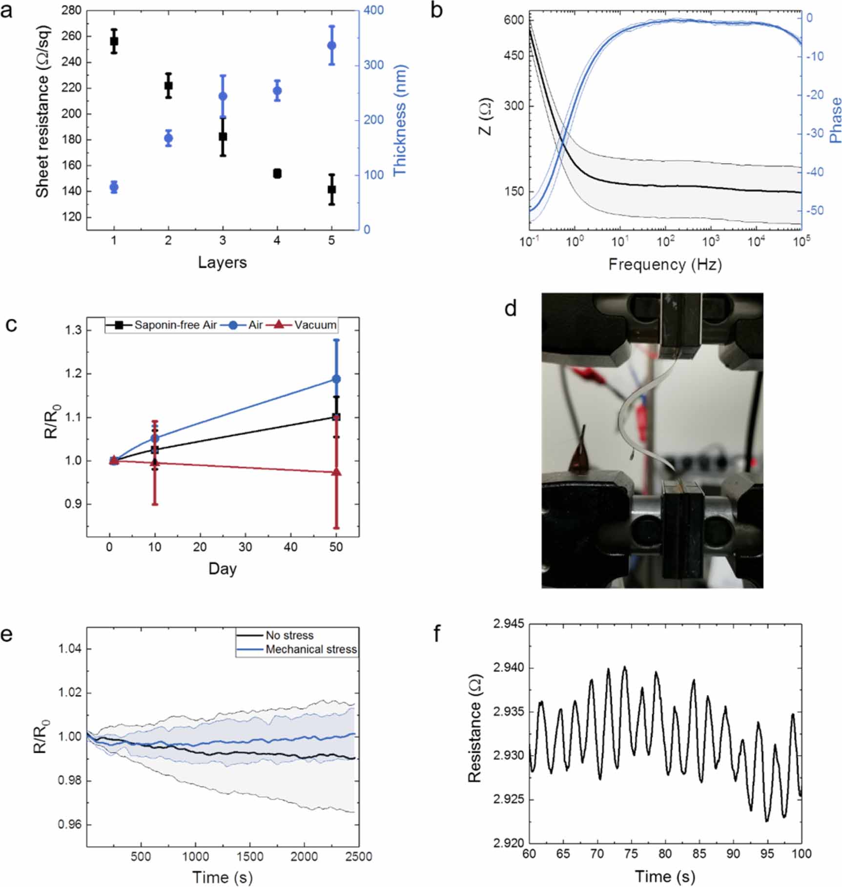
Standard image High-resolution imageAs we will use PEDOT as the flexible carrier for the printed MXene film in biosensing applications, we first evaluated the conductivity of the films printed on PEDOT coated glass substrate. We found that there is a linear trend of film thickness and the sheet resistance determined using a four-point-probe configuration (figure 2(a)). The decrease in sheet resistance of the system with MXene film thickness suggests that each layer of printed MXene is similar to its successive layer in its electrical performance. The system of three layers printed MXene on PEDOT:PSS was found to have a conductivity of 162.2 ± 24.2 S cm−1. This value is lower than that shown in figure 1(d). This may be due to the method we use to estimate conductivity which also takes the conductivity of the PEDOT layer underneath into account. Printed films may also have a decrease in conductivity due to small gaps between successively printed layers. These layers of printed MXene film could be identified in the cross-sectional scanning electron microscope (SEM) image of the film printed with five layers directly on silicon wafer or on PEDOT:PSS coating (figures S3 and S4). Figure 2(b) shows the electrochemical impedance spectrum of such a MXene film printed on the flexible PEDOT substrate. The capacitance of the system was calculated to be 3.76 ± 0.3 mF leading to an areal capacitance of 29.9 ± 2.5 mF cm−2 (figure S5).

Figure 2. (a) Thickness and sheet resistance of MXene film printed on PEDOT:PSS coated glass substrates with different number of layers (n = 3). (b) Bode plot of MXene film printed on flexible PEDOT substrate (n = 3). The impedance characteristics were recorded in PBS. The line represents the mean and the shaded region represents the standard deviation. (c) Electrical stability of drop-casted MXene/saponin films in air and in vacuum over time (n = 3). Saponin-free film was evaluated in air. (d) Image of a MXene film printed on PEDOT:PSS covered polyimide substrate that is under compressive strain with a maximum bending radius of 2 cm. (e) The change in film resistance over 1000 bending cycles at a frequency of 0.4 Hz (n = 3). The line represents the mean and the shaded region represents the standard deviation. (f) Resistance change of MXene film during bending cycles showing minimal change between cycles.
Download figure:
Standard image High-resolution imageNext, we evaluated the shelf-life stability of MXene films in air and in vacuum over 50 d. The films were deposited on paper and allowed to dry at 60 °C. Once the films dried, two PEDOT films were connected on either end to form a two-electrode probe. The films were then placed under vacuum to remove any excess water on the surface. Figure 2(c) shows that the printed films had only a small increase in their resistance over 50 d when left at room temperature in air (R/R0 = 1.19 ± 0.09). MXene films have thus a remarkable shelf-life, which can be further enhanced with vacuumed packaging. We also compared the stability of printed films to the film that did not contain saponin. This saponin-free film had air stability similar to the films with saponin, suggesting that the presence of saponin does not have a particular effect on the electrical stability of the film in air. The following experiments aimed to evaluate the electrical performance of printed MXene films under deformation. We recorded the electrical resistance of the MXene film printed on the flexible PEDOT substrate while the film was subject to compressive load/unload cycles, causing a maximum buckling amplitude of 2 cm (figure 2(d)). A schematic of the mechanical test setup in figure 2(d) is shown in figure S6. This mechanical load was applied up to 1000 cycles at a frequency of 0.4 Hz. The films showed a negligible increase in their resistance while bending (figure 2(e) and 1(f)), validating their suitability for the intended on-skin use where the electrodes may be exposed to repeated contractions.
Since the printed MXene will directly interface the skin and its surface will be used for biofunctionalization, we carried out SEM and X-ray photoelectron spectroscopy (XPS) to examine surface physicochemical properties. The SEM images shown in figures 3(a) and 3(b) compare the surfaces of a drop-casted, saponin-free MXene film with that of a printed MXene film. For the saponin-free MXene film, the individual MXene flakes are distinct and easily distinguished, and they layer on top of each other, creating a dense film. For the printed MXene film, we see a similar morphology, but we also observe additional circular features (marked with red arrows in figure 3(b)) that surround the edges of each flake. Similar features could be identified in atomic force microscopy (AFM) images, which also reveal a higher root-mean-squared (RMS) roughness for the printed MXene film compared to the saponin-free one (37.46 nm vs. 28.74 nm) (figure S7). We sought after the origins of these features in the printed films through XPS experiments. In the high-resolution XPS spectra of the C 1s region recorded for the printed MXene film, we detected a distinct peak at 285.8 eV (figure 3(c)). This peak was absent in the saponin-free, drop-casted film and attributed to the C–N bond present in the saponin structure. Moreover, the relative intensity of the C–O component was higher for the printed film compared to the saponin-free film, accompanied by a decrease in the ratio of C–Ti to C–C bond intensity. These differences arise from the contribution of C–O and C–C bonds in saponin molecules on the surface. In the O 1s XPS spectra of all MXene films (figure S8), we also note the presence of –OH groups on the surface, independent of the presence of saponin in the film.

Figure 3. SEM image of (a) drop-casted (saponin-free) MXene film and (b) printed MXene film. The scale bar is 500 nm. The arrows in (b) point to some surfactant aggregates. (c) XPS characterization of a drop-casted (saponin-free) MXene and a printed MXene film. (d) Fluorescent live/dead imaging of HeLa cells cultured on a printed MXene film. Green label is for live cells while the red is for dead ones. Inset is a bright field image of the same area. Shadow is due to a scratch. The scale bar is 100 µm. (e) XPS depth profiling of the printed MXene film analyzing the Ti and N atomic distribution throughout the film. Inset is the zoom-in of N atom line. (f) Ti 2p XPS spectra depth profiling of printed MXene film. The intensity of the TiC peak decreases and the TiO2 peak goes up as etching time increases. All printed films contain saponin.
Download figure:
Standard image High-resolution imageConsidering that XPS investigates the composition of the surface of the film up to ca. 10 nm in-depth and the features observed in SEM and AFM images, we conclude that printed MXene film has saponin aggregates on its surface. These aggregates seem to bind preferentially to the edges of MXene flakes, which are composed of C–C bonds that attract the hydrophobic heads of saponin and are otherwise hardly observed on the surface of the flakes. To investigate the biocompatibility of this particular chemical composition, we carried out an in vitro cell viability test by growing epithelial cells on printed MXene films. We found that the cells adhere well to MXene film and adapt a similar morphology to cells that are adhering to glass. Moreover, the majority of cells remain viable after 48 h in contact with the film (figure 3(d)).
After verifying the composition of the printed MXene surface and its compatibility with biological tissue, we next sought to understand whether saponin is only accumulated on the surface of printed MXene films and investigate the molecular composition of the bulk. Figure 3(e) shows the atomic distribution measured from wide XPS spectra obtained with etching treatment throughout the film. As we examined the film composition from the surface on towards the substrate, we found that the surface coverage ratio of Ti increases rapidly as we etch the film for 1 min, then remains constant after 2 min while there is a continuous reduction in N content, and no N atoms can be observed after 3 min. The analysis suggests that saponin is indeed accumulated predominantly on the surface, and the bulk of the film is formed of MXene only. Ti 2p spectra of the film after iterative etching evidenced that the TiC peak goes down and the TiO2 peak goes up the farther we etch into the film (figure 3(f)), indicating the oxidation of MXene. We reason that the oxidized content in the bulk arises because of trapped water molecules inside the film, presumably between the flakes. Nevertheless, this printable MXene formulation is highly conductive (831.87 ± 76.06 S cm−1) while the shelf-life stability, mechanical stability, surface functional groups, and biocompatibility highlight the potential of the material for cutaneous biosensing.
2.2. MXene electrodes for ECG recordings
The conventional electrodes for recording ECG signals are the commercially available, gel integrated Ag/AgCl electrodes. The gel serves to lower the impedance of the Ag/AgCl at the skin contact, hence improving the signal-to-noise ratio (SNR) of the recordings. Despite their good performance, typical problems with gel electrodes are associated with their durability (as gel evaporates over time) and the fact that the adhesive causes skin rashes and discomfort for some patients. We hypothesized that the printed MXene film could measure ECG without the need for a gel thanks to its conformability, high conductivity and sufficient capacitance in aqueous media. However, printed MXene films are too thin to form freestanding films on their own. A popular approach for attaching electronic materials directly on skin is via a water dissolvable tattoo paper [9, 14]. MXene could be printed on tattoo papers and then transferred on the skin using a water-soluble sacrificial layer (figure S9(a)). The tattoo-transferred, skin attached MXene platform exhibits excellent adherence and conformability on the skin. However, the integration of the tattoo with the acquisition system is often an undermined challenge, requiring adhesives or metallic interconnects that must be placed between the tattooed film and the skin, which severely deteriorates the integrity of the electrode as we observed for this platform as well (figure S9(b)).
To address the issue of connection of the MXene interfacing the skin with the acquisition system, we used PEDOT:PSS as a conductive flexible substrate. Mecerreyes et al have recently introduced a biocompatible cross-linker to stabilize PEDOT:PSS films in water [41]. This cross-linker, divinyl sulfone (DVS), also allows to detach PEDOT:PSS from the substrate and acts as a plasticizer promoting the chain mobility in the film and thus rendering the film flexible [42]. Hence, the freestanding, DVS modified PEDOT:PSS is an excellent substrate for our skin conformable electronics platform. We printed MXene (on a square-shaped area of 10 × 10 mm) on the back-side of conductive and flexible PEDOT substrates (25 × 25 mm), and insulated the remaining geometry by printing a dielectric layer (recall scheme 1). The PEDOT film acted as both a substrate and a conductive line to the acquisition system connected with an Au contact with ease, as illustrated in figure 4(a). Figure 4(b) is a photograph of the MXene film printed on the stand-alone, flexible PEDOT which adheres and conforms well around the finger and can be easily applied to any other parts of the body to measure electrophysiological signals. In our measurement set-up, MXene is the working electrode, and the reference and counter electrodes are commercial Ag/AgCl electrodes (figure 4(c)). These measurements used Ag/AgCl counter and reference electrodes to make sure that we only compare the working electrode which is the electrode of interest, and remove any additional differences that might occur due to a change in the reference or counter electrode material. Despite the absence of a gel between the electrode and the skin, the printed MXene electrode showed lower impedance on the skin over a broad frequency range (from 0.1 Hz to 10 000 Hz) compared to Ag/AgCl electrode (figure 4(d)), which we attribute to the enhanced electrode conformability. The MXene electrodes were also reusable, evidenced by the similar impedance values measured over 3 d of excessive use (figure 4(e)). The combination of MXene with PEDOT allowed intimate interfacing with the skin and an easy connection to the acquisition system, envisaged to reduce the motion artifacts arising from external connectors and prevent the failure caused by metal adhesives. Figure 4(f) shows a trace of ECG signals that the platform on the wrist of a volunteer recorded. When we placed both commercial and MXene electrodes on the skin to measure ECG simultaneously, both electrodes showed similar ECG signal profiles, as shown in figure 4(g), with comparable SNR values (SNRMXene = 7.1, SNRAg/AgCl = 6.7). As we gently removed the electrode from the skin and placed it back, the measurements recovered rapidly with no loss in the signal fidelity, showing the robustness and user-friendly application of the electrode (movie S2). The air stability, reusability, gel-free application, and easy removal and re-attachment of MXene electrodes printed on flexible PEDOT substrates highlight the advantages of this technology compared to the conventional, single-use, wet Ag/AgCl electrodes.

Figure 4. (a) Schematic of the connection between the printed MXene electrode and acquisition system. (b) Photograph of the skin conformable, printed MXene based ECG electrode patch placed on the index finger of the volunteer. (c) Electrochemical impedance spectroscopy (EIS) and ECG measurement set up. For EIS, the working electrode (WE) and the counter electrode (CE) are placed on the left arm 3 cm apart, while the reference electrode (RE) is placed on the right arm. For ECG, the electrodes were placed on the left arm (LA), right arm (RA), and left leg (LL). (d) Skin-contact impedance spectra of MXene electrodes as well as commercial wet Ag/AgCl electrodes (n = 3). (e) Bode plot of the same MXene electrodes used on skin for three days (n = 3). (f) A trace of ECG recordings acquired using the skin-attached MXene electrode signal. (g) ECG signals recorded simultaneously with a MXene electrode and a commercial Ag/AgCl electrode placed nearby.
Download figure:
Standard image High-resolution image2.3. MXene electrodes for biochemical sensing
The hydrophilicity associated with the surface species of the MXene is expected to allow these printed electrodes to be easily functionalized with biorecognition units. Biofunctionalized printed MXene films can then be used for the detection of disease-associated biochemical molecules present in the sweat, creating a highly modular sensing system designed for specific applications. Our first application concerns the detection of metal cations from sweat. Abnormalities of the metal cation content of sweat can be an indicator for various dysfunctions related to dehydration, fatigue, and kidney or heart failures [43]; hence their analysis is a routine part of clinical evaluations [44]. Na+ is one of these target cations, which are typically detected using ion-selective electrodes relying on ion-selective membranes (ISMs) [45]. To render our MXene electrode sensitive to Na+, we drop-casted a Na+ ISM on top of the printed pattern (recall scheme 1, middle). The sensitivity of the sensor to Na+ was evaluated by recording its open circuit potential (OCP) vs. an Ag/AgCl electrode while changing the concentration of Na+ stepwise from low to high molarities in the artificial sweat solution. Figure 5(a) shows the linear behavior of MXene sensors (with a slope of 40 mV per decade) within the physiological range of Na+ in sweat [46], and their good device-to-device reproducibility. When exposed to solutions containing potassium (K+) at its physiologically relevant concentrations [46], the same MXene electrodes exhibited an irregular and less significant change in OCP with respect to an increase in K+ content (figure S10), showcasing the specificity of the ISM.

Figure 5. (a) The response of the Na+ ISM integrated printed MXene electrode to various Na+ concentrations in artificial sweat. (b) The response of the antibody modified MXene electrode to varying concentrations of IFNγ. All measurements were performed in artificial sweat solution to evaluate the performance of the electrodes for skin electronics.
Download figure:
Standard image High-resolution image
Our second MXene biosensor example involves the functionalization of the printed electrode with an antibody (recall scheme 1, right). Interferon gamma (IFNγ) is a pro-inflammatory cytokine that is directly involved in the immune response of the body to pathogens [47]. Increased levels of IFNγ have been related to several diseases such as tuberculosis [48], HIV [49], and, more recently, to the presence of the highly infectious virus, SARS-CoV-2 [50]. IFNγ is typically measured in serum using colorimetric assays in clinical settings, and there have been only a few reports on point-of-care sensors for IFNγ [51–53]. Recently, the detection of IFNγ as an immune biomarker in sweat has been shown [54], however, no correlations between sweat and serum levels have been made. IFNγ sensing in sweat is nevertheless an exciting alternative for non-invasive and fast diagnosis of many of these conditions. To create the IFNγ biosensor, we functionalized the printed MXene surface with the IFNγ antibody. Thanks to the functional groups on the MXene surface, the protein could physically adsorb therein and remain immobilized upon subsequent washing steps. We preferred physical adsorption over chemical bonding to reduce the impact of covalent bonding on antibody function. As the target protein was captured by the antibody on MXene surface, we recorded a change in the OCP of the electrode. These changes scaled linearly with the concentration of the protein, with a sensitivity of 3.9 mV per decade (figure 5(b)). Notably, without the antibody functionalization, the MXene electrode was not sensitive to the protein (figure S11), evidencing the successful incorporation of the recognition unit. These results demonstrate the multi-modality of printed MXene electrodes in a biosensor platform and suggest that the surface functional groups of MXene can be utilized for further functionalization with different biorecognition units selective to disease biomarkers.
3. Conclusion
In this work, we reported a modular biosensing platform based on inkjet-printed MXene films. We were able to reduce the surface tension of the aqueous MXene solution down to a printable level of 40.19 ± 1.54 mN m−1 using a nonionic surfactant, while maintaining a high film conductivity of 831 ± 76 S cm−1. Such MXene formulation has yielded a set of uniform films with the surfactant mostly aggregating on the surface. The obtained films render a high level of biocompatibility owing to surface-terminated –OH species of MXene nanosheets. Using inkjet printing, MXene films were printed on a self-standing, conducting PEDOT that acted as both the substrate and the conductive line to the recording system. While PEDOT conformed well to the skin, its integration simplified the device fabrication and lowered the motion artifacts. The printed MXene patch was used to record electrocardiograms (ECG) from a volunteer with a performance on par with commercial electrodes despite the absence of a gel. Incorporating an ion-selective layer on printed MXene surface rendered the electrode selective to Na+ sensor in sweat-like media. Furthermore, immobilizing the IFNγ antibodies on the surface enabled to use the films as a sensor of the cytokine protein. To our knowledge, this is the first report of using inkjet-printed MXene electrodes for recording ECG signals alongside the detection of sweat-related biochemical species. The results of our proof-of-concept platform aim to underline the promising utility of printed MXenes for various sensing applications.
4. Experimental section
4.1. Materials and ink-formulation
4.1.1. Substrates
Microscope glass slides were purchased from Thermo Scientific (10144633B), paper was purchased from Arjowiggins (Powercoat HD230), polyimide was purchased from DuPont (Kapton), silicon wafer was purchased from University Wafers (S4N02SP).
4.1.2. MXene preparation
Ti3C2Tx MXene suspension was prepared by etching the Ti3AlC2 MAX phase (Carbon Ukraine) in HF/HCl for 15 h. A lithium chloride (LiCl) solution was added to this mixture and allowed for mixing for one hour. The obtained Ti3C2Tx aqueous dispersion contains MXene flakes with an average lateral dimension of 3.2 µm and within a range of 900 nm to 6 µm. The dispersion was then sonicated in a water bath for 1 h to obtain flakes of size ca. 500 nm. The resulting MXene suspensions were diluted to 2 mg ml−1 with DI water. The diluted suspensions were then stored at 4 °C after flushing with N2 or Ar to avoid oxidation. MXene/surfactant solutions were prepared at the time of use.
4.1.3. Ink preparation
All printing was done using a Dimatix DMP-2831 inkjet printer with 10 pl cartridges with a nozzle size of 21.5 µm. For the MXene ink, 2 mg ml−1 MXene solution in water was mixed with 0.5 mg ml−1 of saponin. The ink was vortexed before filling the cartridges. MXene was printed on a heated substrate at 50 °C which was then dried at 60 °C overnight. All printed MXene films in this work are done with the MXene/saponin formulation, as MXene could not be printed otherwise. The dielectric ink was purchased from SunJet (EMD6201); the nozzle was heated to 50 °C with the substrate being at room temperature. The film was then cross-linked with UV-Ozone for 15 min. To compare different classes of surfactants, 20 µl of ink was placed on a glass slide with a coverslip placed on top. The films were imaged while still in suspension, i.e. before drying. Digital photographs were taken with a mobile phone. Microscope images were taken with a Leica microscope with top illumination.
4.1.4. Flexible PEDOT preparation
PEDOT:PSS (PH1000, Heraeus) was mixed with DVS, dodecylbenzenesulfonic acid (DBSA), and ethylene glycol (EG) in the following ratios for a 10 ml solution: 9.45 ml PEDOT:PSS, 500 µl EG, 50 µl DVS, and 0.4% v v−1 DBSA. The dispersion (before adding the DVS) was sonicated for 15 min then filtered through a 0.45 µm fiberglass filter. The solution was then drop-casted on a glass substrate and dried at 60 °C for 2 h, then at 140 °C for 2 h. The film was then submerged in DI water overnight.
4.2. Physicochemical and surface characterization
XPS analysis was conducted using an AMICUS/ESCA 3400 KRATOS instrument equipped with an achromatic Al Kα x-ray source (1468.6 eV). The source was operated at a voltage of 10 kV and a current of 10 mA. The elemental narrow scan region was acquired with a step of 0.1 eV. The pressure in the analysis chamber was 10–7 Pa during measurements. The obtained spectra were calibrated to the reference C 1s at 284.8 eV. The spectra were deconvoluted using Gaussian and Lorentzian methods, and background subtraction was carried out using the Tougaard method. For XPS etching, the Kratos AXIS XPS Supra instrument was equipped with a Ga+ ion source, which was used to etch the surface sample. Sputter-etching was performed every 1 min, and the wide scan and high-resolution spectra were obtained subsequently.
SEM images were obtained using a Merlin SEM enhanced with a Gemini II column. Surface images were obtained at 5k×, 10k×, 30k×, and 50k×, with an electron high tension of 5 kV, probe current of 120 pA. A working distance (WD) of 5.2 mm was used for saponin-free MXene films and 4.7 mm for MXene with the surfactant. AFM images were obtained with a Veeco Dimension 3100 Scanning Probe System. Bruker RTESPA 300 probes were used to probe the film surface. All images were post-treated using Gwyddion software.
4.3. Mechanical and electrical characterization
The conductivity measurements were done on MXene printed on a PEDOT:PSS-covered glass, which was prepared by spin-coating PEDOT:PSS/DVS at 1500 rpm for 45 s on glass wafer then drying at 140 °C for 2 h. Sheet resistance was measured with a four-point probe using a Jandel RM3000 + Test Unit with an Rµ 100 probe. The thicknesses of the same MXene samples used for the sheet resistance measurement were determined using a Tencor profilometer. The force applied was 1 mg, and the sampling frequency was 50 Hz. Surface tension measurements were conducted using a Krüss Drop Shape Analyzer DSA100. The surface tension was measured with the pendant drop mode. First, the device was calibrated with water until the surface tension was measured to be 72 ± 2 mN m−1. The measurement was then carried out by suspending a drop from the syringe without dropping it. The droplet was made to be as large as possible. The measurement was repeated three times. The mechanical test was performed by applying compressive load/unload on the film specimen (comprising three layers of MXene printed on a flexible PEDOT substrate, while both were supported by a thin polyimide layer), causing a buckling mode, while the resistance of MXene film was measured. Universal testing machine Instron 5944 (2 kN load cell) was used to apply a maximum displacement of 20 mm (under compression) at the speed of 500 mm min−1. The cyclic loading was performed up to 1000 cycles, sufficient to gauge the mechanical durability of the film. The dimension of the film specimen was 110 mm length (gauge length = 60 mm), 25 mm width, 100 µm thickness. Chronoamperometry on the films was done with a potentiostat (PalmSens 4) at −0.1 mV by connecting a wire to the printed MXene film on one side, and to the PEDOT substrate on the other side. The electrochemical impedance spectrum was recorded using a potentiostat (Metrohm) in PBS with a saturated Ag/AgCl electrode as a reference and Pt wire as a counter electrode. Capacitance was then calculated using the following equation considering the frequency of AC pulses (f) and the imaginary part of impedance ( ):
):

4.4. Biocompatibility
MXene was partially printed (three layers) on clean coverslips and sterilized with ethanol 70% for 30 min. HeLa cells were cultured in Dulbecco's modified eagle media supplemented with 10% fetal bovine serum, 2 × 10⁻³ M Glutamine, and 1% PenStrep (Invitrogen). Cells were seeded on a coverslip and incubated at 37 °C for 48 h. A mixture of Calcein AM (green)/Propidium Iodide (red) (Sigma Aldrich) was added for 5 min at 37 °C to the cells to evaluate the biocompatibility of the printed ink. HeLa cells were imaged with a fluorescent microscope (Nikon). Green color represents the living cells, and red labels are for the dead cells.
4.5. Skin impedance and ECG measurements
Two able-bodied volunteers (two males, aged between 25 and 40 years) participated in this study with informed consent acquired before conducting the experiments. All the volunteers participated in the ECG recordings and the measurement of the electrode–skin impedance. All protocols and procedures were approved by the direction of research of the KAUST Institutional Biosafety and Bioethics Committee. A potentiostat (PalmSense 4) was used to obtain the electrical impedance spectra of electrodes in contact with the skin of the volunteer. Gel Ag/AgCl electrodes (Covidien) were used as counter and reference electrodes. To connect the MXene electrode to the acquisition system, a gold electrode on polyimide was glued to the back of the electrode (to the flexible PEDOT substrate) using silver paste. The working and counter electrodes were placed 3 cm apart. The impedance spectra were acquired between 0.1 Hz and 10 kHz with a VDC of open-circuit conditions, and VAC of 0.01 V. The ECG signal was acquired using a Shimmer3 system. The electrodes were attached to the left arm (printed MXene electrode, three layers), right arm (Ag/AgCl electrode), and left leg (Ag/AgCl electrode). All data were filtered using the MATLAB Signal Analyzer through a high pass filter at 1 Hz with 0.85 steepness and a 60 dB attenuation. SNR was calculated using the equation below:

where RMSsignal is the RMS value of ECG data filtered with a 1 Hz high-pass filter; RMSnoise is the RMS value of the raw data minus RMSsignal.
4.6. Fabrication of the ion-selective membrane and ion sensing measurements
The ion sensors were fabricated by printing MXene (three layers) on the flexible PEDOT substrate then drop-casting the ion-selective membrane on the MXene pattern. The ISM was prepared by mixing Na+ ionophore VI 71739 (Sigma), bis(2-ethylehexyl) sebacate (DOS), sodium tetrakis[3,5-bis(trifluoromethyl)phenyl] borate (Na-TFPB), high-molecular-weight polyvinyl chloride, valinomycin (potassium ionophore), and sodium tetraphenylborate (NaTPB) in tetrahydrofuran. The active area was defined by a PDMS well (4 mm diameter). Before starting the measurement, the MXene electrodes were conditioned in a 10 mM aqueous NaCl solution overnight. The OCP measurements were performed using a potentiostat (Biologic VSP-300, Science Instruments) in a two-electrode configuration with a saturated Ag/AgCl in 3 M KCl as the reference electrode. The electrolytes to be tested were placed on the ion-selective MXene electrodes immediately before measurement, and the OCP was recorded with different electrolytes concentration. The measurements were done in artificial sweat, which had the following composition: 100 × 10−3 M NaCl, 5 × 10⁻³ M NH4Cl, and 10 × 10−3 M MgCl2.
4.7. Immobilization of the antibody and immunosensor experiments
The sensor was prepared by printing MXene (three layers) on a flexible PEDOT substrate and incubating the active area defined by a PDMS well (4 mm diameter) for 30 min with the IFNγ antibody (Abcam) in 10 × 10−3 M PBS. The surface was then incubated with 10 g l−1 of bovine serum albumin (BSA) in 10 × 10−3 M PBS for 1 h, acting as a blocker molecule. Each concentration of the recombinant IFNγ protein (Abcam) was sequentially added to the artificial sweat from low to high (100 fg ml−1 to 100 ng ml−1). The electrodes were exposed to each solution for 30 min before rinsing with PBS three times. OCP was then measured in 10 × 10−3 M PBS using a two-electrode setup as described above for the ion sensor. In the negative control experiments without the IFNγ antibody functionalization, the electrode was only incubated with BSA for 1 h before the measurements.
Acknowledgments
The authors thank Nimer Wehbe at Imaging and Characterization facilities at KAUST Core Labs for assisting with the XPS etching measurements. Scheme 1 was created by Heno Hwang, a scientific illustrator at King Abdullah University of Science and Technology (KAUST).