Article

Obtaining, Evaluation, and Optimization of Doxycycline-Loaded Microparticles Intended for the Local Treatment of Infectious Arthritis

by
Maria Minodora Marin
1,2,
Mădălina Georgiana Albu Kaya
2,*,
Horia Iovu
1,3,
Cristina Elena Stavarache
1,
Ciprian Chelaru
2,
Rodica Roxana Constantinescu
2,
Cristina-Elena Dinu-Pîrvu
4 and
Mihaela Violeta Ghica
4
1
Advanced Polymer Materials Group, University Politehnica of Bucharest, 1-7 Polizu Street, Bucharest 011061, Romania
2
Department of Collagen, Division Leather and Footwear Research Institute, National Research and Development Institute for Textile and Leather, Bucharest 031215, Romania
3
Chemical Sciences Section, Academy of Romanian Scientists, 54 Splaiul Independentei, Bucharest 50085, Romania
4
Department of Physical and Colloidal Chemistry, Faculty of Pharmacy, University of Medicine and Pharmacy “Carol Davila”, Bucharest 20956, Romania
*
Author to whom correspondence should be addressed.
Coatings 2020, 10(10), 990; https://doi.org/10.3390/coatings10100990
Submission received: 4 September 2020 / Revised: 4 October 2020 / Accepted: 15 October 2020 / Published: 17 October 2020

Abstract

:
Compared to the classical systemic administration, the local drug release has some advantages, such as lack of systemic toxicity and associated side effects, increased patient compliance, and a low rate of bacterial resistance. Biopolymers are widely used to design sustained drug delivery systems and biomaterials for tissue engineering. Type II collagen is the indispensable component in articular cartilage and plays a critical role in the growth and proliferation process of chondrocytes. Thus, type II collagen has drawn more attention and interest in the treatment and research of the cartilage regeneration. The aim of this study was to obtain, characterize, and optimize the microcapsules formulation based on type II collagen, sodium alginate, and sodium carboxymethyl cellulose loaded with doxycycline as an antibiotic model drug that could be incorporated further in hydrogels to improve the localized therapy of septic arthritis. The new synthesized microcapsules were assessed by spectral (FT-IR), morphological (optical microscopy), and biological analysis (enzymatic biodegradation, antimicrobial activity). The size distribution of the obtained microcapsules was determined using optical microscopy. The drug encapsulation efficiency was also determined. To optimize the microcapsules’ composition, some physical-chemical and biological analyses were subjected to an optimization technique based on experimental design, response surface methodology, and the Taguchi technique, and the adequate formulations were selected. The results obtained recommend these new microcapsules as promising drug systems to be further incorporated in type II collagen hydrogels used for septic arthritis.

1. Introduction

Septic arthritis (SA) represents an inflammatory disease of the joints which is most often caused by an infectious agent. Usually, SA affects joints such as the knee or hip, but it can also involve another joint. The incidence of this pathology is higher for people under 15 and over 55 years old [1]. The most crucial risk factors for SA are rheumatoid arthritis and prosthetic joint surgery. For patients who present these effects, the frequency of SA is increased [2]. Additionally, SA is commonly seen as a resulting infection because the bacteria get away from the bloodstream and arrive in adjacent tissues [3,4]. Several approaches such as endothelial attachment and bacterial passing by specialist phagocytes have been defined as mechanisms that allow the infectious agent to spread from the blood to the joint or further tissues [5,6,7]. The factors that facilitate bacterial growth are perceived to be of pathogenic significance for SA [8]. The first step of the joint destruction is generally caused through the cartilage-synovium friction, with inflammation development followed by cartilage and bone damage [9]. The entire process may cause permanent damage to joint role and it correlated with the creation of a large quantity of cytokines [10,11]. Rapid and precise therapy is critical in order to obtain good results. The delay for only some days in the healing process might initiate definitive joint destruction and an enhancement of the mortality percentage [12]. Commonly, the bacteria which cause SA are Staphylococcus aureus (S. aureus) [13]. S. aureus is a Gram-positive bacteria [14], and it is the main source of septic arthritis appearances in the majority of cases [15,16]. Other agents which can cause SA are Streptococcus pneumoniae, Escherichia coli, and Salmonella [17]. Because the gravity of this pathology involves rapid therapy, instead of the isolation of the infective agent the most useful thing in these cases is the drug delivery system [18,19].
Natural polymers offer significant benefits in biomedical applications such as drug delivery and tissue engineering due to properties such as biocompatibility, non-toxicity, and biodegradability [20]. Various drug encapsulation techniques using natural polymers have been developed over the years [21].
The widely preferred natural biopolymers for drug encapsulation applications are collagen, alginate, chitosan, carrageenans, and cellulose [22]. Alginate represents a great material for drug delivery systems. The usage of alginate is preferred because it is a cheap biomaterial and presents a high biocompatibility [23,24].
Collagen has been perceived as the most representative protein existing in the extracellular matrix of vertebrates and forms a considerable proportion of connective tissue [25].
Collagen is a natural polymer extensively used in tissue engineering due to its superior properties such as biocompatibility; rate of biodegradation, correlated with the formation of the new tissue; low antigenicity; and promoting extraordinary biological properties (cell viability and proliferation) [26,27,28]. Type II collagen has been considered as an essential biomaterial to treat articular cartilage damage due to its low antigenicity and great cell attachment properties [29]. Type II collagen represents the main component of cartilage (more than 90% of the collagen produced by chondrocytes in the extracellular matrix) and it helps proteoglycans to form fibrils. Additionally, type II collagen in a mixture with other biomaterials has drawn more significance in the healing process of cartilage [30].
Sodium carboxymethylcellulose (NaCMC) is an important product obtained from cellulose and presents excellent properties such as biocompatibility, non-toxicity, and great biodegradation behavior [31].
Doxycycline is a representative of the tetracycline category of antibiotics used in the therapy of infections produced by Gram-positive, Gram-negative, or specific organisms. Doxycycline has a huge usage in the treatment of infectious diseases due to its broad-spectrum efficiency [32].
In the last few years, biodegradable polymeric microparticles used in medical applications for drug release have been developed [33]. For example, Yadav et al. [34] reported microspheres based on chitosan and alginate loaded with doxycycline and ornidazolfor chronic infections. Additionally, Uyen et al. [33] synthesized curcumin-loaded alginate microspheres for drug delivery. Currently, no report has been published on the fabrication of a formulation based on type II collagen, sodium alginate, and sodium carboxymethyl cellulose loaded with doxycycline. Additionally, the presence of the type II collagen plays a major role in SA treatment.
The aim of this study was to obtain, characterize, and optimize the microcapsule formulation based on type II collagen, sodium alginate, and sodium carboxymethyl cellulose loaded with doxycycline as an antibiotic model drug that could be incorporated further in hydrogels to improve the localized therapy of septic arthritis. The new synthesized microcapsules were assessed by spectral (FTIR), morphological (optical microscopy), and biological analysis (enzymatic biodegradation, antimicrobial activity). The size distribution of the obtained microcapsules was determined using optical microscopy. The drug encapsulation efficiency was also determined. To optimize the microcapsule composition, some physical-chemical and biological analyses were subjected to an optimization technique based on experimental design, response surface methodology, and the Taguchi technique, and the adequate formulations to be further incorporated in type II collagen hydrogels were selected.

2. Materials and Methods

2.1. Materials

Type II fibrillar collagen gel with a concentration of 2.54% (w/w) was obtained from bovine cartilage using a new technology for collagen extraction [35]. Sodium carboxymethyl cellulose (NaCMC) and doxycycline hyclate (Doxy) were purchased from Fluka; sodium alginate (Alg) was purchased from Sigma-Aldrich, Germany, and calcium chloride (CaCl2) was purchased from Merck.

2.2. Methods

2.2.1. Preparation of Microcapsules

Type II collagen, sodium alginate, and NaCMC were solubilized in distilled water, and doxycycline previously solubilized in distilled water was added and homogenized together, at 24 °C, to obtain 100 mL of gel for the microcapsule preparation in accordance with the composition given in Table 1 and Table 2. The gel was dropped with a syringe in a 1 M CaCl2 solution for crosslinking, and spherical capsules were obtained.

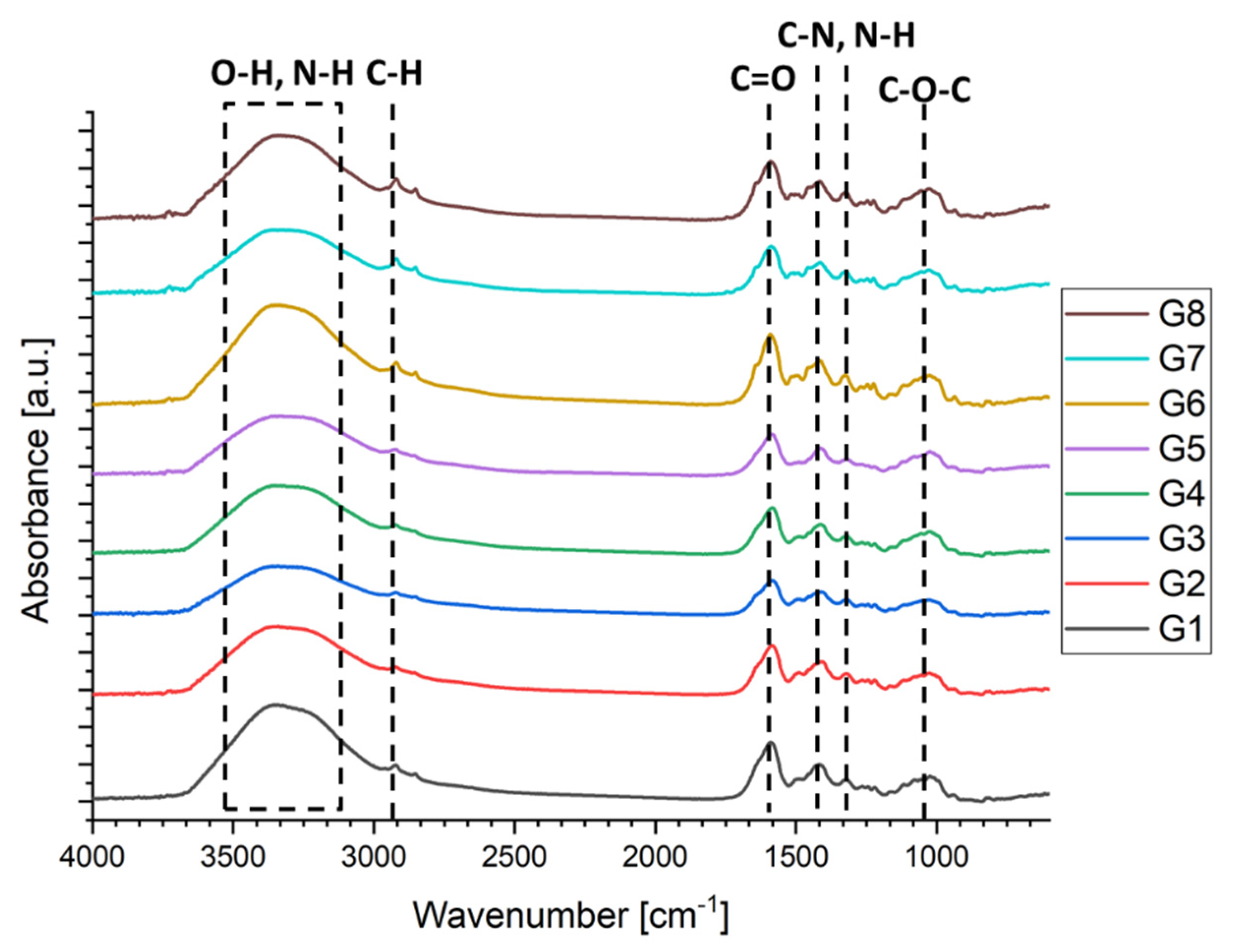
2.2.2. Fourier-Transform Infrared Spectroscopy (FTIR)

FTIR measurements were achieved on a Vertex 70 Bruker FTIR spectrometer (Billerica, MA, USA) using an attenuated total reflectance (ATR) accessory. For all formulations, the FTIR spectra were registered in the ATR-FTIR mode (in triplicate) at a resolution of 4 cm−1 in the 600–4000 cm−1 wavenumber region.

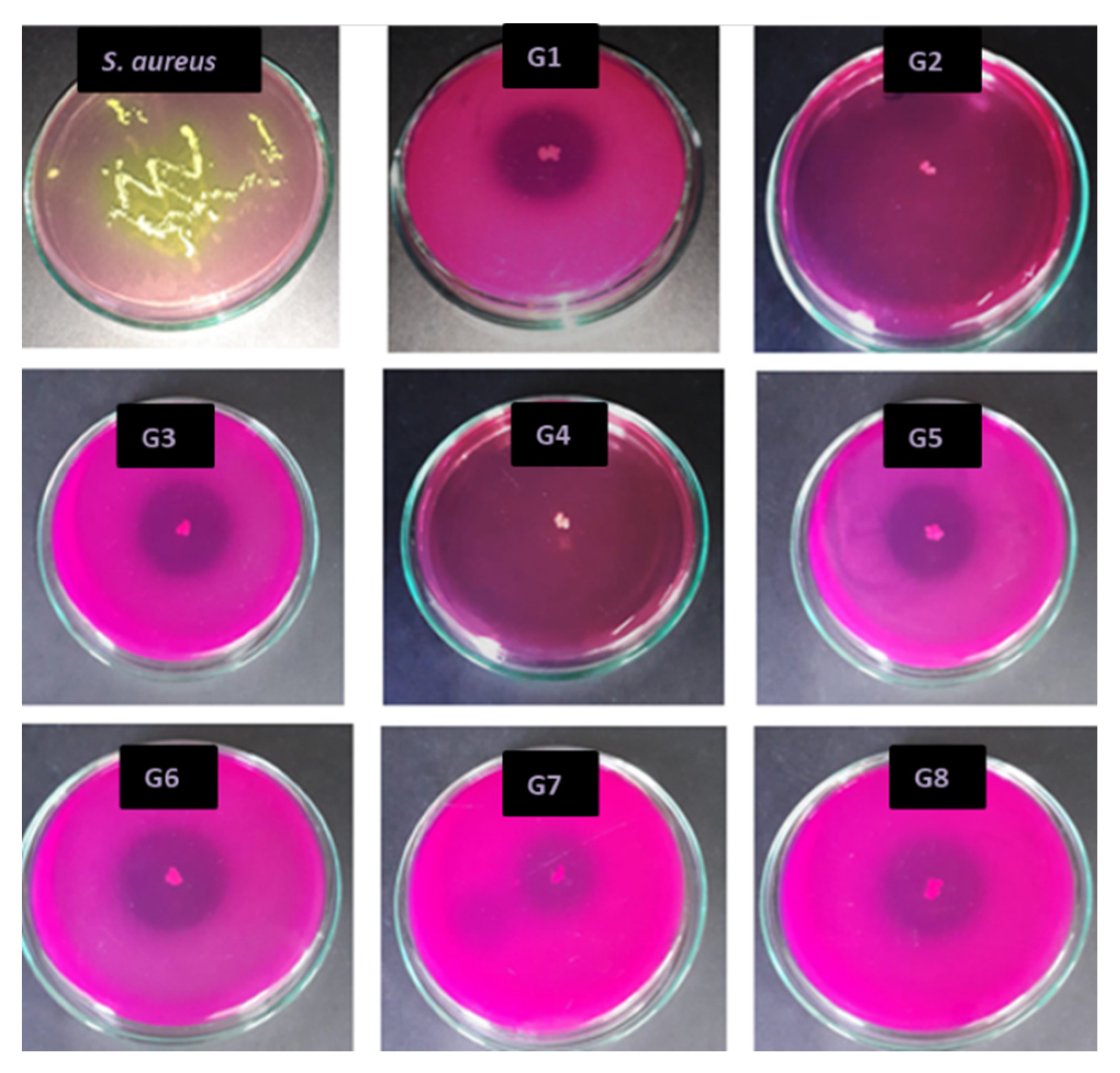
2.2.3. Antimicrobial Activity against Staphylococcus Aureus (S. aureus) Using “Agar Diffusion Method”

The control of the antimicrobial activity was tested against the S. aureus (gram positive) strain. Firstly, the gel (Manitol Salt Agar) for the lower layer without bacteria was prepared. A total of 10 mL of gel was placed into each sterilized Petri dish and allowed to solidify. Secondly, gel for the upper layer was prepared and cooled down to 45 °C in a water bath. The inoculation of 150 mL of gelose with 1 mL of bacterial working solution ((1–5) × 108 μfc/mL) was performed. The samples were placed on the surface of the nutrient medium and then incubated at 37 °C between 18 and 24 h. Bioactivity was determined by determining the diameter of inhibition zones in mm. Each measurement was repeated three times and the mean of the diameter of the inhibition areas was calculated.

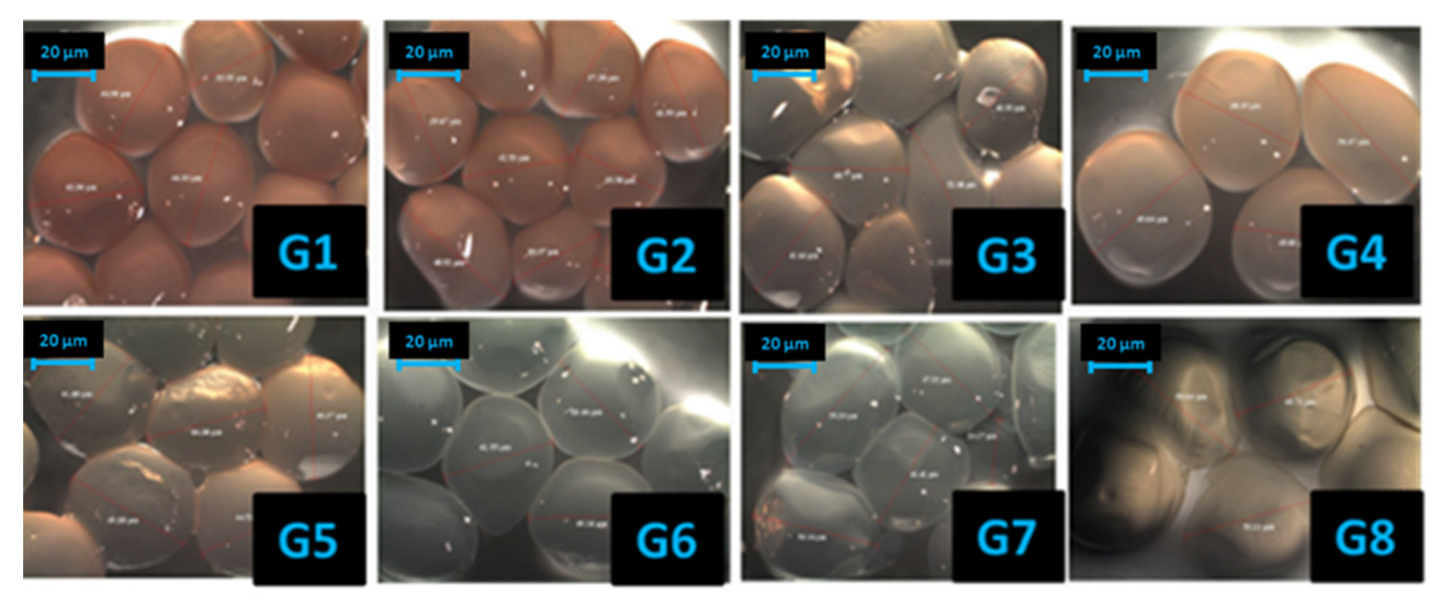
2.2.4. Optical Microscopy Images

All the images were obtained with a Leica Stereomicroscope model S8AP0, with a 20×–160× magnification capacity (Leica Microsistems, Wetzlar, Germany). For a superior evaluation of the samples, a 100× magnification and incident external cold light were used. Each experiment was performed in triplicate.

2.2.5. Enzymatic Degradation

The enzymatic degradation of microcapsules was investigated by measuring the weight loss depending on the contact time with the collagenase solution. At specific intervals, the swollen scaffold was weighed. The percentage of scaffold degradation was determined by the relation (Equation (1)):
%   Weight   Loss = W i W t W i ×   100
where Wi is the initial weight and Wt is the weight after time t. The experiments were carried out in triplicate.

2.2.6. Drug Encapsulation Efficiency (EE)

The EE of doxycycline was determined by evaluating the amount of drug encapsulated in the microcapsules by the UV-VIS spectrophotometer UV 3600 Shimadzu provided with a quartz cell with a light path of 10 mm. The doxycycline assay was obtained using a calibration curve prepared with standard samples with concentrations between 0.05 and 0.001 mg/mL. Three determinations were performed for each sample, and the results were averaged.
The EE was calculated using the following equation (Equation (2)):
EE   ( %   ) = Loaded   amount   of   doxycycline Theoretical   doxycycline   amount ×   100

2.2.7. Experimental Design and Optimization Technique

The obtaining of the microcapsules was carried out based on the 4-factor, 2-level Taguchi factorial design (Table 1 and Table 2). The formulation factors considered as independent variables were selected as follows: collagen, NaCMC, sodium alginate, and doxycycline hyclate concentrations (g%) (Table 1). The variation levels for the input parameters were coded 1 for the inferior and 2 for the superior level, respectively. The physical-chemical and biological parameters affecting the drug delivery from the designed microcapsules further incorporated in type II collagen hydrogels and considered as dependent variables (system responses, Yi) were selected as follows: microcapsule diameter (μm), enzymatic degradation at 2 h, expressed as weight loss (%), and drug encapsulation efficiency (%). The coded forms of the dependent parameters are given in Table 1 along with the specific constraints imposed by the design of such formulations loaded in hydrogels that ensure an adequate drug delivery for the localized therapy of septic arthritis. The statistical analysis used for the optimization of the above independent variables was based on different routines of the Statistical Stat Soft Release software package. The steps of this analysis were detailed in our previous works [36,37,38,39,40]. Briefly, a second-degree polynomial equation was set for each dependent parameter using a stepwise regression analysis, for which only the significant terms (p < 0.05) were kept. The adequacy of the reduced quadratic models was determined based on the correlation (R) and determination (R2) coefficients, analysis of variance (ANOVA), and residual analysis. To assess the influence of the input variables on the dependent parameters, 3D response surface graphs were further drawn. As a final step of the optimization technique, the Taguchi method through the performance indicator Signal/Noise ratio was applied.

3. Results and Discussion

3.1. FTIR

The microcapsules prepared according to a Taguchi fractional factorial plan with four factors and two levels were firstly spectrally analyzed. Thus, the comparison between the polymers and the drug microcapsule formulations were determined by FT-IR studies (Figure 1). The FTIR investigations were performed on the dried microcapsules.
The infrared frequencies were observed at 3424, 2876, 1655, 1599, 1423, 1381, and 1030 cm−1, corresponding to the –NH2, –OH stretching, and carbonyl (C=O) stretching of the secondary amide (amide I); the bending vibrations of the N–H (amide II); and the C–N stretching of the amide and ether and N–H bonds (amide III band), respectively. The strong bands at 1030 cm−1 are attributed to the C–O–C stretching vibrations, which are considered as the fingerprinting area characterized for the natural polysaccharides [41]. Having the same components with different concentrations, the differences between the samples do not appear significant.

3.2. Antimicrobial Activity Against S. aureus Using the “Agar Diffusion Method”

The antimicrobial activity of the obtained microcapsules was evaluated after 24 h of incubation. The results showed that all the tested samples exhibited a good antibacterial activity against S. aureus. In all cases, a significant zone of inhibition was observed, confirming the antimicrobial potency of the microspheres against S. aureus (Figure 2).
The highest inhibition area is shown by samples G2 and G4, which present a total inhibition. No significant differences in the inhibition zone were obtained for the other formulas, as can be observed in Table 3.
The inhibition zone produced by all the formulas, except G2 and G4, showed the size of the inhibition zone ranging between 13 and 20 mm. Additionally, it can be observed that the largest areas of inhibition were presented by the samples with the highest drug concentration.
The antimicrobial results highlight that all the microcapsules can be used successfully in systems for the treatment of septic arthritis.

3.3. Optical Microscopy Images

The surface morphology of the microcapsules was observed by optical microscopy (Figure 3). As shown in Figure 3, the microcapsule that was prepared has a regular near-spherical shape, is uniform, has an appropriate particle size, and has a good dispersibility.
During optimization studies, the particle size of various microcapsule formulations was determined also by optical microscopy. The average diameter for each microcapsule can be seen in Table 4.
It was found that the particle size distribution of each formulation was adequate (<1000 μm) for drug delivery systems [42], with the mean particle size ranging between 35.62 and 53.19 μm, depending on their composition.

3.4. Enzymatic Degradation

Biodegradability is extremely necessary for microcapsules used as drug delivery systems in biomedical applications. The control of the biodegradation rate of the microspheres is a crucial parameter, as the in vivo resorption affects the drug release and tissue regeneration ability. The collagen structure can be destroyed only by collagenases, which are distinctive enzymes as they are able to destroy the collagen triple helical region under the biological conditions of pH and temperature [43]. Alg, and NaCMC are also biodegradable polymers (Figure 4).
The enzymatic degradation was expressed as weight loss. The enzymatic degradation can be observed for a period about 48 h. All the samples are stable for the first 2 h. After 24 h, a total degradation was observed for samples G1, G2, G5, and G6.
During 48 h, the lowest mass lost was presented by sample G4, which is the most stable duo to the high content of the alginate. Another sample which presents a good stability was G8, with a double content of collagen, NaCMC, and doxycycline compared with G1. Even though both G1 and G8 had the same amount of alginate, which is responsible for crosslinking, G8 is more stable than G1. This can be explained by the inhibition of collagenase by doxycycline or the crosslinking between collagen and doxyxycline.

3.5. Drug Encapsulation Efficiency

The capacity of the microcapsules to entrap the drug was to establish the factor for the selection of the preparation method and composition. Accordingly, the effect of microcapsule composition on the entrapment efficiency and drug content was investigated. Table 4 shows the results of the drug encapsulation efficiency.
All the formulations exhibited good encapsulation efficiencies of 67.57% and 80.14%.

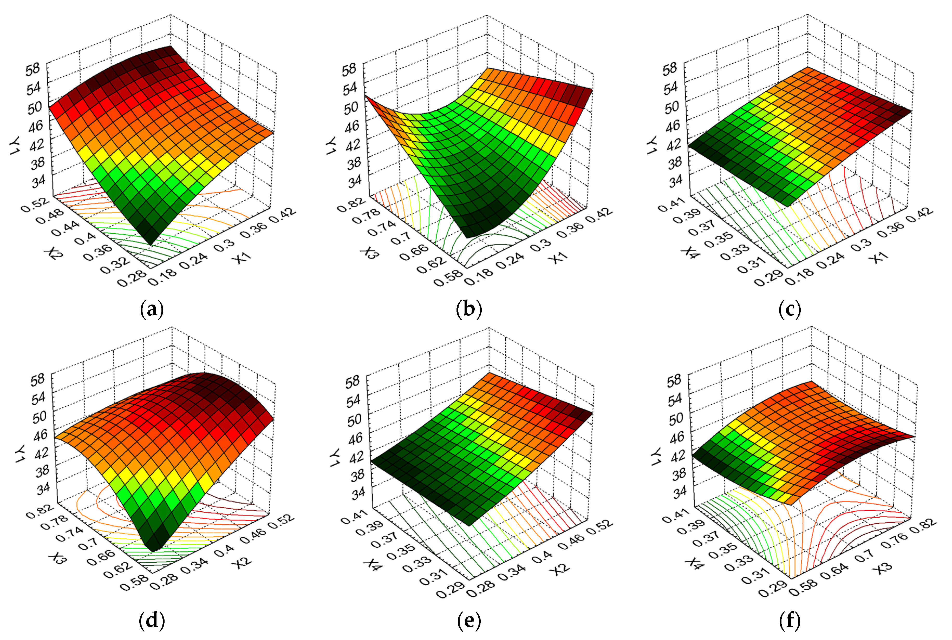
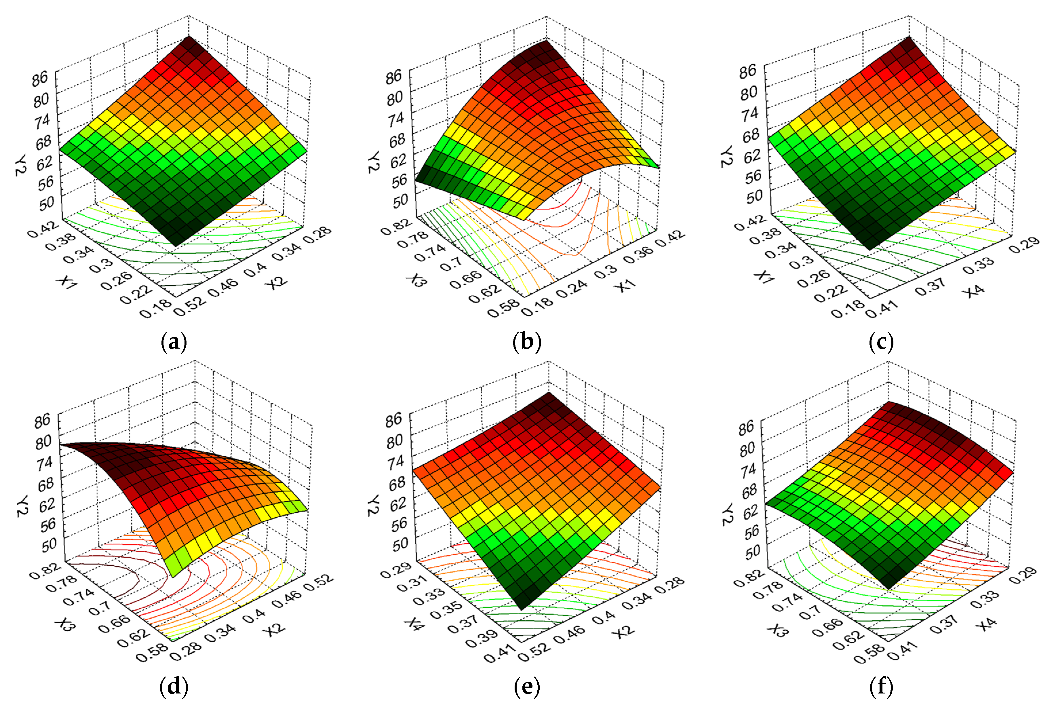
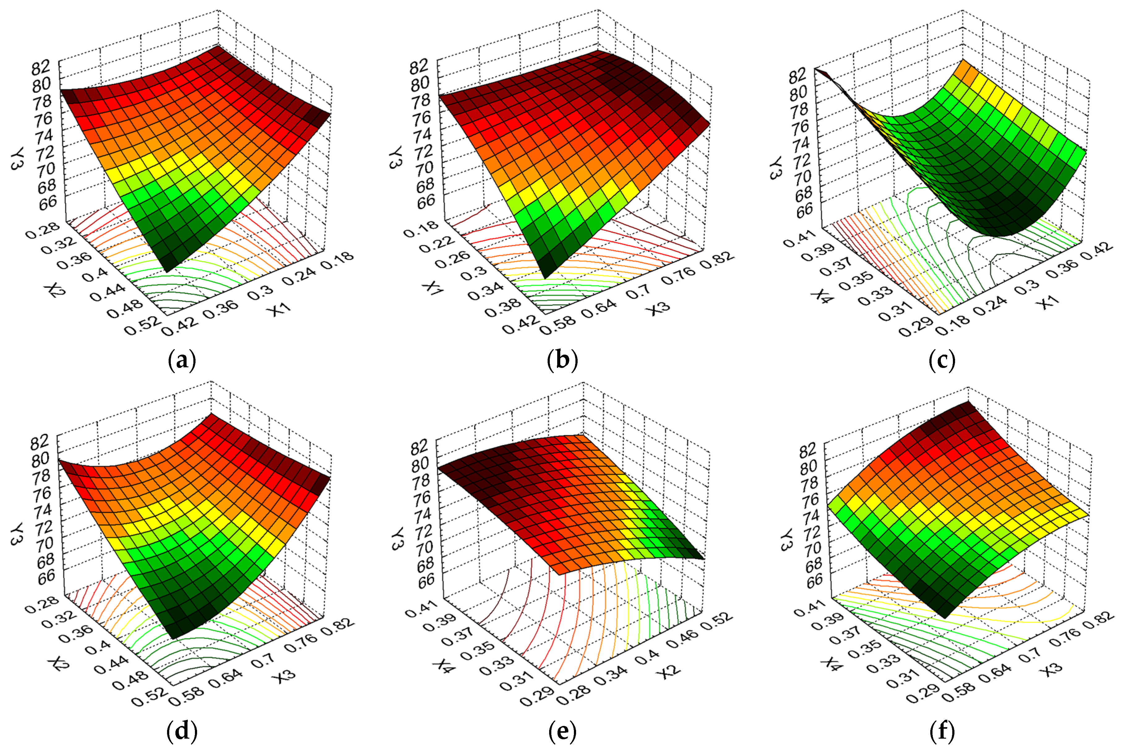
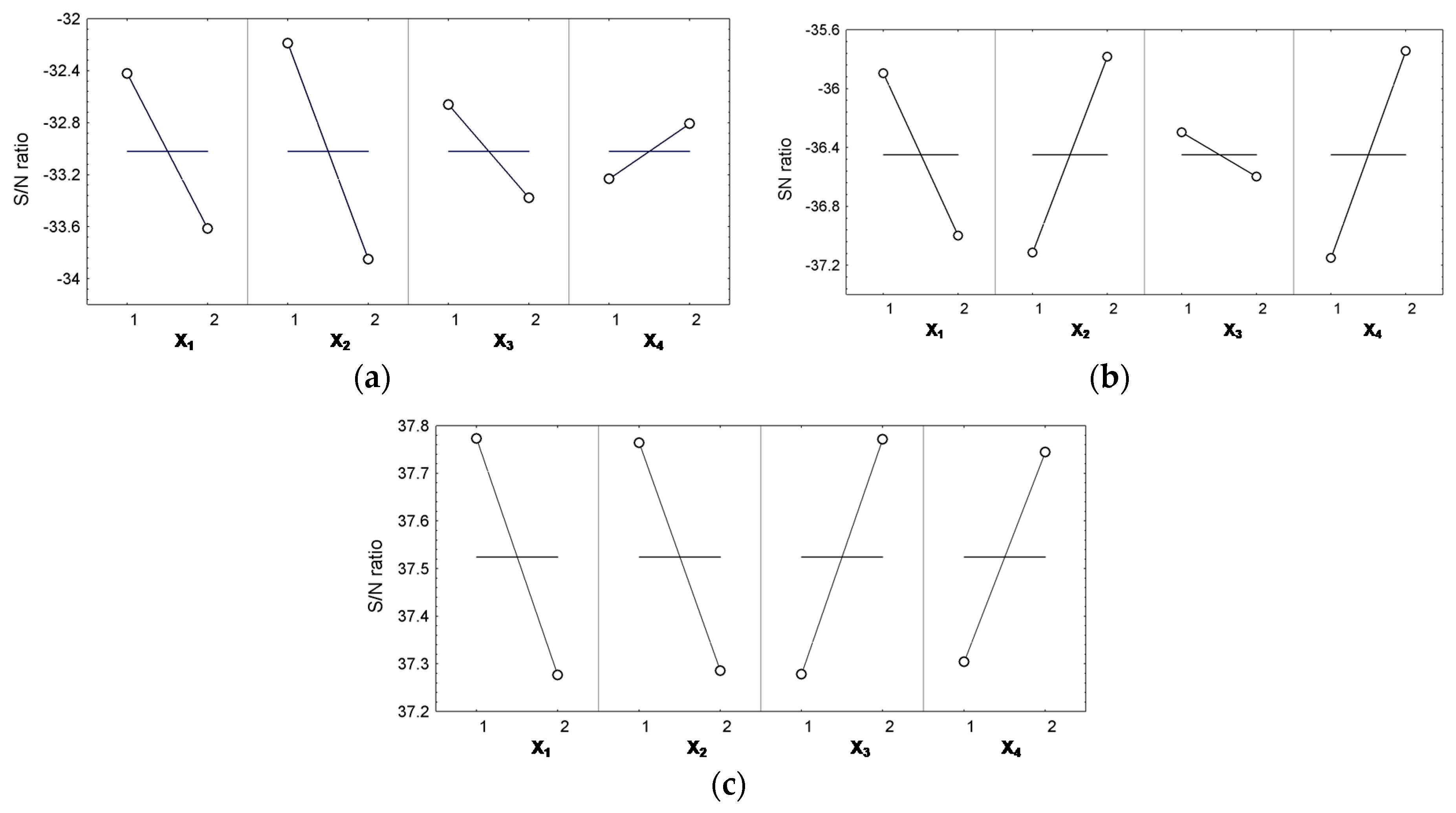
3.6. The Optimization Technique

In order to study the influence of the formulation factors on the above physical-chemical, biological, and biopharmaceutical parameters (diameter, drug encapsulation efficiency andenzymatic degradation at 2 h) involved in the drug delivery from the designed microcapsules and to optimize the microcapsule composition to ensure an adequate drug release for the localized therapy of septic arthritis, the experiments resulting from the Taguchi factorial design were subjected to an optimization technique based on experimental design, response surface methodology, and the Taguchi technique, presented in our previous works [36,37,38,39,40]. In Taguchi fractional design, the number of experiments is reduced from 16 (corresponding to the full factorial design) to 8, as summarized in Table 4.
The reduced quadratic models indicating the effect of the formulation factors (Xi) and their interactions on the dependent variables (Yi, Table 4) are illustrated in Equations (3)–(5), where only the significant terms (p < 0.05) were considered.
Y 1 = 28.10 + 85.28 X 1 X 2 + 56.69 X 2 X 3 64.94 X 2 X 4 ,
Y 2 = 72.02 + 49.86 X 2 + 60.31 X 1 X 3 268.58 X 2 X 4 ,
Y 3 = 70.14 56.98 X 1 X 2 + 49.07 X 3 X 4 .
From Equation (3), a positive impact on the Y1 response is noticed for the interaction between X2 and X4, while the interactions between X1 and X2, and also between X2 and X3 negatively influence this response.
Equation (4) shows a negative effect of X2 (linear) and of the interaction between X1 and X3 on Y2, with this response being influenced in a positive manner by the interaction between X2 and X4.
For Y3, an antagonist effect of the interaction between X1 and X2 and a synergistic effect of the interaction between X3 and X4 can be remarked from Equation (5).
The predictive power of the reduced second polynomial equations is well sustained through high values of correlation, R (0.9920, 0.9942, 0.9672, respectively), and determination, R2 coefficients (0.9842, 0.9884, 0.9355, respectively), as well as by the ANOVA results, summarized in Table 5.
The results of the residual analysis, listed in Table 4, also indicated a good correlation between the observed and predicted values.
To evaluate the influence of the formulation factors on the system responses, the 3D response surface graphs were further plotted (Figure 5, Figure 6 and Figure 7).
Figure 5a indicates that a small microcapsules diameter is obtained for the smallest concentrations of collagen and carboxymethylcellulose, the maximum level of the two formulation factors leading to an important increase of Y1. Thus the microcapsules diameter increases with 42.64% (from 37.29 to 53.19 µm) when X1 and X2 are varying from minimum to maximum level.A small microcapsules diameter is also favored by small concentrations of collagen and sodium alginate (Figure 5b). A diameter reduction from 53.19 to 45.75 µm (13.98%) is noticed for maximum collagen concentration and a variation of the sodium alginate concentration from the minimum to maximum level. The collagen concentration or glutaraldehyde concentration at the minimum level in combination with any amount of doxycycline (Figure 5c,e) are leading to low Y1 values, this response having practically no variation in these cases. Figure 5d, showing the dependance of the microcapsules diameter on the carboxymethylcellulose and sodium alginate concentrations, indicates that small concentrations of X2 and X3 result in favorable Y1. Variation between the minimum and maximum level for carboxymethylcellulose when sodium alginate concentration is kept at the minimum level is resulting in an important Y1 increase, of 35.26%, between 35.62 and 48.18 µm.The interaction between sodium alginate and doxycycline is less affecting Y1, the microcapsules diameter variation being reduced for the modification of X3 and X4 levels (Figure 5f).
Concerning the enzymatic degradation at 2 h, collagen and glutaraldehyde concentrations, collagen and doxycycline concentrations respectively are determining similar variation patterns for Y2, as it results from Figure 6a,c. A small enzymatic degradation can be noticed for small amounts of collagen, but high amounts of glutaraldehyde and doxycycline. An opposite behaviour of the enzymatic degradation is observed for the minimum and maximum levels of collagen concentration when the sodium alginate concentration is varying between the minimum and maximum levels (Figure 6b). While for the minimum level of X1 the enzymatic degradation decreases with X3 increase (from 63.21% to 52.96%, so a 16.21% decrease, the maximum level of X1 is determining a stronger increase of Y2 for X3 variation between minimum and maximum levels (from 56.99% to 81.88%, i.e., a 43.67% increase). According to Figure 6d, the glutaraldehyde concentration variation is not influencing the enzymatic degradation for small concentrations of sodium alginate.In turn, for high values of X3, the X2 variation between minimum and maximum level is resulting in a strong reduction in the enzymatic degradation (35.32%), from 81.88% to 52.96%.High concentrations of glutaraldehyde and doxycycline determine a favorable enzymatic degradation (Figure 6e) with a linear evolution of Y2 for X2 and X4 between their minimum and maximum levels. Y2 evolution is less influenced by the variation of sodium alginate and doxycycline concentrations (Figure 6f). Thus, the enzymatic degradation sees a 10.98% decrease (81.88% to 72.89% when the sodium alginate concentration is decreasing from maximum to minimum level and the doxycycline concentrations is kept at the minimum level.
Figure 7a–c show that the collagen concentration at minimum level is determining the most favorable drug encapsulation efficiency when glutaraldehyde concentration is at minimum level and sodium alginate and doxycycline concentrations are kept at minimum levels. According to Figure 7d, Y3 values are decreasing when glutaraldehyde concentration is varying from minimum to maximum level and the sodium alginate is at minimum level (a decrease from 79.25% to 67.68%) and remains at practically the same levels for the X2 variation when X3 is at maximum level. Concerning the interaction between the glutaraldehyde and doxycycline concentrations, Figure 7e indicates that the best Y3 values are recorded for small amounts of X2 and high amounts of X4, with an almost linear evolution of drug encapsulation efficiency for glutaraldehyde and doxycycline concentrations modification between minimum and maximum levels.High concentrations of sodium alginate and doxycycline are determining the best drug encapsulation efficiency (81.04%), Y3 value decreasing to 67.68% (a 16.48%) decrease when X3 and X4 are at minimum level, as resulting from Figure 7f.This is due to the greater availability of active calcium binding sites in the polymeric chains and consequently the greater degree of crosslinking.
To evaluate the microcapsule formulation determining the dependent parameters affected to the minimum extent by the noise factors, the final step of the optimization technique consists of the application of the performance indicator Signal/Noise ratio (S/N) introduced by Taguchi.
Depending on the constraints imposed on system responses (Table 1), the “larger-the-better” criterion was applied for S/N ratios related to drug entrapment efficiency, while the “smaller-the-better” criterion was selected for the microcapsules diameters and enzymatic degradation at 2 h.
The influence of the control parameters (input variables X1X3) on the S/N ratio for the system responses resulting in the optimal combination of the microcapsule formulation factors is given in Table 6 and Figure 8a–c.
The variation levels of the independent variables for all responses are illustrated in Figure 8. From this figure, it can be remarked that all the formulation factors highly influence the drug encapsulation efficiency.
The carboxymethylcellulose concentration has a primary influence on all responses, with the optimum coded levels being 1 for Y1, 2 for Y2, and 1 for Y3.
A moderate influence of collagen concentration is noticed for the Y1 and Y2 responses, with a primary influence on the Y3 response. The optimum coded levels for this formulation factor are 1 for all responses.
The variation levels for the sodium alginate concentration indicate a small influence of this formulation factor on the Y1 and Y2 responses and a high influence on Y3. The optimum coded levels of X3 leading to a reduction in the noise factors to the minimum extent are 1 for Y1 and Y2 and 2 for Y3, respectively.
Where the doxycycline concentration is concerned, it has a small influence on Y1 and a primary influence on Y2 and Y3, with the optimum coded level being 2 for all responses.
A similar influence of all formulation factors is remarked for the drug encapsulation efficiency, with the effect size varying between 0.22 for X4 and 0.248 for X1.
The carboxymethylcellulose has also a strong influence on the diameter and enzymatic degradation. The effect size for this formulation factor is about 1.4 times higher compared to X1, 2.3 times higher compared to X3, and 4 times higher compared to X4 for the diameter.
The enzymatic degradation is influenced in a similar manner by X2 and X4, with the highest influence recorded for doxycycline concentration, the effect size of this formulation factor being 1.27 times higher than for the collagen concentration and 4.72 times higher in comparison with the sodium alginate concentration.
Applying the Taguchi S/N performance indicator, three combinations of formulation factors leading to the optimal microcapsule compositions that were the most stable, robust, and insensitive to the noise factors responses were selected. Their compositions in a coded form is as follows: 1:1:1:2 was included in the 24 Taguchi fractional factorial design (Trial 2) leading to a microcapsule diameter was affected to the minimum extent by the noise factors, while the other two combinations, 1:2:1:2 and 1:1:2:2, leading to enzymatic degradations expressed as weight loss and respective entrapment efficiency, were affected to the minimum extent by the noise factors and did not belong to the initial experimental plan.

4. Conclusions

A new microcapsule formulation based on type II collagen, sodium alginate, and sodium carboxymethyl cellulose loaded with doxycycline as an antibiotic model drug was obtained, characterized, and optimized. The antimicrobial results suggested that all the microcapsules could be used successfully in systems designed for the treatment of septic arthritis. The particle size for each formulation varied between 35.62 and 53.19 μm, depending on the composition. The obtained microcapsules exhibited good encapsulation efficiencies of 67.57% and 80.14%. A modern optimization technique was applied to reduce the experiment number and consequently the time and costs, while maintaining the process quality and robustness, and three combinations of microcapsules were finally selected. The results obtained recommend these new optimized microcapsules as promising drug systems to be further incorporated in type II collagen hydrogels used for septic arthritis.

Author Contributions

Conceptualization, M.M.M., H.I., M.G.A.K., and M.V.G.; Investigation, M.M.M., C.C., R.R.C., and C.E.S.; Methodology, M.M.M., C.C., C.-E.D.-P., and M.V.G.; Supervision, H.I.; Visualization, H.I.; Writing—original draft, M.M.M., M.G.A.K., and M.V.G.; Writing—review and editing, M.M.M., M.G.A.K., M.V.G., and H.I.; Funding acquisition, C.-E.D.-P. All authors have read and agreed to the published version of the manuscript.

Funding

This work was performed through the project No. PN 19 17 03 02/2019. This paper was financially supported by University of Medicine and Pharmacy “Carol Davila” through contract No. 23PFE/17.10.2018 funded by the Ministry of Research and Innovation within PNCDI III, Program 1—Development of the National RD System, Subprogram 1.2—Institutional Performance—RDI excellence funding projects.

Conflicts of Interest

The authors declare no conflict of interest.

References

  1. Nade, S. Septic arthritis. Best Pract. Res. Clin. Rheumatol. 2003, 17, 183–200. [Google Scholar] [CrossRef]
  2. Kaandorp, C.J.; Van Schaardenburg, D.; Krijnen, P.; Habbema, J.D.; van de Laar, M.A. Risk factors for septic arthritis in patients with joint disease. A prospective study. Arthritis Rheum. 1995, 38, 1819–1825. [Google Scholar] [CrossRef] [Green Version]
  3. Edwards, A.M.; Massey, R.C. How does Staphylococcus aureus escape the bloodstream? Trends Microbiol. 2011, 19, 184–910. [Google Scholar] [CrossRef] [PubMed]
  4. Bremell, T.; Abdelnour, A.; Tarkowski, A. Histopathological and serological progression of experimental Staphylococcus aureus arthritis. Infect. Immun. 1992, 60, 2976–2985. [Google Scholar] [CrossRef] [PubMed] [Green Version]
  5. Verdrengh, M.; Carlsten, H.; Ohlsson, C.; Tarkowski, A. Rapid systemic bone resorption during the course of Staphylococcus aureus-induced arthritis. J. Infect. Dis. 2006, 194, 1597–1600. [Google Scholar] [CrossRef] [Green Version]
  6. Kong, Y.Y.; Feige, U.; Sarosi, I.; Bolon, B.; Tafuri, A.; Morony, S.; Capparelli, C.; Li, J.; Elliott, R.; McCabe, S.; et al. Activated T cells regulate bone loss and joint destruction in adjuvant arthritis through osteoproteger in ligand. Nature 1999, 402, 304–309. [Google Scholar] [CrossRef] [PubMed]
  7. Campagnuolo, G.; Bolon, B.; Feige, U. Kinetics of bone protection by recombinant osteoproteger in therapy in Lewis rats with adjuvant arthritis. Arthritis Rheum. 2002, 46, 1926–1936. [Google Scholar] [CrossRef]
  8. Shirtliff, M.E.; Mader, J.T. Acute septic arthritis. Clin. Microbiol. Rev. 2002, 15, 527–544. [Google Scholar] [CrossRef] [Green Version]
  9. Sokolove, J.; Lepus, C. Role of inflammation in the pathogenesis of osteoarthritis: Latest findings and interpretations. Ther. Adv. Musculoskelet. Dis. 2013, 5, 77–94. [Google Scholar] [CrossRef]
  10. Wojdasiewicz, P.; Poniatowski, Ł.A.; Szukiewicz, D. The Role of Inflammatory and anti-inflammatory cytokines in the pathogenesis of osteoarthritis. Mediat. Inflamm. 2014, 2014, 561459. [Google Scholar] [CrossRef] [Green Version]
  11. Vangsness, C.T., Jr.; Burke, W.S.; Narvy, S.J.; MacPhee, R.D.; Fedenko, A.N. Human knee synovial fluid cytokines correlated with grade of knee osteoarthritis—A pilot study. Bull. NYU Hosp. Jt. Dis. 2011, 69, 122–127. [Google Scholar] [PubMed]
  12. Colavite, P.M.; Sartori, A. Septic arthritis: Immunopathogenesis, experimental models and therapy. J. Venom. Anim. Toxins Incl. Trop. Dis. 2014, 20, 19–27. [Google Scholar] [CrossRef] [Green Version]
  13. Clerc, O.; Prod’hom, G.; Greub, G.; Zanetti, G.; Senn, L. Adult native septic arthritis: A review of 10 years of experience and lessons for empirical antibiotic therapy. J. Antimicrob. Chemother. 2011, 66, 1168–1173. [Google Scholar] [CrossRef] [Green Version]
  14. Howard-Jones, A.R.; Isaacs, D.; Gibbons, P.J. Twelve-month outcome following septic arthritis in children. J. Pediatr. Orthop. B 2013, 22, 486–490. [Google Scholar] [CrossRef]
  15. Le Dantec, L.; Maury, F.; Flipo, R.M.; Laskri, S.; Cortet, B.; Duquesnoy, B.; Delcambre, B. Peripheral pyogenic arthritis. A study of one hundred seventy-nine cases. Rev. Du Rhum. (Engl. Ed.) 1996, 63, 103–110. [Google Scholar]
  16. Morgan, D.S.; Fisher, D.; Merianos, A.; Currie, B.J. An 18 year clinical review of septic arthritis from tropical Australia. Epidemiol. Infect. 1996, 117, 423–428. [Google Scholar] [CrossRef] [PubMed] [Green Version]
  17. Ryan, M.J.; Kavanagh, R.; Wall, P.G.; Hazleman, B.L. Bacterial joint infections in England and Wales: Analysis of bacterial isolates over a four year period. Br. J. Rheumatol. 1997, 36, 370–373. [Google Scholar] [CrossRef] [Green Version]
  18. Manjanna, K.M.; Shivakumar, B.; Kumar, T.M.P. Microencapsulation: An acclaimed novel drug-delivery system for NSAIDs in arthritis. Crit. Rev. Ther. Drug Carr. Syst. 2010, 27, 501–532. [Google Scholar] [CrossRef] [PubMed]
  19. Birnbaum, D.T. Microparticulate drug delivery systems. Drug Dev. Ind. Pharm. 2008, 12, 117–122. [Google Scholar]
  20. Song, R.; Murphy, M.; Li, C.; Ting, K.; Soo, C.; Zheng, Z. Current development of biodegradable polymeric materials for biomedical applications. Drug Des. Dev. Ther. 2018, 12, 3117–3145. [Google Scholar] [CrossRef] [PubMed] [Green Version]
  21. Gasperini, L.; Mano, J.F.; Reis, R.L. Natural polymers for the microencapsulation of cells. J. R. Soc. Interface 2014, 11, 20140817. [Google Scholar] [CrossRef] [Green Version]
  22. De Vos, P.; Lazarjani, H.A.; Poncelet, D.; Faas, M.M. Polymers in cell encapsulation from an enveloped cell perspective. Adv. Drug Deliv. Rev. 2014, 67, 15–34. [Google Scholar] [CrossRef] [PubMed]
  23. Lee, K.Y.; Mooney, D.J. Alginate: Properties and biomedical applications. Prog. Polym. Sci. 2012, 37, 106–126. [Google Scholar] [CrossRef] [PubMed] [Green Version]
  24. Hoad, C.L.; Rayment, P.; Cox, E.; Wright, P.; Butler, M.; Spiller, R.C.; Gowland, P.A. Investigation of alginate beads for gastro-intestinal functionality, Part 2: In vivo characterisation. Food Hydrocoll. 2009, 23, 833–839. [Google Scholar] [CrossRef]
  25. Chattopadhyay, S.; Raines, R.T. Review collagen-based biomaterials for wound healing. Biopolymers 2014, 101, 821–833. [Google Scholar] [CrossRef] [Green Version]
  26. Dong, C.; Lv, Y. Application of collagen scaffold in tissue engineering: Recent advances and new perspectives. Polymers 2016, 8, 42. [Google Scholar] [CrossRef] [PubMed] [Green Version]
  27. Chevallay, B.; Herbage, D. Collagen-based biomaterials as 3D scaffold for cell cultures: Applications for tissue engineering and gene therapy. Med Biol. Eng. Comput. 2000, 38, 211–218. [Google Scholar] [CrossRef]
  28. Wolf, K.; Alexander, S.; Schacht, V.; Coussens, L.M.; Von Andrian, U.H.; Van Rheenen, J.; Deryugina, E.; Friedl, P. Collagen-based cell migration models in vitro and in vivo. Semin. Cell Dev. Biol. 2009, 20, 931–941. [Google Scholar] [CrossRef] [Green Version]
  29. Wang, K.H.; Wan, R.; Chiu, L.H.; Tsai, Y.H.; Fang, C.L.; Bowley, J.F.; Chen, K.C.; Shih, H.N.; Lai, W.F.T. Effects of collagen matrix and bioreactor cultivation on cartilage regeneration of a full-thickness critical-size knee joint cartilage defects with subchondral bone damage in a rabbit model. PLoS ONE 2018, 13, e0199567. [Google Scholar] [CrossRef] [Green Version]
  30. Pulkkinen, H.; Tiitu, V.; Valonen, P.; Jurvelin, J.; Rieppo, L.; Töyräs, J.; Silvast, T.S.; Lammi, M.; Kiviranta, I. Repair of osteochondral defects with recombinant human type II collagen gel and autologous chondrocytes in rabbit. Osteoarthr. Cartil. 2013, 21, 481–490. [Google Scholar] [CrossRef] [Green Version]
  31. Sannino, A.; Demitri, C.; Madaghiele, M. Biodegradable cellulose-based hydrogels: Design and applications. Materials 2009, 2, 353–373. [Google Scholar] [CrossRef]
  32. Raval, J.P.; Naik, D.R.; Amin, K.A.; Patel, P.S. Controlled-release and antibacterial studies of doxycycline-loaded poly (e-caprolactone) microspheres. J. Saudi Chem. Soc. 2014, 18, 566–573. [Google Scholar] [CrossRef] [Green Version]
  33. Uyen, N.T.T.; Hamid, Z.A.A.; Thi, L.A.; Ahmad, N. Synthesis and characterization of curcumin loaded alginate microspheres for drug delivery. J. Drug Deliv. Sci. Technol. 2020, 58, 101796. [Google Scholar] [CrossRef]
  34. Yadav, S.K.; Khan, G.; Bonde, G.V.; Bansal, M.; Mishra, B. Design, optimization and characterizations of chitosan fortified calcium alginate microspheres for the controlled delivery of dual drugs. Artif. Cells Nanomed. Biotechnol. 2017, 46, 1180–1193. [Google Scholar] [CrossRef]
  35. Marin, M.M.; Albu Kaya, M.; Marin, S.; Danila, E.; Bumbeneci, G.; Aldea, C.; Coara, G.; Albu, L. Process for Obtaining Collagen Extracts From Bovine Cartilage For Medical Applications. CBI OSIM A 00840/26.10.2018. 26 October 2018. [Google Scholar]
  36. Ghica, M.V.; Albu, M.G.; Leca, M.; Popa, L.; Moisescu, S.T. Design and optimization of some collagen-minocycline based hydrogels potentially applicable for the treatment of cutaneous wound infections. Die Pharm. 2011, 66, 853–861. [Google Scholar]
  37. Ghica, M.V.; Popa, L.; Saramet, G.; Leca, M.; Lupuliasa, D.; Moisescu, S. Optimization of the pharmaceutical products and process design applying Taguchi quality engineering principles. Farmacia 2011, 59, 321–328. [Google Scholar]
  38. Ghica, M.V.; Albu, M.G.; Popa, L.; Moisescu, S. Response surface methodology and Taguchi approach to assess the combined effect of formulation factors on minocycline delivery from collagen sponges. Die Pharm. 2013, 68, 340–348. [Google Scholar]
  39. Ghica, M.V.; Kaya, M.G.A.; Dinu-Pîrvu, C.-E.; Lupuleasa, D.; Udeanu, D.I. Development, optimization and in vitro/in vivo characterization of collagen-dextran spongious wound dressings loaded with flufenamic acid. Molecules 2017, 22, 1552. [Google Scholar] [CrossRef] [PubMed]
  40. Kaya, D.A.; Ghica, M.V.; Dănilă, E.; Öztürk, Ş.; Türkmen, M.; Kaya, M.G.A.; Dinu-Pîrvu, C.-E. Selection of optimal operating conditions for extraction of Myrtus Communis L. essential oil by the steam distillation method. Molecules 2020, 25, 2399. [Google Scholar] [CrossRef]
  41. Wiercigroch, E.; Szafraniec, E.; Czamara, K.; Pacia, M.Z.; Majzner, K.; Kochan, K.; Kaczor, A.; Baranska, M.; Malek, K. Raman and infrared spectroscopy of carbohydrates: A review. Spectrochim. Acta Part A Mol. Biomol. Spectrosc. 2017, 185, 317–335. [Google Scholar] [CrossRef]
  42. Lengyel, M.; Kállai-Szabó, N.; Antal, V.; Laki, A.J.; Antal, I. Microparticles, microspheres, and microcapsules for advanced drug delivery. Sci. Pharm. 2019, 87, 20. [Google Scholar] [CrossRef] [Green Version]
  43. Del Prado Audelo, M.L.; Gómez Lizárraga, K.K.; Gómez, D.M.G.; Martínez Hernández, H.; Rodríguez Fuentes, N.; Castell Rodríguez, A.E.; Montufar, E.B.; Piña Barba, M.C. Development of collagen-EDC scaffolds for skin tissue engineering: Physicochemical and biological characterization. Int. J. Eng. Res. Sci. 2016, 2, 73–83. [Google Scholar]
Figure 1. FT-IR spectra of the obtained microcapsules.
Coatings 10 00990 g001
Figure 2. Antimicrobial activity against S. aureus.
Coatings 10 00990 g002
Figure 3. Optical microscopy image of the obtained microcapsules (100×).
Coatings 10 00990 g003
Figure 4. Enzymatic stability of the designed microcapsules; each value represents the mean ± SD for three determinations.
Coatings 10 00990 g004
Figure 5. The 3D response surface and contour plot showing the effect of different formulation factors on the microcapsule diameter (Y1): (a) collagen concentration (X1) and NaCMC concentration (X2); (b) collagen concentration (X1) and sodium alginate concentration (X3); (c) collagen concentration (X1) and doxycycline concentration (X4); (d) NaCMC concentration (X2) and sodium alginate concentration (X3); (e) NaCMC concentration (X2) and doxycycline concentration (X4); (f) sodium alginate concentration (X3) and doxycycline concentration (X4).
Coatings 10 00990 g005
Figure 6. The 3D response surface and contour plot showing the effect of different formulation factors on the microcapsule enzymatic degradation at 2 h (Y2): (a) collagen concentration (X1) and NaCMC concentration (X2); (b) collagen concentration (X1) and sodium alginate concentration (X3); (c) collagen concentration (X1) and doxycycline concentration (X4); (d) NaCMC concentration (X2) and sodium alginate concentration (X3); (e) NaCMC concentration (X2) and doxycycline concentration (X4); (f) sodium alginate concentration (X3) and doxycycline concentration (X4).
Coatings 10 00990 g006
Figure 7. The 3D response surface and contour plot showing the effect of different formulation factors on the microcapsule drug encapsulation efficiency (Y3): (a) collagen concentration (X1) and NaCMC concentration (X2); (b) collagen concentration (X1) and sodium alginate concentration (X3); (c) collagen concentration (X1) and doxycycline concentration (X4); (d) NaCMC concentration (X2) and sodium alginate concentration (X3); (e) NaCMC concentration (X2) and doxycycline concentration (X4); (f) sodium alginate concentration (X3) and doxycycline concentration (X4).
Coatings 10 00990 g007
Figure 8. Control factors effects on S/N ratio for: (a) diameter (Y1), (b) enzymatic degradation at 2 h (Y2), (c) drug entrapment efficiency (Y3).
Coatings 10 00990 g008
Table 1. Process variables and experimental conditions in the 24 Taguchi design.
Independent Variables *Coded SymbolCoded and Uncoded Variation Levels
Inferior (1)Superior (2)
Collagen (%)X10.20.4
NaCMC (%)X20.30.5
Alg (%)X30.60.8
Doxycycline hyclate (%)X40.30.4
Dependent VariablesCoded SymbolConstraints
Diameter (μm)Y1Minimize
Enzymatic degradation—2 h (%)Y2Minimize
Drug encapsulation efficiency (%)Y3Maximize
* The amounts are reported for 100 g of hydrogel.
Table 2. 24 Taguchi design: coded values for the independent variables.
Trials. No.Exp. NameIndependent Variables (Coded Level)
X1X2X3X4
1G11111
2G21112
3G31221
4G41222
5G52121
6G62122
7G72211
8G82212
Table 3. Evaluation of the antimicrobial activity of the microcapsules.
SampleInhibition Area Diameter (mm)Bacterial StrainEvaluationSD (Standard Deviation)
for Three Determinations
G116Staphylococcus aureusSatisfactory effect0.1
G2Total inhibitionStaphylococcus aureusSatisfactory effect0
G317Staphylococcus aureusSatisfactory effect0.4
G4Total inhibitionStaphylococcus aureusSatisfactory effect0
G515Staphylococcus aureusSatisfactory effect0.3
G620Staphylococcus aureusSatisfactory effect0.4
G713Staphylococcus aureusSatisfactory effect0.3
G818Staphylococcus aureusSatisfactory effect0.3
Table 4. 24 Taguchi design: physical values for the independent variables; the observed and predictive values for different microcapsules.
Trials. No.Exp. NameIndependent Variables
(Coded Level/Physical Level)
Dependent Variables
X1 (g%)X2 (g%)X3 (g%)X4 (g%)Y1
(μm)
Y2
(%)
Y3
(%)
Obs.Pred.Obs.Pred.Obs.Pred.
1G11 (0.2)1 (0.3)1 (0.6)1 (0.3)37.2937.5869.2370.0375.4475.55
2G21 (0.2)1 (0.3)1 (0.6)2 (0.4)35.6235.6363.2161.9779.2578.49
3G31 (0.2)2 (0.5)2 (0.8)1 (0.3)48.7749.5665.1666.3074.0276.21
4G41 (0.2)2 (0.5)2(0.8)2 (0.4)47.0946.3152.9652.8781.0480.14
5G52 (0.4)1 (0.3)2 (0.8)1 (0.3)45.7546.0981.8882.1076.4875.07
6G62 (0.4)1 (0.3)2 (0.8)2 (0.4)45.0644.1573.8474.0478.1079.01
7G72 (0.4)2 (0.5)1 (0.6)1 (0.3)53.1952.4272.8971.1267.6867.57
8G82 (0.4)2 (0.5)1 (0.6)2 (0.4)48.1849.1756.9957.6970.5770.52
Table 5. Analysis of variance (ANOVA) for reduced regressional polynomial models.
ResponsesSources of VariationSum of SquaresdfMean SquaresF-Valuep-Value
Y1Regression239.808379.936
0.960
83.1970.000464 < 0.01
Residual3.8434
Total243.6517
Y2Regression615.7583205.252
1.793
114.4220.000248 < 0.01
Residual7.1754
Total622.9337
Y3Regression130.864265.432
5.180
36.310.001053 < 0.01
Residual9.0095
Total139.8737
Table 6. Optimal combinations of input variables coded levels, their effect size on the S/N ratio for the system responses, the expected and observed S/N value.
Control Factors (Input Variables)Y1Y2Y3
“Smaller-the-Better”Effect Size“Smaller-the-Better”Effect Size“Larger-the-Better”Effect Size
X110.59610.55210.248
X210.83120.66510.239
X310.35810.14920.246
X420.21220.70320.220
S/N ratio expected (dB)−31.021−34.37738.479
Publisher’s Note: MDPI stays neutral with regard to jurisdictional claims in published maps and institutional affiliations.

