Article

Staphylococcus aureus and Methicillin Resistant S. aureus in Nepalese Primates: Resistance to Antimicrobials, Virulence, and Genetic Lineages

1
Department of Environmental and Occupational Health, School of Public Health, University of Washington, Seattle, WA 98105, USA
2
Nepalese Farming Institute, Maitidevi, Kathmandu 44600, Nepal
3
Leibniz Institute for Photonic Technologies Leibniz-IPHT), 07745 Jena, Germany
4
InfectoGnostics Research Campus Jena, 07743 Jena, Germany
5
Institute for Medical Microbiology and Hygiene, Medical Faculty “Carl Gustav Carus”, Technische Universität Dresden, 01062 Dresden, Germany
6
Institute of Physical Chemistry, Friedrich-Schiller University, 07743 Jena, Germany
7
PTC—Phage Technology Center GmbH, 59199 Bönen, Germany
8
Central Department of Zoology, Institute of Science and Technology, Tribhuvan University, Kathmandu 44613, Nepal
9
Center for Postgraduate Studies, Nepal Engineering College, Pokhara University, Lalitpur 44800, Nepal
10
Department of Psychology, University of Washington, Seattle, WA 98195, USA
11
Nepal Biodiversity Research Society and Central Department of Zoology, Tribhuvan University, Kirtipur, Kathmandu 44613, Nepal
12
Washington National Primate Research Center, Center for Global Field Study, Departments of Psychology, Global Health, Anthropology, University of Washington, Seattle, WA 98195, USA
*
Author to whom correspondence should be addressed.
Antibiotics 2020, 9(10), 689; https://doi.org/10.3390/antibiotics9100689
Submission received: 11 September 2020 / Revised: 30 September 2020 / Accepted: 10 October 2020 / Published: 13 October 2020

Abstract

:
Staphylococcus aureus is a ubiquitous pathogen and colonizer in humans and animals. There are few studies on the molecular epidemiology of S. aureus in wild monkeys and apes. S. aureus carriage in rhesus macaques (Macaca mulatta) and Assam macaques (Macaca assamensis) is a species that has not previously been sampled and lives in remote environments with limited human contact. Forty Staphylococcus aureus isolates including 33 methicillin-susceptible S. aureus (MSSA) and seven methicillin-resistant S. aureus (MRSA) were characterized. Thirty-four isolates were from rhesus macaques and six isolates (five MSSA, one MRSA) were from Assam macaques. Isolates were characterized using StaphyType DNA microarrays. Five of the MRSA including one from Assam macaque were CC22 MRSA-IV (PVL+/tst+), which is a strain previously identified in Nepalese rhesus. One MRSA each were CC6 MRSA-IV and CC772 MRSA-V (PVL+). One MSSA each belonged to CC15, CC96, and CC2990. Six MRSA isolates carried the blaZ, while ten known CC isolates (seven MRSA, three MSSA) carried a variety of genes including aacA-aphD, aphA3, erm(C), mph(C), dfrA, msrA, and/or sat genes. The other 30 MSSA isolates belonged to 17 novel clonal complexes, carried no antibiotic resistance genes, lacked Panton–Valentine Leukocidin (PVL), and most examined exotoxin genes. Four clonal complexes carried egc enterotoxin genes, and four harbored edinB, which is an exfoliative toxin homologue.

Graphical Abstract

1. Introduction

Staphylococcus aureus is a ubiquitous pathogen and colonizer in humans and in a variety of wild and domestic animals [1,2]. However, there are only a few studies on its presence, and molecular epidemiology, in monkeys and apes. In earlier studies, we characterized methicillin-resistant S. aureus (MRSA) isolates from nasal samples in a United States Primate Center [2]. Three species of macaques [Macaca mulatta, M. fascicularis, and M. nemestrina] were identified with an MRSA as well as a few methicillin-susceptible S. aureus (MSSA) isolates [2,3]. Two different MRSA clones were identified as having a previously uncharacterized sequence type (ST3268) and ST188, which is a rare ST in the United States but more common among humans from Southeast Asia [2,3] and wild animals [1]. Both clones have been identified in other United States primate facilities as well as commercial facilities [1,3 and unpublished observations]. The isolates were characterized by PCR, multilocus sequence typing (MLST), whole genome sequencing, single -nucleotide polymorphism [SNP] analysis, pulsed-field gel electrophoresis (PFGE) and microarray-based assays [2,3,4]. The data suggested an importation of the MRSA strains together with the primates from outside rather than an introduction by local staff members [2,3].
Therefore, it was of interest to look at wild Macaca spp. to determine if either ST188 or ST3268 were carried by these primates as well. We did two studies in Nepal characterizing MRSA isolates cultured from wild rhesus macaques (Macaca mulatta) saliva samples [4,5]. The animals were living in and around temple areas of the Kathmandu valley in Nepal, where human–macaque interaction is common. In this first study, we found four MRSA belonging to two sequence types (ST), 22 SCCmec type IV and CC239 SCCmec type III. Both lineages are known pandemic MRSA lineages. ST239-MRSA-III is a hospital-associated strain that spread globally for the last five decades. CC22-MRSA-IV comprises several related strains with different SCCmec IV subtypes and toxin genes profiles [4,5,6]. This includes EMRSA-15, which is a common and widespread strain in hospital and community settings especially in Western Europe. It also includes a strain that harbors PVL and tst1 genes emerging in Arabian Gulf countries that appear to be identical by microarray with regard to a rare combination of Panton–Valentine Leukocidin (PVL) and the toxic shock (tst-1) gene and by SCCmec IV subtype to the Nepalese isolates. CC22-MRSA were first identified in Nepal from hospitalized patients in 2012 [7], although other studies did not identify CC22 in hospital samples [8]. The data from the previous work led us to hypothesize that humans were a likely source of the CC22-MRSA and CC239-MRSA isolates in the wild Nepalese macaques. In the second study, we collected saliva samples from Macaca mulatta as well as environmental samples from five surrounding areas, including the Bajrayogini temple site outside the Kathmandu valley [4] (Figure 1). Thirteen MRSA were isolated from primates, 19 were isolated from the environment, and 5 were taken, for comparison, from humans. Four (31%) of the primate isolates, 14 (74%) of the environmental, and the five human MRSA isolates were all CC22 SCCmec IV, as found in the first study [5]. Sixteen (89%) carried both the PVL and the tst-1 gene, which is an unusual combination that according to the microarray profiles is the same as in the previously characterized strains from both Nepalese macaques and pigs [5]. Most of the strains from both the primates and environment are known from earlier studies to be associated with humans [4,5].
In the current study, we wanted to focus on MSSA isolates from these primates in order to elucidate if their native MSSA population also was directly related to those in humans. This has been previously the case of MSSA isolated from great apes and lemurs in a wildlife sanctuary in Africa, where human interaction was significant compared to that found typically in the US Primate Centers and zoos [1,9]. We also wanted to look at a primate population with limited human interaction and a different host species than has previously been sampled and thus, we chose to sample Assam macaques (Macaca assamensis). This would allow us to determine if MSSA and MRSA isolates are most likely acquired from humans in the Assam macaque’s population or if there is a potential to have primate-specific MSSA and/or MRSA. We included a few MRSA from areas previously sampled to verify that the clones had not changed over the years, since our first and second set of isolates were taken as well as new areas not previously sampled.

2. Results

2.1. CC for MSSA Typing and CC and SCCmec Typing for MRSA Ancestral Lineage

The sampling locations, the primate host species, and the SCCmec typing data are provided for the seven MRSA, which includes one MRSA from an Assam macaque sample and six isolates from rhesus macaque samples. The same information without the SCCmec typing data are provided for the 33 MSSA isolates, including five MSSA sampled from the Assam macaques and 28 sampled from the rhesus macaques (Table 1).
There were ten (25%) isolates that could be assigned to known lineages (Table 1). This includes all seven MRSA isolates including one isolated from the Assam macaque and three MSSA isolates [2 rhesus and 1 Assam] (Table 1). The other 30 MSSA isolates were not able to be clonally characterized using the StaphyType DNA microarrays and the underlying database, although experiments were valid. This indicated affiliation to previously unknown lineages (Supplementary Table S1 MLST Sequences). The presence of genes of the Staphyloxanthin operon (crtM, crtN, crtO, crtP, as established by a second array-based assay for one isolate of each strain) ruled out an identification as Staphylococcus argenteus. Strong positive reactivity with probes for coA, nuc1, and sbi ruled out Staphylococcus schweitzeri.

2.2. Antibiotic Resistance Genes

Antibiotic resistance gene carriage, based on the microarray, is shown in Table 2 for all 40 isolates. The antibiotic susceptibility patterns are shown in Table 3 with the actual MIC data in Supplementary Table S2. The microarray detection of specific antibiotic resistance genes and the phenotypic susceptibility patterns are correlated with the MSSA isolates that could not be assigned to known lineage while also being susceptible to all 20 antibiotics tested. Thirty of 33 (91%) MSSA isolates, which includes all of the isolates for which we could not determine their clonal lineage, did not carry any of the 33 detected antibiotic resistance genes often associated with MSSA and MRSA isolates (aacA-aphD, aadD, aphA3, blaZ, cat, cfr, dfrA, erm(A), erm(B), erm(C), far1, fexA, fusC, lnu(A), mecA, mecC, mef(A), msr(A), mph(C), mupR, sat, tet(K), tet(M), qacA, qacC, vga(A), vanA, vanB, vanZ, vga(B), vgb(A), vat(A), and vat(B)), which were detected by the StaphyType DNA microarrays. The remaining three MSSA and all seven MRSA carried a variety of antibiotic resistance genes. All MRSA isolates carried one to six different antibiotic resistance genes besides the mecA gene. The CC722-MRSA-V carried six of the eight antibiotic resistance genes identified in the MRSA and MSSA isolates.
CC06 MRSA-IV isolate carried, besides mecA, only the erm(C) macrolide–lincosamide–streptogramin B resistance gene (Table 2). The CC22-MRSA-IV isolates carried blaZ (beta-lactamase) and the bifunctional aminoglycoside resistance gene aacA-aphD. The dfrA gene coding for co-trimoxazole resistance was present in all five isolates, but it was not always expressed; two isolates were susceptible. The erm(C) gene was found in four out of five isolates; it translated into inducible clindamycin resistance. The MRSA isolate #97 from the Assam macaque had the same antibiotic resistance profile as three of the four other CC22 MRSA-IV [isolates #93, #94, #C] isolated from rhesus macaque samples in areas with more human contact. (Table 2 and Table 3).
The CC772-MRSA carried aacA-aphD, the mph(C) macrolide resistance gene, and the msr(A) macrolide and streptogramin B resistance gene. It also harbored both aph3, coding aminoglycoside resistance, and sat, coding for streptothricin resistance, which is a combination that is frequently present in CC772 “Bengal Bay Clone” isolates [10].
Three MSSA isolates [CC15 Rhesus, CC96 Assam, CC2990 Rhesus] carried between one and two different antibiotic resistance genes. The CC15 [#83] carried the blaZ and the erm(C) genes, while CC96 [#89] carried the msr(A) and the CC2990 isolate [H] was positive for the aacA-aphD gene. The remaining 30 MSSA regardless of where they were collected and whether they were from Assam or rhesus macaques carried none of the antibiotic resistance genes covered by the microarray assays, and apart from intermediate resistance to nitrofurantoin, they did not show phenotypic resistance to the compounds tested (Table 3).

2.3. Accessory and Virulence Factors

The presence of important virulence factors is summarized in Table 4. The five CC22 MRSA-IV carried most notably the PVL and tst-1 genes (Table 1 and Table 4). Thus, it is the same clone as identified in previous work on MRSA carriage in Nepalese rhesus macaques [4,5]. The CC772-MRSA-V harbored not only PVL but also the egc enterotoxin cluster genes, enterotoxins sea, sec, and sel as well as an enterotoxin homologue ORF CM14. This is in accordance with previous descriptions of that strain [10,11,12]. The CC96 MSSA from the Assam macaque had virulence-associated genes lukD/E, cna, and sasG. The CC15 MSSA carried virulence-associated genes lukD/E, chp, and scn, which is in accordance with previously described human isolates [13].
CC2990 is a poorly known clonal complex. One isolate, from a rhesus, was found to harbor sec+sel, lukD/E, sak, chp, scn, edinB+etD2, cna, and sasG. The other MSSA isolates from both primate species had different sets of virulence-associated genes (Table 4); two of these 17 lineages carried the egc cluster; four harbored edinB (epidermal cell differentiation inhibitor B/ADP-ribosyltransferase; GenBank AB057421.1, 7154 to 7897) and an exfoliative toxin homologue etD2 (GenBank HF563069), which is a rare combination among previously studied strains (see Discussion).

3. Discussion

The S. aureus isolates from free-ranging Nepalese macaques could roughly be divided into three categories. First, there were MRSA strains that frequently have been observed also in humans, in Nepal, the Indian subcontinent, and in the Arabian Gulf states [6,7]. The CC6-MRSA-IV and CC22-MRSA-IV (PVL+/tst+) are human MRSA with strong Middle Eastern connections [6]. CC772-MRSA-V (PVL+), “Bengal Bay Clone”, is an epidemic strain emerging from the Indian subcontinent, being common in India, Pakistan, and Bangladesh, as well as in the Gulf States where there are many Indian/Pakistani and Nepalese working migrants [10,11,12,13]. It might be speculated that Nepalese expatriates brought these strains from the Arabian Gulf states into Nepal, triggering local outbreaks among humans and, via food and sacrificial offering at temples, among macaque populations [14].
Second, there are MSSA strains that might come from humans or from animals/livestock, although data for some of these lineages are not sufficient to assess their host specificity and geographic distribution. The MSSA CC15 is a common lineage among humans [15]. MSSA CC96 is a rare lineage among humans, with a few reports on MRSA from Malaysia, Central Asia, and Middle East [16]. It has also previously been isolated from rabbits [17], so that it cannot be decided if it was primarily a zoonotic or anthropozoonotic lineage. One isolate was assigned to CC2990 based on both the microarray profile and MLST, matching some Western European isolates of this lineage; however, these were PVL-positive (unpublished observation by the authors).
Third, the remaining 17 lineages comprising 30 MSSA isolates did not match with any known array profile, neither human nor known animal ones; thus, no CC could be assigned (Supplementary Table S1 MLST Sequences) and no information on origin, host specificity, and geographic distribution is available. In contrast to the other lineages, from this as well as from the previous studies [4,5], they did not carry commonly found antibiotic resistance genes and did not display phenotypic resistance. With regard to virulence factors, there was a conspicuous presence of edinB and etD2 in four of the unknown lineages as well as in CC2990. The edinB gene is rare among human isolates, and it is usually linked to an exfoliative toxin homologue, etD (GenBank AB057421.1, 5409 to 6254). For instance, they can be found in the European community-acquired CC80-MRSA-IV clone. The divergent etD2 exfoliative toxin homologue was previously identified in European sheep and hedgehogs (GenBank HF563069) [18,19]. The role of these virulence factors in macaques is not yet elucidated. These 17 lineages can preliminarily be considered as colonizers native to wild macaque populations. However, since virtually nothing is known on MSSA population structures among humans and livestock in remote Nepalese regions, this cannot currently be assessed. Further investigations on geographic distribution, host specificity, and possible relevance for animal/livestock and human health are warranted.
We found the same strains circulating among Assam macaques and rhesus macaques (Table 1, Table 2, Table 3 and Table 4). Sample numbers are too low to detect possible differences in carriage between the two macaque species. However, this suggests that there could be a transmission between macaque species, and it strongly indicates a transmission of “human”, anthropozoonotic strains (such as CC22 and CC772-MRSA) from contaminated environments and occasional human or livestock contact to the Assam and rhesus macaques. How the colonization of MRSA impacts the carriage of MSSA in the same host should also be investigated, as well as the impact on the health of the primate hosts. It is not clear whether exposure to such anthropozoonotic MRSA strains might pose a danger to wild monkey populations, especially given the fact that Assam macaques are much more rare than rhesus. Luckily, PVL seems to be less effective as a cytotoxin in macaques (long-tailed macaque, Macaca fascicularis) than in humans [20]. However, there are no data on the ability to cause disease in wild monkeys, neither for these “human” and/or PVL-positive strains nor for the 17 novel macaque lineages. Conversely, it is not known if macaque-specific lineages can be passed to humans and if “human” strains, in Nepal and elsewhere, evolved from macaque strains. This can be elucidated only by genome-wide analysis of less known, local, human strains and those macaque strains. Furthermore, it should be determined if the MSSA lineages identified in the current study are primarily found in primates from Nepal or also in wild primates elsewhere and/or in other wild animals. It would also be of interest to determine if these MSSA isolates are carried with or without causing disease in either primates or humans.

4. Materials and Methods

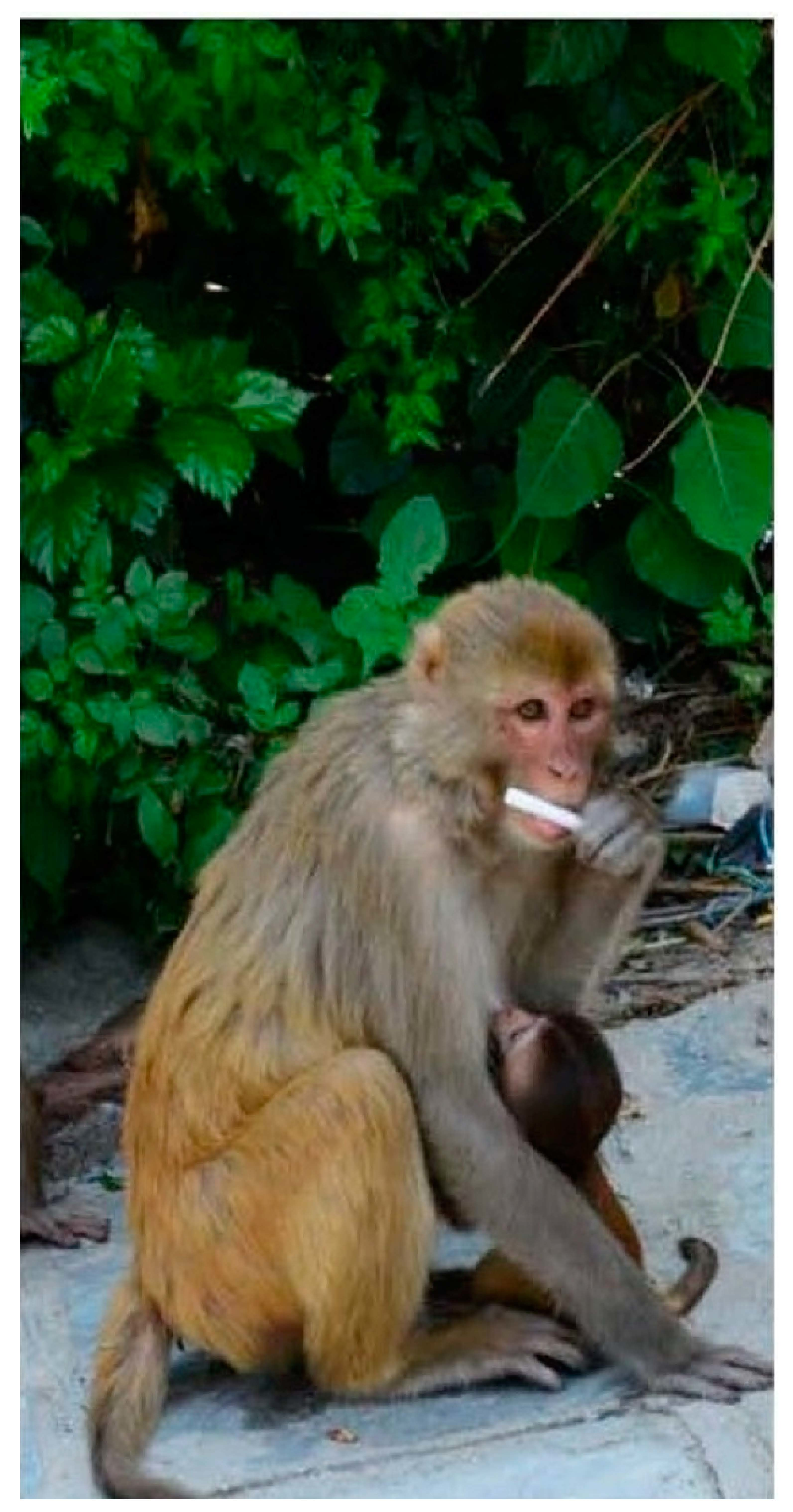
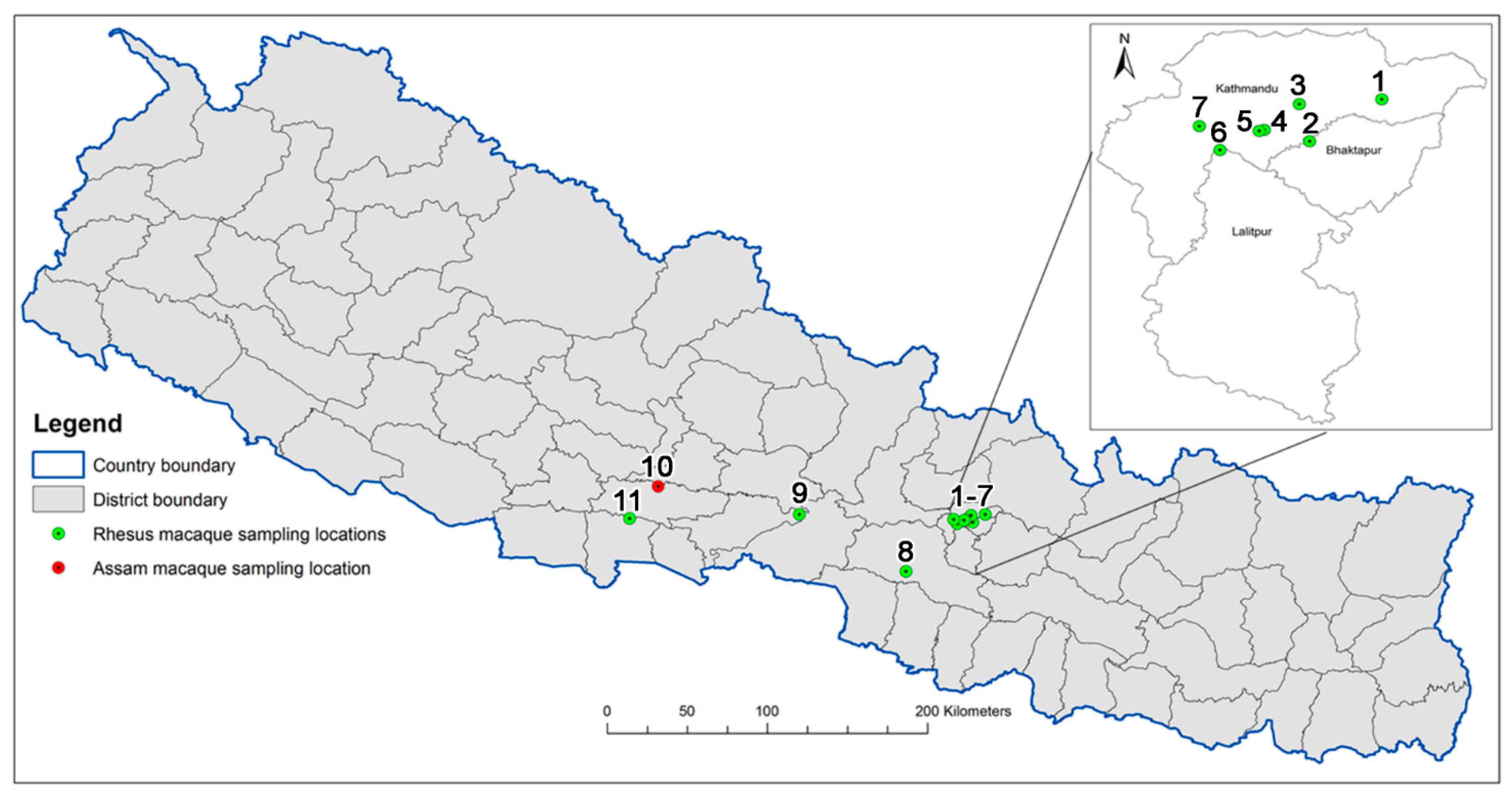
4.1. Primate Sampling

A total of 11 locations were sampled in the current study (Figure 1). The locations, all of which represent religious/temples sites, included Bajrayogini, Nilbarahi, Pashupati, Swayambhu [21], and Thapathali (these five locations were sampled in 2018 [5] and 2019 [4]). Additionally, six new locations were sampled including, Chitwan, Guheswari, Gokarna, Hetauda, Rupandehi, and Ramdi. All of the locations involved the sampling of rhesus macaques with the exception of Ramdi, where we sampled a resident group of Assam macaques consisting of about 32 individuals living around the Ramdi temple (West-Ramdi) [22]. Human dwellings are situated a few hundred meters from the temple. The diet of these macaques consists of food from the forest, but they also are fed fruits and other household scraps by the local people and pilgrims visiting the temple. These macaques are habituated to humans and have been reported to visit and raid human settlements and crop fields in the area [23]. The sampled macaques appeared healthy based on physical appearance and behavior.
The collection technique involved an adaptation of the non-invasive oral sampling method previously described [4,5,14] using SalivaBio Children’s Swabs (Salimetrics LLC, State College, PA, USA) [4,24]. Swabs were soaked in a sterile glucose solution (10% w/v) and thrown to the macaques. A new pair of disposable gloves was used before taking the swab out of the tube and providing it to the monkey. After chewing for several seconds/minutes, the monkey realized the swab was not food and discarded it. The storage tube contained enrichment broth Bacto® m Staphylococcus Broth (Difco Laboratories, Sparks, MD, USA) supplemented with a final concentration of 75 mg/L of polymyxin B, 0.01% potassium tellurite, and either with or without 12.5 mg/L nystatin to prevent fungal growth (Sigma-Aldrich, St Louis, MO, USA). The tubes were returned to the laboratory the same day when possible and incubated at 37 °C until turbid (24–96 h), as previously described [25]. The broth was streaked for isolation onto mannitol salt agar plates (HiMedia Laboratories, Mumbai, India), and yellow colonies were sub-cultured onto blood agar plates (HiMedia Laboratories, Mumbai, India). Colonies that had β−hemolysis were verified as S. aureus as described below. Forty isolates, including 7 MRSA and 33 MSSA, were randomly selected from a total of 97 isolates (85 MSSA and 13 MRSA). One MRSA was selected from each region that had an MRSA isolate. All five of the MSSA from the Assam were included, while from the other regions, 28 randomly selected MSSA were included in the current study.

4.2. Ethical Statement

The research protocol for the sampling of free-ranging primates in Nepal was approved by the Department of Forest and Soil Conservation under the Ministry of Forest and Environment, Government of Nepal (Reference Letter Number: 075/076/663). This research also complied with the animal use protocol for primates (#3143-04) approved by the Institutional Animal Care and Use Committee at the University of Washington, USA, and the American Society of Primatologists (ASP) Principles for the Ethical Treatment of Nonhuman Primates.

4.3. Identification of S. aureus from Primates

Colonies that had β-hemolysis on blood agar plates were verified as S. aureus by Gram stain and with the Staphaurex test as previously described (Thermo Fisher Scientific Remel Products, Lenexa, KS [25]. MRSA isolates were identified by their ability to grow on Mueller–Hinton agar (HiMedia Laboratories, India) supplemented with 4 mg/L of oxacillin (HiMedia Laboratories, India). The MRSA isolates were confirmed using the Thermo Scientific PBP2′ latex agglutination test kit according to the manufacturer’s instructions (Thermo Fisher Scientific Remel Products, Lenexa, KS, USA) [25].

4.4. Antibiotic Susceptibility Testing

The antimicrobial testing was done for 20 antibiotics: benzylpenicillin, oxacillin, cefoxitin, gentamicin, tobramycin, ciprofloxacin, levofloxacin, moxifloxacin, erythromycin, clindamycin, linezolid, teicoplanin, vancomycin, tetracycline, fosfomycin, nitrofurantoin, fusidic acid, mupirocin, rifampicin, and trimethoprim/sulfamethoxazole using automated microdilution, VITEK 2 by Biomerieux according to VITEK 2 by Biomerieux using the manufacturer’s instructions and utilizing European Committee on Antimicrobial Susceptibility Testing EUCAST breakpoints Table 2 [26].

4.5. DNA Microarray Analysis

The StaphyType (Abbott/Alere Technologies, Jena, Germany) DNA microarray-based assay was used to screen for a presence or absence of a multitude of genes, including antibiotic resistance markers, virulence factors, species-specific controls, and typing markers (Table 1, Table 3 and Table 4). This system has previously been used for a variety of studies on MRSA [3,4,11,19,20,27,28,29]. The microarray typing includes 334 target sequences and ≈170 separate genes and allelic variants including species markers, SCCmec, capsule, and agr group typing markers, which commonly carried staphylococcal antibiotic resistance genes, toxins, and microbial surface components recognizing adhesive matrix molecules [MSCRAMM] genes. Isolates were assigned to clonal complexes (CCs) by an automated comparison of the microarray hybridization profiles to a large database of previously characterized isolates [11]. Then, strains were assigned to clonal complexes when possible. However, 30 of the isolates could not be assigned to a clonal complex and were assumed to be novel (Supplementary Table S1). The detailed protocol as well as the sequences of primers and probes have previously been published [15]. A second array was used on representative isolates for the detection of Staphyloxanthin genes [30].

4.6. Multilocus Sequence Analysis

Genomic DNA was isolated from an overnight culture grown at 37 °C on Columbia Blood Agar plates (Becton Dickinson GmbH, Heidelberg, Germany) using a Macherey and Nagel NucleoSpin® Microbial DNA kit (MACHEREY-NAGEL GmbH & Co. KG, Dueren, Germany). The Oxford Nanopore MinION platform was used for sequencing the whole genome of twelve isolates. Briefly, size selection and DNA clean-up were performed using Agencourt AMPure XP beads (Beckman Coulter GmbH, Krefeld, Germany) in a ratio of 1/1 (v/v). The DNA library was generated using the Nanopore native barcoding genomic DNA kit SQK-LSK109 in combination with the native barcoding expansion kit EXP-NBD104 (Oxford Nanopore Technologies, Oxford, UK) according to the manufacturer’s instructions. The used flow cell FLO-MIN106 (revD R9.4.1) was primed by the flow cell priming kit EXP-FLP001 (Oxford Nanopore, Oxford, UK). The protocol named “Native barcoding genomic DNA” was used in version NBE_9065_v109_revV_14Aug2019 (Last update: 21/02/2020).
The Guppy basecaller (version 4.0.14+8d3226e, Oxford Nanopore Technologies, Oxford, UK) translated the MinION raw reads (FAST5) into quality tagged sequence reads (4000 reads per FASTQ-file) using the barcode trimming option. Flye (v2.8-b1674) was used to assemble the quality tagged sequence reads of each strain to one big circular contig. The polishing of assemblies was split into two steps. At first, racon (v1.4.17) was iteratively used four times with the following parameter: match 8; mismatch 6; gap 8; and windows-lengths 500. Afterwards, medaka (v1.0.3) (https://nanoporetech.github.io/medaka/) ran on the last racon polished assembly using the model r941_min_high_g360. Corrected assemblies were used for further MLST analysis by upload to the S. aureus pubMLST database (https://pubmlst.org/bigsdb?db=pubmlst_saureus_seqdef&page=sequenceQuery).

5. Conclusions

The MRSA isolated from both Assam and rhesus macaques were linked to other countries including India and Middle East as the previous study illustrated for MRSA isolated in 2017–2018 [4,5]. The same cannot be said for the majority of the MSSA. Thirty isolates representing 17 distinct lineages did not belong to previously characterized clonal complexes nor did they carry antibiotic resistance genes commonly found in S. aureus. This suggests that these isolates may not commonly circulate in humans and may be adapted for macaques. We did not find comparable MRSA to these novel MSSA, suggesting that the two may use different transmission routes in the primates. Future studies will need to expand the knowledge of MSSA in primates and determine if these isolates can be identified in domestic or other types of wild animals. The number of isolates from Assam macaques that have limited contact with humans suggest that they carry the same type of MRSA and MSSA as the rhesus. Therefore, it will be of interest to determine if both MRSA and MSSA are transmitted from Assam adults to their infants and how contaminated their environment is with MSSA.

Supplementary Materials

The following are available online at https://www.mdpi.com/2079-6382/9/10/689/s1, Table S1: MLST Sequences. Table S2 MICs of the Isolates.

Author Contributions

M.C.R. and R.C.K. coordinated the study and supervised the original field and data collection. P.R.J. coordinated the in-country collection of the primate samples and writing the manuscript. S.P. and M.A. helped with sample collection and processing. S.M., R.E., E.M. and D.G. did data analyses and helped with writing the manuscript along with P.R.J. and L.K., L.K., N.P.K. and M.C. helped with facilitating access to isolates. E.M. and D.G. did array experiments, E.M. did the susceptibility tests, C.D. and S.D.B. did the sequencing. All authors have read and agreed to the published version of the manuscript.

Funding

The project was supported in part by the One Earth Institute and the Office of Research Infrastructure Programs (ORIP) of the National Institutes of Health through Grant Number P51OD010425 to the WaNPRC.

Acknowledgments

We would like to thank the Department of Forest and Soil Conservation, Kathmandu, Nepal for permission to conduct sampling. We also thank Nima Singh who helped with the laboratory preparation of the samples.

Conflicts of Interest

The authors declare they have no conflict of interest.

References

  1. Heaton, C.J.; Gerbig, G.R.; Sensius, L.D.; Patel, V.; Smith, T.C. Staphylococcus aureus epidemiology in wildlife: A systematic review. Antibiotics 2020, 9, 89. [Google Scholar] [CrossRef] [PubMed] [Green Version]
  2. Soge, O.O.; No, D.; Michael, K.; Dakoff, J.; Lane, J.; Vogel, K.; Smedley, J.; Roberts, M.C. Transmission of MDR MRSA between primates personnel and environment at a United States primate center. J. Antimicrob. Chemother. 2016, 71, 2798–2803. [Google Scholar] [CrossRef] [PubMed] [Green Version]
  3. Roberts, M.C.; Feßler, A.T.; Monecke, S.; Ehricht, R.; No, D.; Schwarz, S. Molecular analysis of two different MRSA clones ST188 and ST2368 from primates (Macaca spp.) in a United States primate center. Front. Microbiol. Antimicrob. Resist. Chemother. 2018, 9, 2199. [Google Scholar] [CrossRef]
  4. Roberts, M.C.; Joshi, P.R.; Monecke, S.; Ehricht, R.; Muller, E.; Gawlik, D.; Paudel, S.; Acharya, M.; Bhattarai, S.; Pokharel, S.; et al. MRSA strains among Nepalese Rhesus macaques (Macaca mulatta) and their environment. Front. Microbiol. Antimicrob. Resist. Chemother. 2019, 10, 2505. [Google Scholar] [CrossRef] [Green Version]
  5. Roberts, M.C.; Joshi, P.R.; Greninger, A.L.; Melendez, D.; Paudel, S.; Acharya, M.; Bimali, N.K.; Koju, N.P.; No, D.; Chalise, M.; et al. The human clone ST22 SCCmec IV methicillin-resistant Staphylococcus aureus isolated from swine herds and wild primates in Nepal: Is man the common source? FEMS Microbiol. Ecol. 2018, 1, 94. [Google Scholar] [CrossRef] [PubMed] [Green Version]
  6. Senok, A.; Somily, A.; Raji, A.; Gawlik, D.; Al-Shahrani, F.; Baqi, S.; Boswihi, S.; Skakni, L.; Udo, E.E.; Weber, S.; et al. Diversity of methicillin-resistant Staphylococcus aureus CC22-MRSA-IV from Saudi Arabia and the Gulf region. Int. J. Infect. Dis. 2016, 51, 31–35. [Google Scholar] [CrossRef] [PubMed] [Green Version]
  7. Pokhrel, R.H.; Aung, M.S.; Thapa, B.; Chaudhary, R.; Mishra, S.K.; Kawaguchiya, M.; Urushibara, N.; Kobayashi, N. Detection of ST772 Panton-Valentine leukocidin-positive methicillin-resistant Staphylococcus aureus (Bengal Bay clone) and ST22 S. aureus isolates with a genetic variant of elastin binding protein in Nepal. New Microbes New Infect. 2016, 11, 20–27. [Google Scholar] [CrossRef] [Green Version]
  8. Joshi, P.R.; Acharya, M.; Aryal, R.; Thapa, K.; Kakshapati, T.; Seng, R.; Singh, A.; Sitthisak, S. Emergence of staphylococcal cassette chromosome mec type I with high-level mupirocin resistance among methicillin-resistant Staphylococcus aureus. Asian Pac. J. Trop. Biomed. 2017, 7, 193–197. [Google Scholar] [CrossRef]
  9. Schaumburg, F.; Mugisha, L.; Kappeler, P.; Fichtel, C.; Kock, R.; Kondgen, S.; Becker, K.; Leendertz, F.H.; Peters, G. Evaluation of non-invasive biological samples to monitor Staphylococcus aureus colonization in great apes and lemurs. PLoS ONE 2013, 8, e78046. [Google Scholar] [CrossRef]
  10. Monecke, S.; Baier, V.; Coombs, G.W.; Slickers, P.; Ziegler, A.; Ehricht, R. Genome sequencing and molecular characterisation of Staphylococcus aureus ST772-MRSA-V, “Bengal Bay Clone”. BMC Res. Notes 2013, 6, 548. [Google Scholar] [CrossRef] [Green Version]
  11. Steinig, E.J.; Duchene, S.; Robinson, D.A.; Monecke, S.; Yokoyama, M.; Slickers, P.; Andersson, P.; Williamson, D.; Kearns, A.; Goering, R.V.; et al. Evolution and Global Transmission of a Multidrug-Resistant, Community-Associated Methicillin-Resistant Staphylococcus aureus Lineage from the Indian Subcontinent. mBiotechnology 2019, 10, e01105-19. [Google Scholar] [CrossRef] [PubMed] [Green Version]
  12. Prabhakara, S.; Khedkar, S.; Loganathan, R.M.; Chandana, S.; Malali, G.; Arakere, G.; Seshasayee, A.S.N. Draft genome sequence of Staphylococcus aureus 118 (ST772), a major disease clone from India. J. Bacteriol. 2012, 194, 3727–3728. [Google Scholar] [CrossRef] [PubMed] [Green Version]
  13. Monecke, S.; Luedicke, C.; Slickers, P.; Ehricht, R. Molecular epidemiology of Staphylococcus aureus in asymptomatic carriers. Eur. J. Clin. Microbiol. Infect. Dis. 2009, 28, 1159–1165. [Google Scholar] [CrossRef] [PubMed]
  14. Labor Report. Labor Migration in Nepal. Available online: https://www.ilo.org/kathmandu/areasofwork/labour-migration/lang--en/index.htm (accessed on 1 September 2020).
  15. Monecke, S.; Coombs, G.; Shore, A.C.; Coleman, D.C.; Akpaka, P.; Borg, M.; Chow, H.; Ip, M.; Jatzwauk, L.; Jonas, D.; et al. A field guide to pandemic, epidemic and sporadic clones of methicillin-resistant Staphylococcus aureus. PLoS ONE 2011, 6, e17936. [Google Scholar] [CrossRef]
  16. Azis, N.M.; Pung, H.P.; Bachman, A.R.A.; Nordin, S.A.; Sarchio, S.N.E.; Suhaili, Z.; Desa, M.N.M. A persistant antimicrobial resistance pattern and limited methicillin-resistance-associated genotype in a short-term Staphylococcus aureus carriage isolated from a student population. J. Infect. Publ. Health 2017, 10, 156–164. [Google Scholar] [CrossRef] [Green Version]
  17. Penades, M.; Viana, D.; Garcia-Quiros, A.; Munoz-Siverstre, A.; Moreno-Grua, E.; Perez-Fuentes, S.; Pascual, J.; Corpa, J.M.; Selva, L. Differences in virulence between the two more prevalent Staphylococcus aureus clonal complexes in rabbitries (CC121 and CC96) using an experimental model of mammary gland infection. Vet. Res. 2020, 51, 11. [Google Scholar] [CrossRef] [Green Version]
  18. Harrison, E.M.; Paterson, G.K.; Holden, M.T.G.; Larsen, J.; Stegger, M.; Larsen, A.R.; Petersen, A.; Skov, R.L.; Christensen, J.M.; Zeuthen, A.B.; et al. Whole genome sequencing identifies zoonotic transmission of MRSA isolates with the novel mecA homologue mecC. EMBO Mol. Med. 2013, 5, 509–515. [Google Scholar] [CrossRef]
  19. Monecke, S.; Gavier-Widen, D.; Mattsson, R.; Rangstrup-Christensen, L.; Lazaris, A.; Coleman, D.C.; Shore, A.C.; Ehricht, R. Detection of mecC-positive Staphylococcus aureus (CC130-MRSA-XI) in diseased european hedgehogs (Erinaceus europaeus) in Sweden. PLoS ONE 2013, 8, e66166. [Google Scholar] [CrossRef] [Green Version]
  20. Loffler, B.; Hussain, M.; Grundmeier, M.; Bruck, M.; Holzinger, D.; Varga, G.; Roth, J.; Kah, B.C.; Proctor, R.A.; Peters, G. Staphylococcus aureus panton-valentine leukocidin is a very potent cytotoxic factor for human neutrophils. PLoS Pathog. 2010, 6, e1000715. [Google Scholar] [CrossRef]
  21. Kyes, R.C.; Jones-Engel, L.; Chalise, M.K.; Engel, G.; Heidrich, J.; Grant, R.; Bajimaya, S.S.; McDonough, J.; Smith, D.G.; Ferguson, B. Genetic characterization of rhesus macaques (Macaca mulatta) in Nepal. Am. J. Primatol. 2006, 68, 445–455. [Google Scholar] [CrossRef]
  22. Khanal, L.; Chalise, M.K.; He, K.; Acharya, B.K.; Kawamoto, Y.; Jiang, X. Mitochondrial DNA analyses and ecological niche modeling reveal post-LGM expansion of the Assam macaque (Macaca assamensis) in the foothills of Nepal Himalaya. Am. J. Primatol. 2018, 80, e22748. [Google Scholar] [CrossRef] [PubMed]
  23. Adhikari, K.P.; Khanal, L.; Chalise, M.K. Status and effects of food provisioning on ecology of Assamese monkey (Macaca assamensis) in Ramdi area of Palpa, Nepal. J. Inst. Sci. Technol. 2018, 22, 183–190. [Google Scholar] [CrossRef]
  24. Evans, T.S.; Barry, P.A.; Gilardi, K.V.; Goldstein, T.; Deere, J.D.; Fike, J.; Yee, J.-A.; Ssebide, B.J.; Karmacharya, D.; Cranfield, M.R.; et al. Optimization of a novel non-invasive oral sampling technique for zoonotic pathogen surveillance in nonhuman primates. PLoS Negl. Trop. Dis. 2015, 9, e0003813. [Google Scholar] [CrossRef]
  25. Roberts, M.C.; Soge, O.O.; Horst, J.A.; Ly, K.A.; Milgrom, P. Methicillin-resistant Staphylococcus aureus from dental school clinic surfaces and students. Am. J. Infect. Control. 2011, 39, 628–632. [Google Scholar] [CrossRef]
  26. ISO 20776-1 (2006). Clinical Laboratory Testing and In Vitro Diagnostic Test. Systems—Susceptibility Testing of Infectious Agents and Evaluation of Performance of Antimicrobial Susceptibility Test. Devices—Part. 1: Reference Method for Testing the In Vitro Activity of Antimicrobial Agents against Rapidly Growing Aerobic Bacteria Involved in Infectious Diseases. 2006. Available online: http://www.eucast.org/ast_of_bacteria/mic_determination/ (accessed on 1 September 2020).
  27. Kinneyvey, P.M.; Shore, A.C.; Brennan, G.I.; Sullivan, D.J.; Ehricht, R.; Monecke, S.; Slickers, P.; Coleman, D.C. Emergence of sequence type 779 methicillin-resistant Staphylococcus aureus harboring a novel pseudo staphylococcal cassette chromosome mec (SCCmec)-SCC-SCCCRISPR composite element in Irish hospitals. Antimicrob. Agents Chemother. 2012, 57, 524–531. [Google Scholar] [CrossRef] [Green Version]
  28. Monecke, S.; Jatzwauk, L.; Muller, E.; Nitschke, H.; Pfohl, K.; Slickers, P.; Reissig, A.; Ruppelt-Lorz, A.; Ehricht, R. Diversity of SCCmec elements in Staphylococcus aureus as observed in South-Eastern Germany. PLoS ONE 2016, 11, e0162654. [Google Scholar] [CrossRef]
  29. Monecke, S.; Slickers, P.; Gawilik, D.; Muller, E.; Reissig, A.; Ruppelt-Lorz, A.; Akpaka, P.E.; Bandt, D.; Bes, M.; Boswihi, S.S.; et al. Molecular typing of ST239-MRSA-III from diverse geographic locations and the evolution its SCCmec III element during its intercontinental spread. Front. Microbiol. 2018, 9, 1436. [Google Scholar] [CrossRef] [Green Version]
  30. Monecke, S.; Jatzwauk, L.; Weber, S.; Slickers, P.; Ehricht, R. DNA microarray-based genotyping of methicillin-resistant Staphylococcus aureus strains from eastern saxony. Clin. Microbiol. Infect. 2009, 14, 534–545. [Google Scholar] [CrossRef] [Green Version]
Figure 1. Primate Sampling Locations. Saliva Sampling was conducted at 11 locations in Nepal: 1-Bajrayogini; 2-Nilbarahi; 3-Gokarna; 4-Guheswari; 5-Pashupati; 6-Thapathali; 7-Swayambhu; 8-Hetauda; 9-Chitwan; 10-Ramdi; 11-Rupandehi.
Antibiotics 09 00689 g001
Table 1. Methicillin-resistant S. aureus (MRSA) strains, sample types, and sampling locations as well as species markers, agr groups, and capsule types.
Clonal ComplexStrain Isolate IDNumberSampling LocationHost SpeciesgapA, katA, coA, nuc1, spa, sbi, eno, clfA+B, fnbAStaphyloxanthin Operonagr GroupCapsule Type
CC06CC6-MRSA-IVB1NilbarahiRhesusPOSnot testedI8
CC22CC22-MRSA-IV (PVL+/tst+)93, 94, 05, 97, C52 Gokarna, 1 each Chitwan, Ramdi and Nilbarahi1 Assam,
4 Rhesus
POSnot testedI5
CC772CC772-MRSA-V (PVL+), “Bengal Bay”911HetaudaRhesusPOSnot testedII5
CC15CC15-MSSA831RupandehiRhesusPOSnot testedII8
CC96CC96-MSSA891RamdiAssam POSnot testedIII8
CC2990CC2990-MSSAH1GuheswariRhesusPOSPOSII8
UNKNOWN 01ST(4-13-1-105-11-5-x)-MSSA86, 872RamdiAssam POSPOSI5
UNKNOWN 02ST(1-421-1-1-12-238-11)-MSSA52, 532GokarnaRhesusPOSPOSI5
UNKNOWN 03ST(1-3-1-15-28-x-1)-MSSA31, 32, 71, D42 Nilbarahi, 1 each Chitwan and GokarnaRhesusPOSPOSI8
UNKNOWN 04MSSA011PashupatiRhesusPOSPOSI8
UNKNOWN 05ST(1-1-1-1-28-4-11)-MSSA73, F21 Chitwan,
1 Gokarna
RhesusPOSPOSI8
UNKNOWN 06ST(1-421-1-x-x-1-11)-MSSA541GorkarnaRhesusPOSPOSI8
UNKNOWN 07ST(12-x-1-66-11-x-x)-MSSA29, 30, 5132 Swayambhu,
1 Gokarna
RhesusPOSPOSII8
UNKNOWN 08ST(1-38-1-1-x-238-x)-MSSA81, 8821 Ramdi,
1 Rupandehi
1 Assam,
1 Rhesus
POSPOSII8
UNKNOWN 09ST(3-3-2-66-28-x-x)-MSSA101PashupatiRhesusPOSPOSII8
UNKNOWN 10ST(3-1-1-66-28-1-x)-MSSA82, 9021 Ramdi,
1 Rupandehi
1 Assam,
1 Rhesus
POSPOSIII5
UNKNOWN 11MSSA501BajrayoginiRhesusPOSPOSIII8
UNKNOWN 12ST(4-421-1-105-1-5-x)-MSSA611HetaudaRhesusPOSPOSIV5
UNKNOWN 13ST(1-3-1-x-1-1-11)-MSSA62, 7221 Chitwan,
1 Hetauda
RhesusPOSPOSIV5
UNKNOWN 14ST(3-38-1-15-1-x-40)-MSSAG1GuheswariRhesusPOSPOSIV5
UNKNOWN 15ST(1-421-1-1-12-1-11)-MSSA11, E21 Gokarna,
1 Thapathali
RhesusPOSPOSIV5
UNKNOWN 16ST(3-3-1-66-4-x-x)-MSSA 49, I21 Bajrayogini,
1 Guheswari
RhesusPOSPOSIV8
UNKNOWN 17MSSA131ThapathaliRhesusPOSPOSIV8
Table 2. Susceptibility tests.
Clonal ComplexStrain NumberPENOXAFOXGENTOBCIPLEVMOXERYCLILINTEIVANTETFOSNIFFUSMUPRIFTSU
CC06CC6-MRSA-IV1RRRSSSSSRR aSSSSSSSSSS
CC22CC22-MRSA-IV (PVL+/tst+)5RRRRRRRRVAR bVAR abSSSSSSSSSVAR c
CC772CC772-MRSA-V (PVL+), “Bengal Bay”1RRRRRRRRISSSSSSISSSR
CC15CC15-MSSA1RSSSSRRRRR aSSSSSSSSSS
CC96CC96-MSSA1SSSSSSSSRSSSSSSSSSSS
CC2990CC2990-MSSA1SSSSSSSSSSSSSSSSSSSS
UNKNOWN 01ST(4-13-1-105-11-5-x)-MSSA2SSSSSSSSSSSSSSSISSSS
UNKNOWN 02ST(1-421-1-1-12-238-11)-MSSA2SSSSSSSSSSSSSSSSSSSS
UNKNOWN 03ST(1-3-1-15-28-x-1)-MSSA4SSSSSSSSSSSSSSSSSSSS
UNKNOWN 04MSSA1SSSSSSSSSSSSSSSSSSSS
UNKNOWN 05ST(1-1-1-1-28-4-11)-MSSA2SSSSSSSSSSSSSSSSSSSS
UNKNOWN 06ST(1-421-1-x-x-1-11)-MSSA1SSSSSSSSSSSSSSSSSSSS
UNKNOWN 07ST(12-x-1-66-11-x-x)-MSSA3SSSSSSSSSSSSSSSSSSSS
UNKNOWN 08ST(1-38-1-1-x-238-x)-MSSA2SSSSSSSSSSSSSSSISSSS
UNKNOWN 09ST(3-3-1-66-28-x-x)-MSSA1SSSSSSSSSSSSSSSSSSSS
UNKNOWN 10ST(3-1-1-66-28-1-x)-MSSA2SSSSSSSSSSSSSSSSSSSS
UNKNOWN 11MSSA1SSSSSSSSSSSSSSSSSSSS
UNKNOWN 12ST(4-421-1-105-1-5-x)-MSSA1SSSSSSSSSSSSSSSSSSSS
UNKNOWN 13ST(1-3-1-x-1-1-11)-MSSA2SSSSSSSSSSSSSSSISSSS
UNKNOWN 14ST(3-38-1-15-1-x-40)-MSSA1SSSSSSSSSSSSSSSSSSSS
UNKNOWN 15ST(1-421-1-1-12-1-11)-MSSA2SSSSSSSSSSSSSSSSSSSS
UNKNOWN 16ST(3-3-1-66-4-1-x)-MSSA2SSSSSSSSSSSSSSSSSSSS
UNKNOWN 17MSSA1SSSSSSSSSSSSSSSSSSSS
PEN, Benzylpenicillin; OXA, Oxacillin; FOX, Cefoxitin; GEN, Gentamicin; TOB, Tobramycin; CIP, Ciprofloxacin; LEV, Levofloxacin; MOX, Moxifloxacin; ERY, Erythromycin; CLI, Clindamycin; LIN, Linezolid; TEI, Teicoplanin; VAN, Vancomycin; TET, Tetracycline; FOS, Fosfomycin; NIF, Nitrofurantoin; FUS, Fusidic acid; MUP, Mupirocin; RIF, Rifampicin and TSU, Trimethoprim/Sulfamethoxazole a Inducible clindamycin resistance. b Variable, four out of five isolates resistant (ERY)/inducibly resistant (CLI); one susceptible. c Variable, three out of five isolates resistant, two susceptible. The actual MICs are found in Supplementary Table S2.
Table 3. SCCmec markers and resistance genes a.
Clonal ComplexStrain NumbermecADelta mecRccrA-2ccrB-2ccrAAccrCblaZ/I/Rerm(C)lnu(A)msr(A)mph(C)aacA-aphDaphA3+satdfrA
CC06CC6-MRSA-IV1POSPOSPOSPOSNEGNEGNEGPOSNEGNEGNEGNEGNEGNEG
CC22CC22-MRSA-IV (PVL+/tst+)5POSPOSPOSPOSNEGNEGPOSVAR bNEGNEGNEGPOSNEGPOS
CC772CC772-MRSA-V(PVL+), “Bengal Bay “1POSNEGNEGNEGPOSPOSPOSNEGNEGPOSPOSPOSPOSNEG
CC15CC15-MSSA1NEGNEGNEGNEGNEGNEGPOSPOSNEGNEGNEGNEGNEGNEG
CC96CC96-MSSA1NEGNEGNEGNEGNEGNEGNEGNEGNEGPOSNEGNEGNEGNEG
CC2990CC2990-MSSA1NEGNEGNEGNEGNEGNEGNEGNEGNEGNEGNEGNEGNEGNEG
UNKNOWN 01ST(4-13-1-105-11-5-x)-MSSA2NEGNEGNEGNEGNEGNEGNEGNEGNEGNEGNEGNEGNEGNEG
UNKNOWN 02ST(1-421-1-1-12-238-11)-MSSA2NEGNEGNEGNEGNEGNEGNEGNEGNEGNEGNEGNEGNEGNEG
UNKNOWN 03ST(1-3-1-15-28-x-1)-MSSA4NEGNEGNEGNEGNEGNEGNEGNEGNEGNEGNEGNEGNEGNEG
UNKNOWN 04MSSA1NEGNEGNEGNEGNEGNEGNEGNEGNEGNEGNEGNEGNEGNEG
UNKNOWN 05ST(1-1-1-1-28-4-11)-MSSA2NEGNEGNEGNEGNEGNEGNEGNEGNEGNEGNEGNEGNEGNEG
UNKNOWN 06ST(1-421-1-x-x-1-11)-MSSA1NEGNEGNEGNEGNEGNEGNEGNEGNEGNEGNEGNEGNEGNEG
UNKNOWN 07ST(12-x-1-66-11-x-x)-MSSA3NEGNEGNEGNEGNEGNEGNEGNEGNEGNEGNEGNEGNEGNEG
UNKNOWN 08ST(1-38-1-1-x-238-x)-MSSA2NEGNEGNEGNEGNEGNEGNEGNEGNEGNEGNEGNEGNEGNEG
UNKNOWN 09MSSA1NEGNEGNEGNEGNEGNEGNEGNEGNEGNEGNEGNEGNEGNEG
UNKNOWN 10ST(3-3-1-66-28-x-x)-MSSA2NEGNEGNEGNEGNEGNEGNEGNEGNEGNEGNEGNEGNEGNEG
UNKNOWN 11MSSA1NEGNEGNEGNEGNEGNEGNEGNEGNEGNEGNEGNEGNEGNEG
UNKNOWN 12ST(4-421-1-105-1-5-x)-MSSA1NEGNEGNEGNEGNEGNEGNEGNEGNEGNEGNEGNEGNEGNEG
UNKNOWN 13ST(1-3-1-x-1-1-11)-MSSA2NEGNEGNEGNEGNEGNEGNEGNEGNEGNEGNEGNEGNEGNEG
UNKNOWN 14ST(3-38-1-15-1-x-40)-MSSA1NEGNEGNEGNEGNEGNEGNEGNEGNEGNEGNEGNEGNEGNEG
UNKNOWN 15ST(1-421-1-1-12-1-11)-MSSA2NEGNEGNEGNEGNEGNEGNEGNEGNEGNEGNEGNEGNEGNEG
UNKNOWN 16ST(3-3-1-66-4-1-x)-MSSA2NEGNEGNEGNEGNEGNEGNEGNEGNEGNEGNEGNEGNEGNEG
UNKNOWN 17MSSA1NEGNEGNEGNEGNEGNEGNEGNEGNEGNEGNEGNEGNEGNEG
a The table shows only genes that were found at least once in this study. Genes that were not present in any of the study strains are other ccrA/B genes, mecR, mecI, kdp-operon genes, heavy metal resistance genes, mecC, blaZ SCCmec XI, erm(A), erm(B), lnu(A), mef(A), vat(A), vat(B), vga(A), vga(A), vgb(A), far1, fusC, mupR, tet(K), tet(M), cat, cfr, fexA, qacA, qacC, vanA, vanB, and vanZ. b Variable, present in four out of five isolates.
Table 4. Virulence-associated genes a.
Clonal ComplexStrain Numbertstseasec+selegc GenesORF CM14PVLlukD/E bsakchpscnedinB+etD2cnasasG
CC06CC6-MRSA-IV1NEGNEGNEGNEGNEGNEGPOSPOSNEGPOSNEGPOSPOS
CC22CC22-MRSA-IV (PVL+/tst+)5POSNEGPOSPOSNEGPOSNEGPOSPOSPOSNEGPOSPOS
CC772CC772-MRSA-V (PVL+), “Bengal Bay “1NEGPOSPOSPOSPOSPOSNEGNEGNEGPOSNEGPOSPOS
CC15CC15-MSSA1NEGNEGNEGNEGNEGNEGPOSNEGPOSPOSNEGNEGPOS
CC96CC96-MSSA1NEGNEGNEGNEGNEGNEGPOSNEGNEGNEGNEGPOSPOS
CC2990CC2990-MSSA1NEGNEGPOSNEGNEGNEG(POS) bPOSPOSPOSPOSPOSPOS
UNKNOWN 01ST(4-13-1-105-11-5-x)-MSSA2NEGNEGNEGNEGNEGNEGPOSNEGNEGNEGPOSNEGPOS
UNKNOWN 02ST(1-421-1-1-12-238-11)-MSSA2NEGNEGNEGPOSNEGNEGPOSNEGNEGNEGNEGPOSPOS
UNKNOWN 03ST(1-3-1-15-28-x-1)-MSSA4NEGNEGNEGNEGNEGNEGPOSNEGNEGNEGNEGPOSNEG
UNKNOWN 04MSSA1NEGNEGNEGNEGNEGNEG(POS) bNEGNEGNEGNEGNEGNEG
UNKNOWN 05ST(1-1-1-1-28-4-11)-MSSA2NEGNEGNEGNEGNEGNEGPOSNEGNEGNEGPOSPOSPOS
UNKNOWN 06ST(1-421-1-x-x-1-11)-MSSA1NEGNEGNEGPOSNEGNEGPOSNEGNEGNEGNEGPOSNEG
UNKNOWN 07ST(12-x-1-66-11-x-x)-MSSA3NEGNEGNEGNEGNEGNEGPOSNEGNEGNEGNEGNEGNEG
UNKNOWN 08ST(1-38-1-1-x-238-x)-MSSA2NEGNEGNEGNEGNEGNEGPOSNEGNEGNEGNEGPOSNEG
UNKNOWN 09ST(3-3-1-66-28-x-x)-MSSA1NEGNEGNEGNEGNEGNEG(POS) bNEGNEGNEGNEGNEGNEG
UNKNOWN 10ST(3-1-1-66-28-1-x)-MSSA2NEGNEGNEGNEGNEGNEGPOSNEGNEGNEGNEGNEGPOS
UNKNOWN 11MSSA1NEGNEGNEGNEGNEGNEGPOSNEGNEGNEGNEGNEGPOS
UNKNOWN 12ST(4-421-1-105-1-5-x)-MSSA1NEGNEGNEGNEGNEGNEGPOSNEGNEGNEGNEGNEGPOS
UNKNOWN 13ST(1-3-1-x-1-1-11)-MSSA2NEGNEGNEGNEGNEGNEGPOSNEGNEGNEGPOSNEGVAR c
UNKNOWN 14ST(3-38-1-15-1-x-40)-MSSA1NEGNEGNEGNEGNEGNEGPOSNEGNEGNEGPOSPOSPOS
UNKNOWN 15ST(1-421-1-1-12-1-11)-MSSA2NEGNEGNEGPOSNEGNEGPOSNEGNEGNEGNEGNEGPOS
UNKNOWN 16ST(3-3-1-66-4-1-x)-MSSA2NEGNEGNEGNEGNEGNEGPOSNEGNEGNEGNEGNEGNEG
UNKNOWN 17MSSA1NEGNEGNEGPOSNEGNEGPOSNEGNEGNEGNEGPOSNEG
a Genes that were not present in any of the study strains are seb, sed, sej, ser, seh, see, lukF-P83+lukM, edinA, edinC, eta, etb, and etD. Genes present in all strains were hla, hld, lukF/S-hlg, lukX/Y, aur, and icaA+C+D. See Supplemental File for more details. b (POS) indicates that lukD was detected by array, while the lukE probe yielded no signals. This is more likely due to allelic variation rather than due to true absence. c Variable, present in one out of two isolates.

