Article

Differential Pathogenic Gene Expression of E. histolytica in Patients with Different Clinical Forms of Amoebiasis

by
Enrique González-Rivas
,
Miriam Nieves-Ramírez
,
Ulises Magaña
,
Patricia Morán
,
Liliana Rojas-Velázquez
,
Eric Hernández
,
Angélica Serrano-Vázquez
,
Oswaldo Partida
,
Horacio Pérez-Juárez
and
Cecilia Ximénez
*
Laboratorio de Inmunología, Unidad de Investigación en Medicina Experimental, Facultad de Medicina, UNAM, México City 04510, Mexico
*
Author to whom correspondence should be addressed.
Microorganisms 2020, 8(10), 1556; https://doi.org/10.3390/microorganisms8101556
Submission received: 28 August 2020 / Revised: 30 September 2020 / Accepted: 2 October 2020 / Published: 9 October 2020
(This article belongs to the Special Issue Virulence and Parasitism of Parasitic Protozoa)

Abstract

:
The etiological agent of human amoebiasis is the protozoan parasite E. histolytica; the disease is still an endemic infection in some countries and the outcome of infection in the host infection can range from asymptomatic intestinal infection to intestinal or liver invasive forms of the disease. The invasive character of this parasite is multifactorial and mainly due to the differential expression of multiple pathogenic genes. The aim of the present work was to measure the differential expression of some genes in different specimens of patients with amoebic liver abscess (ALA) and specimens of genital amoebiasis (AG) by RT-qPCR. Results show that the expression of genes is different in both types of samples. Almost all studied genes were over expressed in both sets of patients; however, superoxide dismutase (Ehsod), serine threonine isoleucine rich protein (Ehstirp), peroxiredoxin (Ehprd) and heat shock protein 70 and 90 (Ehhsp-70, EHhsp-90) were higher in AG biopsies tissue. Furthermore, cysteine proteinases 5 and 2 (Ehcp5, Ehcp2), lectin (Ehgal/galnaclectin) and calreticulin (Ehcrt) genes directly associate with pathogenic mechanisms of E. histolytica had similar over expression in both AG and ALA samples. In summary the results obtained show that trophozoites can regulate the expression of their genes depending on stimuli or environmental conditions, in order to regulate their pathogenicity and ensure their survival in the host.

1. Introduction

Entamoeba histolytica (Eh) is a protozoan parasite responsible for gastrointestinal amoebiasis in the human host. The host–parasite relationship in this infection can be very diverse and lead to a commensal relationship between the parasite and the host, as in the case of asymptomatic cyst carriers or a relationship in which the parasite causes different degrees of tissue damage that can lead to the invasion of extra intestinal organs [1,2,3].
E. histolytica infection is mainly acquired by accidental ingestion of mature cysts present in contaminated water and food (intake of raw and poorly washed vegetables or fruits). It can be transmitted from person to person via the fecal–oral route, contact with hands, objects or surfaces contaminated with infected feces, the cysts of the infected person can remain viable for about 10 to 45 min under the nails [2,3,4,5]; in some cases, it can also be sexually transmitted [6,7].
Amoebiasis is usually a relatively frequent intestinal infection in young adults, in most cases it is asymptomatic because trophozoites remain confined to the intestinal lumen (non-invasive luminal amoebiasis). In the event of symptoms, these appear after an incubation period of two to four weeks and are due to trophozoites invading the intestinal mucosa (invasive form) giving rise to the well-known acute invasive colitis or amoebic dysentery, which consists of diarrhea with mucus and/or blood.
In some patients, such as infants, fever may occur [2]. Amoebic colitis in adults is frequently self-limiting and symptoms disappear within 4 to 7 days, certain individuals may develop chronic luminal amoebiasis that persists for a few months and resolves spontaneously. However, in other patients, the infection may progress to invasive colitis with the appearance of ulcers in the intestinal mucosa that may involve the muscularis mucosa and puncture the intestine, the invasion of deep layers of intestinal tissue can lead to the export of amoebic trophozoites to Portal circulation and their implantation in liver tissue with the consequent development of one or multiple amoebic liver abscesses. Very rarely, the brain, pleura, pericardium, genital-urinary tract or skin are also invaded, symptoms vary depending on the affected organ [2,3,4,5]. In the past century, cases of cutaneous amoebiasis in Mexico were not rare, in particular due to the extension of amoebic liver abscess to the abdominal wall, or the presence of intestinal fistulas in cases of intestinal invasive amoebiasis that can reach the abdominal wall [6]. Genital amoebiasis was frequently observed in infants using diapers with dysentery, causing serious damages to the genital organs [7]. More recently the genital cutaneous amoebiasis is detected as a result of unsafe sexual practice, initial lesions might be red and round that rapidly grow giving rise to an ulcer with red gross edges. These lesions evolve rapidly causing inflammation, necrosis and serious damages to the structure of the genital area [8,9].
Multiple mechanisms contribute to the ability of E. histolytica to destroy the host intestinal mucosa and cause disease [10]. Following excystation within the small intestinal lumen, trophozoites attach to host mucous and epithelial cells in the colon, largely through the multisubunit amebic GalNAc lectin [11,12]. Trophozoites also secrete multiple cysteine proteases, which degrade mucin and the extracellular matrix [13,14,15], they kill resident host cells through a contact-dependent process that remains poorly understood [16,17,18]. Host cells appear to suffer from a disruption of the cell membrane with rapid changes in intracellular calcium levels [19]. This leads to changes in the host cell that closely resemble apoptosis with membrane blebbing, DNA digestion, and activation of caspase [18,20,21]. Finally, E. histolytica trophozoites phagocytize red blood cells and nucleated host cells, the ability to phagocytize host cells are strongly associated with virulence [20,21,22,23].
In the present work we analyze the changes in the expression of pathogenicity associated genes of E. histolytica; cysteine proteinases 5 and 2 (Ehcp5, Ehcp2), lectin (Ehgal/galnaclectin), calreticulin (Ehcrt), superoxide dismutase (Ehsod), serine threonine isoleucine rich protein (Ehstirp), peroxiredoxin (Ehprd), heat shock protein 70 and 90 (Ehhsp-70, EHhsp-90) in genital cutaneous amoebiasis and amoebic liver abscess human specimens, taking into account that the type of lesions are different both in their tissue structure and in regeneration capacity.

2. Materials and Methods

2.1. Obtention of Samples

The present work was designed according to the guidelines for the management of human samples for experimental purposes as indicated in the Official Regulation NOM-12SSA3-2007 included in the General Health Law of Mexican Health Ministry. In addition, the project was approved by the Scientific and Ethics Committee of Faculty of Medicine from National Autonomous University of Mexico (project approval number 091/2013).
Samples were obtained from patients admitted in the services of Internal Medicine, Gastroenterology and Infectious Diseases, at the General Hospital of Mexico in Mexico City. The samples then taken for detection of the parasite by serology or PCR at the Immunology laboratory in the Unit of Research in Experimental Medicine, Faculty of Medicine, UNAM.
Patients with clinical diagnosis of amoebic liver abscess (ALA) or genital cutaneous amoebiasis (GA) were included in the study after the signing of a letter of informed consent. Samples of feces and blood were obtained from the two groups of patients; in cases of amoebic liver abscess, a sample of abscess drainage material was also included; in genital cutaneous amoebiasis patients, a biopsy of the lesion was also studied.
The presence of E. histolytica was directly determined by microscopic analysis in the feces, imprint of damage tissue and molecularly in the biopsies specimens or liver abscess material using PCR technique. In both groups, serological analysis for anti-amoebic antibodies detection were performed. A total of 15 patients with amoebic liver abscess diagnosis and 5 with genital cutaneous amoebiasis were studied.

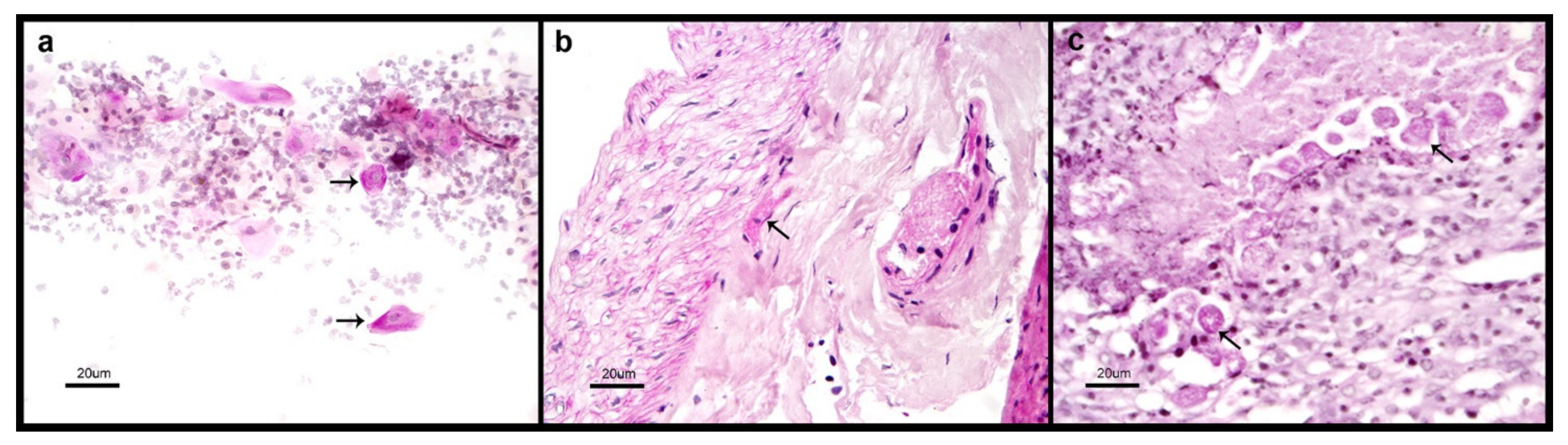
2.2. Microscopic Detection of Entamoeba Trophozoites

Biopsy specimens from patients with genital cutaneous amoebiasis were obtained from ulcer borders located on the penis and scrotum and fixed in (10%) paraformaldehyde for 24 h. The tissue was then segmented, dehydrated, and embedded in paraffin. The tissues (4–5 µm) were cut with a microtome Leica RM 2145 (Buffalo Grove, IL), mounted on slides, and stained with periodic acid-Schiff (PAS) technique [24]. The slides were observed at 10+ and 40+ magnifications with a light microscope. In case of amoebic abscess material, a smear was performed directly over the slide for staining with PAS.

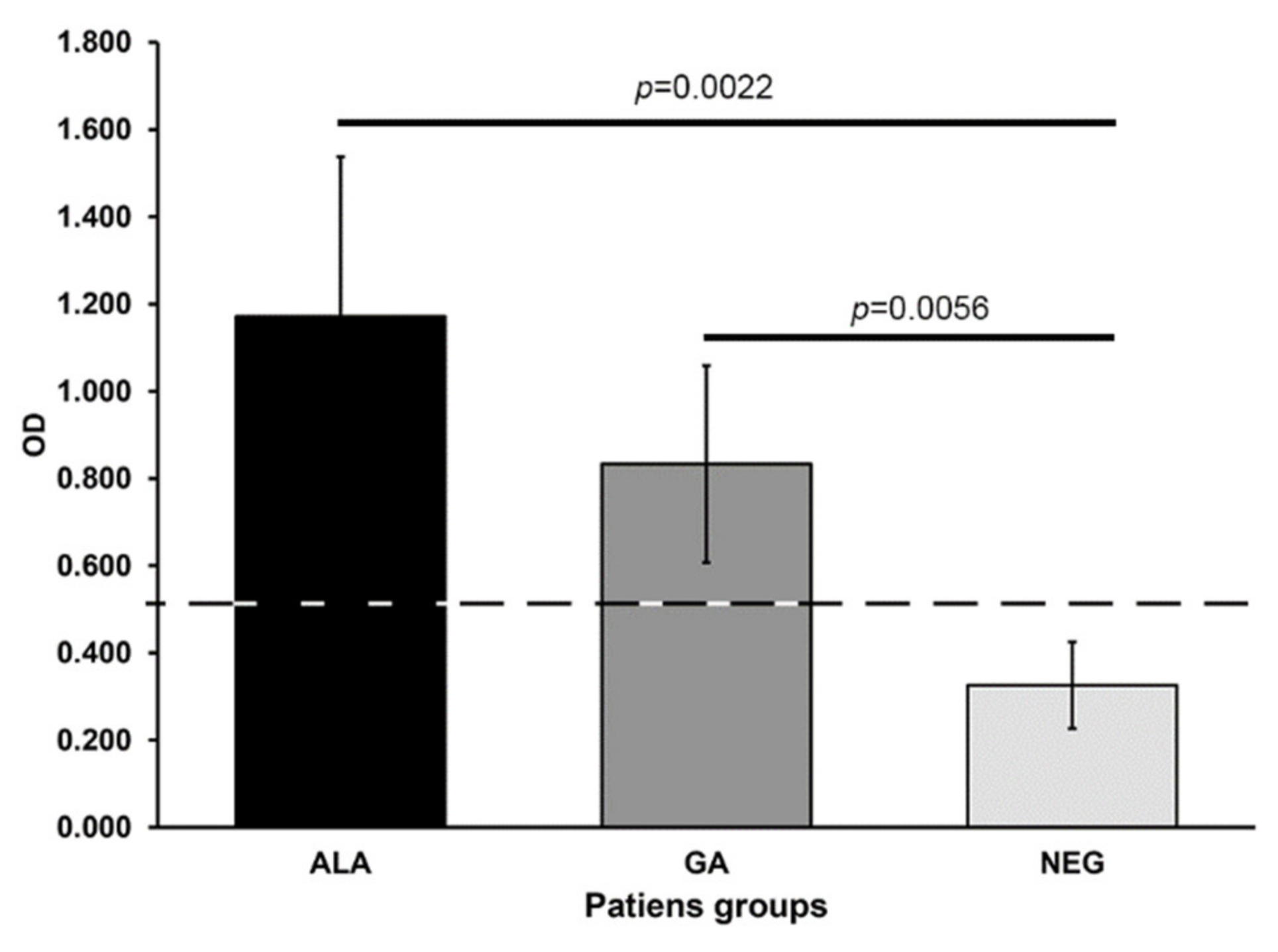
2.3. Anti-Amebic Antibody Detection

Anti-amebic antibody detection was performed in serum samples following the ELISA technique as previously described [25]. For the detection of anti-amebic IgG Serum, samples were diluted 1:1000. The antigen used in ELISA was a membrane-enriched extract (1 µg/well) obtained from axenically cultured E. histolytica HM1:IMSS as previously described [25]. Afterwards, 50 µL/well of serum dilution was added to each microtiter plate, blocking previously the free antigen spaces with 3% PBS-BSA and subsequently incubated for 2 h at room temperature. Plates were then washed once with PBS-BSA, added to 0.5% Tween-20 (Sigma Chemical Co.) (PBS-BSA-Tw), and twice with PBS-Tw. Anti-human IgG heavy chain-specific antibodies coupled with peroxidase (Zymed Laboratories, San Francisco, CA) were used for the detection of the antigen-antibody reaction (50 µL/well of 1: 1000 dilution). The antibodies were incubated overnight at 4 °C with mildly shaken. After three consecutive washes (3 min each) with PBS-BSA-Tw, 50 µL/well of substrate solution was added [0.1 M citrate buffer pH 4.5 (10 mL) added with 10 mg o-phenylenediamine and 4 µL of 30% H2O2 solution and allowed to react for 3 min; then, the reaction was stopped with 1 M H2S04 (200 µL/well). Plates were read at 490 nm in a Micro-ELISA reader. An ELISA test for the presence of anti-amebic antibodies was considered positive when the O.D. value was above the cutoff point defined as the mean of the respective O.D. values obtained from non-parasitized control individuals plus two standard deviations (0.525).

2.4. Obtention of DNA and RNA from Biological Samples

The QIAamp DNA Mini Kit (QIAGEN, Valencia, CA, USA) was used to extract DNA, following the manufacturer’s instructions, from the necrotic and bloody secretion specimens taken from amoebic liver abscesses specimens, or the biopsy fragment of the lesion in genital cutaneous amoebiasis.
Total RNA was extracted from each of the samples (biopsy fragments, abscess material) by Trizol reagent method (Invitrogen; Life Technologies) and the yield and purity determined by the ratio of absorbance at 260/280 nm (NanoDrop, Thermo Scientific). and DNA was digested using DNAse1 (Qiagen) according to the manufacturer’s instructions.

2.5. cDNA Synthesis

cDNA synthesis was performed using Super Script III Reverse Transcriptase system (Thermo Fisher Scientific) following the indications of the supplier. Briefly, 25 µL DPC, treated water containing 2.5 µL of oligo (dT), 10 mM dNTP mix, 5 µL 5× First-Strand buffer, 1.2 µL 0.1 M DTT, 0.25 µL of recombinant ribonuclease inhibitor (40 U/µL), and reverse transcriptase (100 U/sample) were added whit 1 µg of RNA previously obtained and were incubated at 42 °C for 2 h in a moist environment. Thereafter, synthesized cDNA was recovered and quantified by spectrophotometry at 260/280 nm, to await use in the qPCR assays.

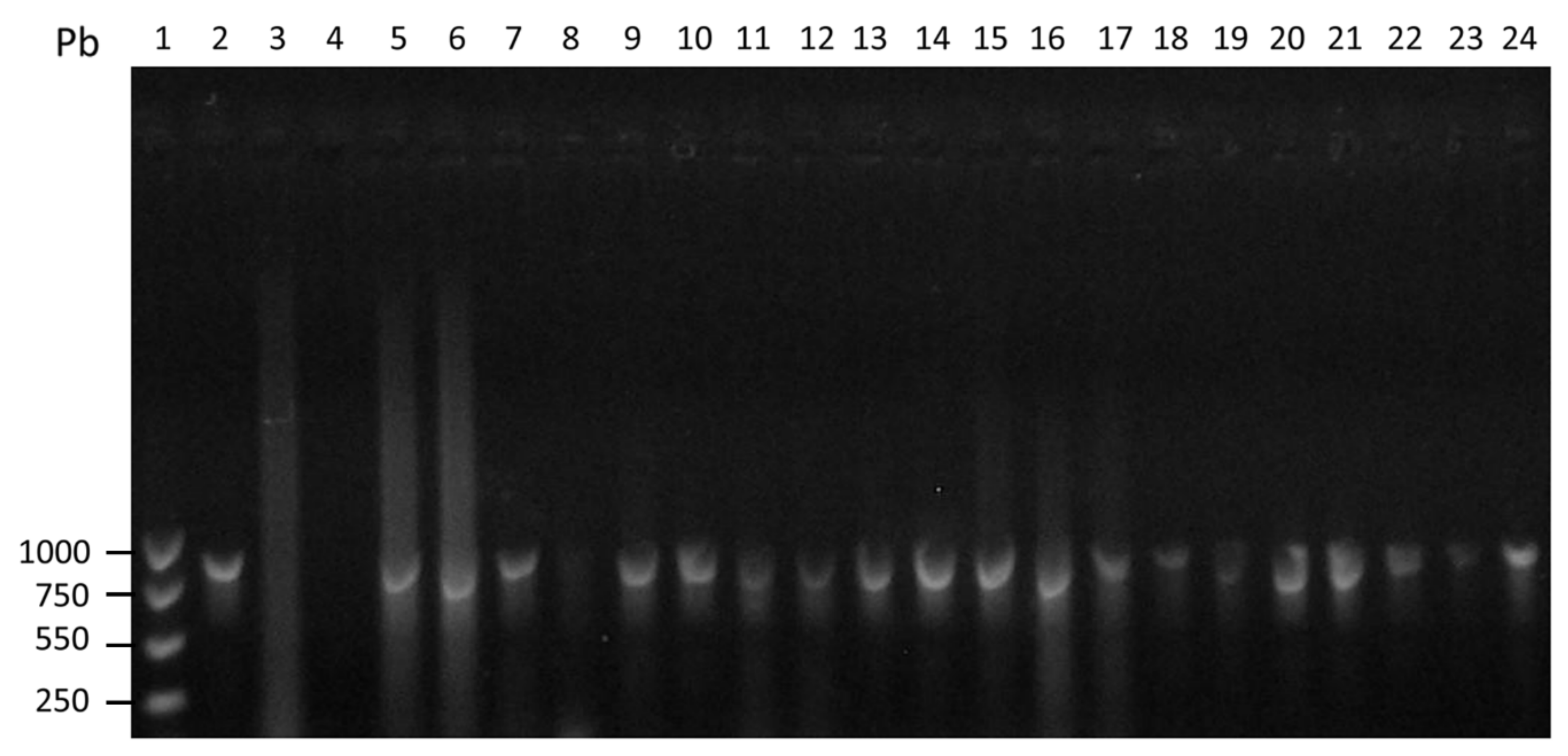
2.6. PCR Identification of E. histolytica

DNA was amplified using specie specific oligonucleotides; Psp5 GGCCAATTCATTCAATGAATTGAG and Psp3 GGGATCCTGATCCTTCCGCAGGTTCACCTAC previously described by Clark and Diamond (1992) [26]. Both oligonucleotides amplified a product of 876 bp. The PCR reaction was performed adding 1 μL of genomic DNA to the reaction mix: 2 μL PCR buffer consisting of Tris-HCl 10 mM pH 8.3, 0.8 μL de MgCl2 (50 μM), 1.6 μL of dNTPs (0.2 mM), 1 μL of ach oligonucleotides (20 μM) and 12.5 µL distillate water. The reaction mix was added with 0.1 μL (0.025 U/μL) of Taq DNA polymerase (AmpliTaq platinum Polymerase, Invitrogen). Amplification was performed in a thermocycler MyCycler Thermal Cycler (BIORAD). Amplification conditions were: DNA denaturation during 5 min at 95 °C followed by 45 cycles of denaturation during 30 sec at 94 °C, 30 sec for alignment at 55 °C; extension takes 1 min at 72 °C and on step of final extension of 10 min at 72 °C; the final volume of reaction was 20 µL.
The PCR products was observed in an electrophoresis in 1% agarose gel. The products were stained with ethidium bromide in a UV chamber. A reference of 1000 bp size and E. histolytica DNA control were included.

2.7. Molecular Analysis of the Expression of Amoebic Genes

To perform studies of quantification and expression of amoebic genes in the two groups of samples studied, we use Reverse Transcriptase quantitative PCR (RT-qPCR). The specific oligonucleotides for the genes studied are shown in Table 1 (Ehcp5, -cp2, -gal/galnaclectin, -crt, -stirp, -prd, -sod, -hsp70 y 90). The cDNAs previously obtained, were used as temperate at (15 ng/µL) in the reactions of qPCR using the Step One-Applied Biosystems and using for the reaction the kit (Quantitec SYBER Green PCR, Qiagen). The amplification was developed with 60 cycles with 3 stages; including a denaturing stage at 95 °C for 10 sec, a phase of alignment to 57 °C for 30 sec and a phase of elongation at 72 °C for 10 sec and finally to 4 °C to finish the reaction. The amplification of each gene was tested by tripled and the differential expression of the genes investigated was calculated by normalized to a reference gene (often referred to as the housekeeping gene) the Eh-α-actin of the trophozoites. The data were analyzed using the method of 2−ΔΔCT described by Schmittgen and Livak (2008) [27] and the validation method reported by Yalcin (2004) [28]. Comparing the differential expression of E. histolytica genes in the samples from both group of patients.

2.8. Statistical Analysis

Results are expressed as the average and the standard deviation of at least two duplicates of each condition and the analysis of independent samples. The statistical analysis was made using the program GraphPad Prism 5 software. We used the Student t tests to determine the statistically significance of differences between the studied patients’ samples. The statistical values were considered significant when the p value was less than p < 0.05.

3. Results

3.1. General Characteristics of Studied Patients

Fifteen patients with amoebic liver abscess were included in the study, 2 females and 13 males between 25 and 60 years old. All patients were submitted to ultrasound guided drainage of the abscess as indicated by the physician in charge. All patients had high levels of serum anti-amoebic antibodies in the ELISA test (OD = 1.171 ± 0.366) (Figure 1 and Supplementary Table S1). The drainage material from the liver abscess was processed for DNA extraction and submitted thereafter to PCR for detection of E. histolytica amplification products, all the abscesses specimens show the presence E. histolytica PCR amplification products (Figure 2).
The five patients with genital cutaneous amoebiasis were all males between 35 to 55 years old. In each case we were able to detect microscopically the presence of trophozoites of Entamoeba (Figure 3). The ELISA test for detection of serum anti-amoebic antibodies was positive in the five cases (OD = 0.833 ± 0.225) (Figure 1), and positive for detection of E. histolytica PCR amplification products (Figure 2).

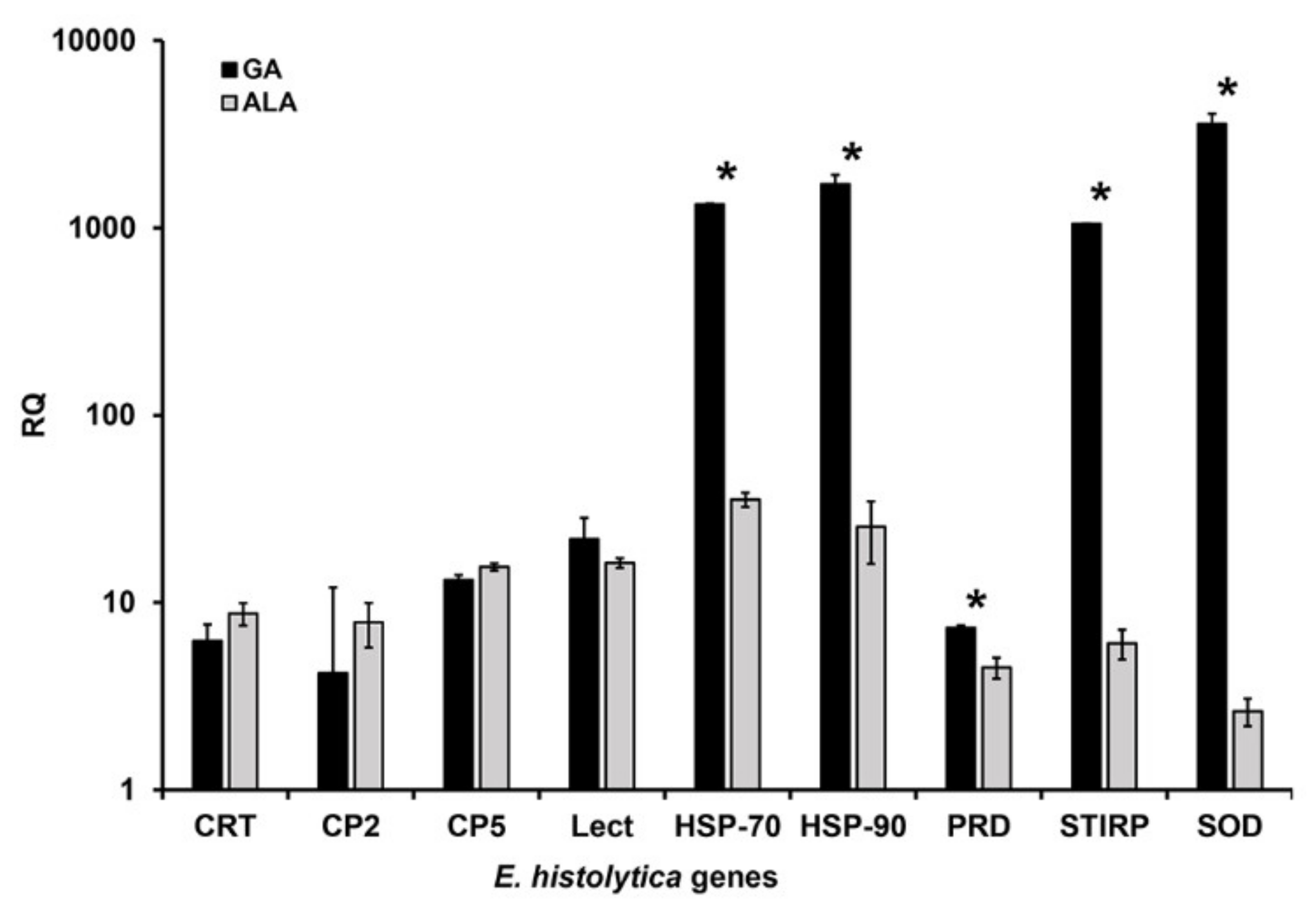
3.2. Analysis of the Expression of Amoebic Genes Associated with Pathogenicity

The qPCR technique was used to determine the differential expression of E. histolytica genes in the two different forms of invasive amoebiasis studied (amoebic liver abscess and genital cutaneous amoebiasis).
The results obtained show a clear overexpression in all the genes studied (Figure 4). However, this over-expression was higher for genes that encode proteins associated with the protection of trophozoites against stressful environmental conditions, such as EhHSP-70, EhHSP-90, EhPRD, and EhSOD in samples obtained from patients with genital cutaneous amoebiasis (p = 0.0027, 0.0013, 0.043, 0.0032). With regard to genes directly associated with pathogenicity factors (EhCP-2 and EhCP-5), a slightly higher overexpression was also observed when compared to patients with amoebic liver abscess but there is no significant difference when compared with the results of the trophozoites present in patients with genital cutaneous amoebiasis (p = 0.072, 0.36, respectively) (Figure 4). While for the EhCRT and EhGal/Galnaclectin genes, the overexpression is mostly the same for both amoebic liver abscess and genital cutaneous amoebiasis infection conditions (p = 0.24, 0.092, respectively).
However, in the case of EhSTIRP the mayor overexpression was observed in the cases of genital cutaneous amoebiasis (p = 0.0038).

4. Discussion

During its life cycle, E. histolytica is challenged by a wide variety of environmental stresses, such as fluctuation in the concentration of oxygen, glucose, changes in the composition of the intestinal microbiota and the release of reactive oxygen species, and oxidative nitrosatives of neutrophils and macrophages as products of the host’s immune response, so that the survivability of this parasite is continuously tested to adapt and survive the dynamic environment of the host [29,30].
The studies of these adaptive mechanisms with which this parasite has been transformed and facilitated due to the development of genomics, proteomics or metabolomics (OMICS sciences) [31,32]. Many transcriptome-level studies in E. histolytica have investigated gene expression patterns to help understand the pathology and biology of the organism. Virulent and avirulent strains have been compared in laboratory culture and after tissue invasion, cells cultured under different stress conditions, and response to anti-amoebic drugs, in order to identify and understand changes in gene expression during the different scenarios [33,34,35,36,37,38].
In this work, we compared the expression level of preselected genes (Ehcp5, Ehcp2, Ehgal/galnac lectin, Ehcrt, Ehstirp, Ehprd, Ehsod, Ehhsp-70 and 90) of E. histolytica, in two of its clinical manifestations the amoebic liver abscess and the genital cutaneous amoebiasis using the qPCR technique, this technique is used as a validation test for gene expression [39,40].
Given the locations of each morphological invasive process, trophozoites will be exposed to physiological and different environmental conditions. The parasite must be able to adapt to the demands of the environment to survive.
In some cases, and after the invading intestinal infection, the trophozoites can reach the bloodstream reaching the Porta circulation travelling to the liver, where it gives rise to one or multiple abscesses. Circulating trophozoites are confronted with the host’s innate immune response, with cytotoxic compounds, as well as with oxygen tensions higher than those usually found in the gut. In addition, trophozoites are exposed to other host defense mechanisms they are notably attacked by reactive oxygen and nitrogen intermediates produced by phagocyte cells, these compounds are highly concentrated in the infected tissue. For example, it has been shown experimentally that trophozoites invading the liver are highly sensitive to blood complement and tissue responses [2,7,41,42].
The invasion in genital cutaneous amoebiasis occurs by contiguity trophozoites from the intestine to the perianal skin, or skin that is around fistulas, colostomy incisions and abscesses drain incisions. This causes epithelial ulcers in this environment, trophozoites are exposed to a higher concentration of oxygen than in the liver [7,41].
In this sense, our results corroborate these findings since the expression of the genes (Eh-stirp, -prd, -sod) underscores the importance of these as they confer the ability of trophozoites to adapt to environments with a higher concentration of oxygen.
Peroxiredoxins of Entamoeba have been shown to play a crucial role for protection against oxidative stress, as in virulence, by helping the parasite to survive host immune response. The decreased gene expression causes the accumulation of ROS, a decrease in parasite viability, as well as in its cytotoxicity [43,44,45]. Moreover, the differential expression and immunolocalization of antioxidant enzymes in E. histolytica isolates during metronidazole stress have been reported [37].
This adaptive parasite response provides protection against the host response, as well as aiding in its survival. In eukaryotic cells, the overall stress is a perfectly orchestrated response mechanism. The first step is the function of a stress protein sensor (heat shock proteins, nutrient detection and antioxidant proteins as well as chromatin-associated proteins) to transmit the message to the cells and adapt to stress [46,47,48]. This coincides with the overexpression of Ehhsp-70 and-90, Ehprd, Ehstirp and Ehsod in patients GA genes that are exposed to greater thermal stress due to lower skin temperature (<37 °C) and higher O2 tension than ALA patients.
Matthiensen et al. (2013), investigate the roles of different papain-like cysteine peptidases (CPs) as pathogenicity factors; they show that the expression of some of the peptidases that are normally expressed at low levels, however, increases during amoebic abscess formation [49].
In this sense, the results obtained in this work show that these cysteine proteinases (Ehcp5, -cp2) are overexpressed in the same way in the two forms of amoebiasis studied, amoebic liver abscess and genital cutaneous amoebiasis. The findings reinforce the importance of CPs as pathogenicity factors of E. histolytica.
Another gene highly expressed in this work, was, Ehstirp (one of a family of E. histolytica serine-threonine- and isoleucine-rich proteins). There is very little information on the importance of this gene; however, it was identified during the process of adherence and then has been described by transcriptomic studies as a virulence factor [33,50,51,52], apparently absent in Rahman [51]. Analyzing the functional role of this protein with the overexpression reported in this work supporting the hypothesis that Ehstirp has a role in amoebic virulence.
It is clear that the expression of these genes (Eh-stirp, prd, sod, Ehhsp-70 and Ehhsp-90) can be regulated by stressful and environmental conditions where the invading process occurs, this environment will be determined not only by the organ invaded, but also by the host’s immune response. As has been observed in other genes, such as the case of AIG1, whose expression contrasts the virulence level of the parasite depending on the genetic background of the parasite, but also on the environmental conditions of the host [53].
The diversity of the compartments that E. histolytica crosses during infection, combined with the variability of induced symptoms, suggest that this parasite expresses different transcriptional programs, although the key components of the regulation of gene expression remain undescribed.
The results obtained in this work, could be the beginning of future investigations that shed light on the participation of environmental conditions in the expression of virulence mechanisms in host pathogenic microorganisms.

Supplementary Materials

The following are available online at https://www.mdpi.com/2076-2607/8/10/1556/s1. Supplementary Table S1: Individual ELISA test results. Supplementary Table S2: Results of differential expression of E. histolytica genes in two forms of extraintestinal infection, (ALA and GA) by RT-qPCR.

Author Contributions

Conceptualization, E.G.-R., M.N.-R., P.M. and C.X.; Formal analysis, E.G.-R., L.R.-V., E.H., A.S.-V., O.P., H.P.-J. and C.X.; Funding acquisition, C.X.; Investigation, E.G.-R., M.N.-R., U.M., P.M., L.R.-V., E.H., A.S.-V., O.P. and H.P.-J.; Methodology, E.G.-R., M.N.-R., U.M., P.M., L.R.-V., E.H., A.S.-V., O.P. and H.P.-J.; Project administration, C.X.; Resources, M.N-R., U.M., P.M., L.R.-V. and A.S.-V.; Supervision, E.G.-R. and C.X.; Validation, E.G.-R. and C.X.; Visualization, E.G.-R., A.S.-V. and C.X.; Writing—original draft, E.G.-R., M.N.-R, P.M. and H.P.-J.; Writing—review & editing, E.G.-R., A.S.-V. and C.X. All authors have read and agreed to the published version of the manuscript.

Funding

This work was partially supported by Grants IN218214 from PAPIIT UNAM and 272601 from CONACyT Mexico.

Acknowledgments

We are indebted to; Marco Gudiño for the art design and appreciate the technical assistance of Martha Zaragoza and Angeles Padilla.

Conflicts of Interest

The authors declare no conflict of interest.

References

  1. WHO; PAN American Health Organization; UNESCO. Expert Consultation on Amoebiasis. WHO Wkly. Epidem. Rec. 1997, 72, 97–100. [Google Scholar]
  2. Mortimer, L.; Chadee, K. The immunopathogenesis of Entamoeba histolytica. Exp. Parasitol. 2010, 126, 366–380. [Google Scholar] [CrossRef] [PubMed]
  3. Haque, R.; Huston, C.D.; Hughes, M.; Houpt, E.; Petri, W.A., Jr. Amebiasis. N. Engl. J. Med. 2003, 348, 1565–1573. [Google Scholar] [CrossRef] [PubMed]
  4. Huston, C.D.; Haque, R.; Petri, W.A., Jr. Molecular-based diagnosis of Entamoeba histolytica infection. Exp. Rev. Mol. Med. 1999, 1999, 1–11. [Google Scholar] [CrossRef]
  5. Ximénez, C.; Cerritos, R.; Rojas, L.; Dolabella, S.; Morán, P.; Shibayama, M.; Gonzalez, E.; Valadez, A.; Hernandez, E.; Valenzuela, O.; et al. Human amebiasis: Breaking the paradigm? Int. J. Environ. Res. Public Health 2010, 7, 1105–1120. [Google Scholar] [CrossRef]
  6. Antony, S.J.; Lopez-Po, P. Genital amebiasis: Historical perspective of an unusual disease presentation. Urology 1999, 54, 952–955. [Google Scholar] [CrossRef]
  7. Ríos-Yuil, J.M.; Mercadillo-Pérez, P.; Yuil-de Ríos, E.; Ríos-Castro, M. Amebiasis Cutánea: Conceptos actuals. Rev. Med. Hosp. Gen. Mex. 2012, 75((2)), 114–122. [Google Scholar]
  8. Schmerin, M.J.; Gelston, A.; Jones, T.C. Amebiasis. An increasing problem among homosexuals in New York City. JAMA 1977, 238, 1386–1387. [Google Scholar] [CrossRef]
  9. Suzuki, J.; Ise, I. Amebiasis as a sexually transmitted disease with special emphasis on the Entamoeba histolytica antibody prevalence in females in Tokyo. Infect. Agents. Surveill. Rep. 2007, 28, 108–109. [Google Scholar]
  10. Chelsea, M.; Petri, W.A., Jr. Regulation of Virulence of Entamoeba histolytica. Annu. Rev. Microbiol. 2014, 68, 493–520. [Google Scholar] [CrossRef]
  11. Petri, W.A., Jr.; Haque, R.; Mann, B.J. The bittersweet interface of parasite and host: Lectin-carbohydrate interactions during human invasion by the parasite Entamoeba histolytica. Annu. Rev. Microbiol. 2002, 56, 39–64. [Google Scholar] [CrossRef] [PubMed]
  12. Petri, W.A., Jr.; Chapman, M.D.; Snodgrass, T.L.-; Mann, B.J.; Broman, J.; Ravdin, J.I. Subunit structure of the galactose and N-acetyl-D galactosamine-inhibitable adherence lectin of Entamoeba histolytica. J. Biol. Chem. 1989, 264, 3007–3012. [Google Scholar] [PubMed]
  13. Bruchhaus, I.; Jacobs, T.; Leippe, M.; Tannich, E. Entamoeba histolytica and Entamoeba dispar: Differences in numbers and expression of cysteine proteinase genes. Mol. Microbiol. 1996, 22, 255–263. [Google Scholar] [CrossRef] [PubMed]
  14. Lidell, M.E.; Moncada, D.M.; Chadee, K.; Hansson, G.C. Entamoeba histolytica cysteine proteases cleave the MUC2 mucin in its C-terminal domain and dissolve the protective colonic mucus gel. Proc. Natl. Acad. Sci. USA 2006, 103, 9298–9303. [Google Scholar] [CrossRef] [PubMed] [Green Version]
  15. Bruchhaus, I.; Loftus, B.J.; Hall, N.; Tannich, E. The intestinal protozoan parasite Entamoeba histolytica contains 20 cysteine protease genes of which only as mall subset is expressed during in vitro cultivation. Eukaryot. Cell. 2003, 2, 501–509. [Google Scholar] [CrossRef] [Green Version]
  16. Ravdin, J.I.; Croft, B.Y.; Guerrant, R.L. Cytopathogenic mechanisms of Entamoeba histolytica. J. Exp. Med. 1980, 152, 377–390. [Google Scholar] [CrossRef] [Green Version]
  17. Petri, W.A., Jr.; Ravdin, J.I. Cytopathogenicity of Entamoeba histolytica: The role of amebic adherence and contact-dependent cytolysis in pathogenesis. Eur. J. Epidemiol. 1987, 3, 123–136. [Google Scholar] [CrossRef] [PubMed]
  18. Huston, C.D.; Houpt, E.R.; Mann, B.J.; Hahn, C.S.; Petri, W.A. Caspase 3-dependent killing of host cells by the parasite Entamoeba histolytica. Cell. Microbiol. 2000, 2, 617–625. [Google Scholar] [CrossRef]
  19. Ravdin, J.I.; Moreau, F.; Sullivan, J.A.; Petri, W.A., Jr.; Mandell, G.L. Relationship of free intracellular calcium to the cytolytic activity of Entamoeba histolytica. Infect. Immun. 1988, 56, 1505–1512. [Google Scholar] [CrossRef] [Green Version]
  20. Ragland, B.D.; Ashley, L.S.; Vaux, D.L.; Petri, W.A., Jr. Entamoeba histolytica: Target cells killed by trophozoites undergo DNA fragmentation which is not blocked by Bcl-2. Exp. Parasitol. 1994, 79, 460–467. [Google Scholar] [CrossRef]
  21. Seydel, K.B.; Stanley, S.L., Jr. Entamoeba histolytica induces host cell death in amebic liver abscess by a non-Fas-dependent, non-tumor necrosis factor alpha-dependent pathway of apoptosis. Infect. Immun. 1998, 66, 2980–2983. [Google Scholar] [CrossRef] [PubMed] [Green Version]
  22. Trissl, D.; Martínez-Palomo, A.; de la Torre, M.; de la Hoz, R.; de Suarez, E.P. Surface properties of Entamoeba: Increased rates of human erythrocyte phagocytosis in pathogenic strains. J. Exp. Med. 1978, 148, 1137–1145. [Google Scholar] [CrossRef] [PubMed] [Green Version]
  23. Orozco, E.; Guarneros, G.; Martínez-Palomo, A. Entamoeba histolytica: Phagocytosis as a virulence factor. J. Exp. Med. 1983, 158, 1511. [Google Scholar] [CrossRef] [PubMed]
  24. Torres, F. Manual de Técnicas en Histología y Anatomía Patológica; Ariel Press: Barcelona, Spain, 2002; pp. 31–74. [Google Scholar]
  25. Morán, P.; Gómez, A.; Valadez, A.; Ramos, F.; González, E.; García, G.; Limón, A.; Valenzuela, O.; Ramiro, M.; Hidalgo, H.; et al. Amebic and pyogenic liver abscess: Importance of differential diagnosis in areas of endemic amebiasis. Trop. Med. Int. Health. 2007, 12, 57–64. [Google Scholar]
  26. Clark, C.G.; Diamond, L.S. Differentiation of pathogenic Entamoeba histolytica from other intestinal protozoa by riboprinting. Arch. Med. Res. 1992, 23, 15–16. [Google Scholar] [PubMed]
  27. Livak, K.J.; Schmittgen, K.J. Analysis of relative gene expression data using real time quantitative PCR and the 2(-Delta Delta Ct) method. Methods. 2001, 25, 402–408. [Google Scholar] [CrossRef]
  28. Yalcin, A. Quantification of thioredoxin mRNA expression in the rat hippocampus by real-time PCR following oxidative stress. Act. Biochem. Pol. 2004, 51, 1059–1065. [Google Scholar] [PubMed]
  29. Vicente, J.B.; Ehrenkaufer, G.M.; Saraiva, L.M.; Teixeira, M.; Singh, U. Entamoeba histolytica modulates a complex repertoire of novel genes in response to oxidative and nitrosative stresses: Implications for amebic pathogenesis. Cell. Microbiol. 2009, 11, 51–69. [Google Scholar] [CrossRef] [Green Version]
  30. Nagaraja, S.; Ankri, S. Utilization of Different Omic Approaches to Unravel Stress Response Mechanisms in the Parasite Entamoeba histolytica. Front. Cell. Infect. Microbiol. 2018, 8, 19. [Google Scholar] [CrossRef] [Green Version]
  31. Naiyer, S.; Bhattacharya, A.; Bhattacharya, S. Advances in Entamoeba histolytica Biology Through Transcriptomic Analysis. Front. Microbiol. 2019, 10, 1921. [Google Scholar] [CrossRef]
  32. Meyer, M.; Fehling, H.; Matthiessen, J.; Lorenzen, S.; Schuldt, K.; Bernin, H.; Zaruba, M.; Lender, C.; Ernst, T.; Ittrich, H.; et al. Overexpression of differentially expressed genes identified in non-pathogenic and pathogenic Entamoeba histolytica clones allow identification of new pathogenicity factors involved in amoebic liver abscess formation. PLoS Pathog. 2016, 12, e1005853. [Google Scholar] [CrossRef]
  33. Gilchrist, C.A.; Houpt, E.; Trapaidze, N.; Fei, Z.; Crasta, O.; Asgharpour, A.; Evans, C.; Martino-Catt, S.; Baba, D.J.; Stroup, S.; et al. Impact of intestinal colonization and invasion on the Entamoeba histolytica transcriptome. Mol. Biochem. Parasitol. 2006, 147, 163–176. [Google Scholar] [CrossRef]
  34. Thibeaux, R.; Weber, C.; Hon, C.C.; Dillies, M.A.; Avé, P.; Coppée, J.Y.; Labruyere, E.; Guillen, N. Identification of the virulence landscape essential for Entamoeba histolytica invasion of the human Colon. PLoS Pathog. 2013, 9, e1003824. [Google Scholar] [CrossRef] [Green Version]
  35. De Cádiz, A.E.; Jeelani, G.; Nakada-Tsukui, K.; Caler, E.; Nozaki, T. Transcriptome analysis of encystation in Entamoeba invadens. PLoS ONE 2013, 8, e74840. [Google Scholar] [CrossRef] [PubMed]
  36. Baumel-Alterzon, S.; Weber, C.; Guillén, N.; Ankri, S. Identification of dihydropyrimidine dehydrogenase as a virulence factor essential for the survival of Entamoeba histolytica in glucose-poor environments. Cell. Microbiol. 2013, 15, 130–144. [Google Scholar] [CrossRef] [PubMed]
  37. Iyer, L.R.; Singh, N.; Verma, A.K.; Paul, J. Differential expression and immunolocalization of antioxidant enzymes in Entamoeba histolytica isolates during metronidazole stress. Biomed. Res. Int. 2014, 2014, 704937. [Google Scholar] [CrossRef] [Green Version]
  38. Weber, C.H.; Koutero, M.; Dillies, M.A.; Varet, H.; Lopez Camarillo, C.; Coppée, J.Y.; Hon, C.; Guillén, N. Extensive transcriptome analysis correlates the plasticity of Entamoeba histolytica pathogenesis to rapid phenotype changes depending on the environment. Sci. Rep. 2016, 6, 35852. [Google Scholar] [CrossRef] [PubMed] [Green Version]
  39. Davis, P.H.; Schulze, J.; Stanley, S.L., Jr. Transcriptomic comparison of two Entamoeba histolytica strains with defined virulence phenotypes identifies new virulence factor candidates and key differences in the expression patterns of cysteine proteases, lectin light chains, and calmodulin. Mol. Biochem. Parasitol. 2007, 151, 118–128. [Google Scholar] [CrossRef]
  40. Taylor, S.; Wakam, M.; Dijkman, G.; Alsarraj, M.; Nguyen, M. A practical approach to RT-qPCR publishing data that conform to the MIQE guidelines. Methods 2010, 50, S1–S5. [Google Scholar] [CrossRef]
  41. Wuerz, T.; Kane, J.B.; Boggild, A.K.; Krajden, S.; Keystone, J.S.; Fuksa, M.; Kain, K.C.; Warren, R.; Kempston, J.; Anderson, J. A Review of Amoebic Liver Abscess for Clinicians in a Non endemic Setting. Can. J. Gastroenterol. Hepatol. 2012, 26, 729–733. [Google Scholar] [CrossRef]
  42. Santos, F.; Nequiz, M.; Hernández-Cuevas, N.A.; Hernández, K.; Pineda, E.; Encalada, R.; Guillen, N.; Luis-Garcia, E.; Saralegui, A.; Saavedra, E.; et al. Maintenance of intracellular hypoxia and adequate heat shock response are essential requirements for pathogenicity and virulence of Entamoeba histolytica. Cell. Microbiol. 2015, 17, 1037–1051. [Google Scholar] [CrossRef] [PubMed]
  43. Rastew, E.; Vicente, J.B.; Singh, U. Oxidative stress resistance genes contribute to the pathogenic potential of the anaerobic protozoan parasite, Entamoeba histolytica. Int. J. Parasitol. 2012, 42, 1007–1015. [Google Scholar] [CrossRef] [PubMed] [Green Version]
  44. Akbar, A.; Chatterjee, N.S.; Sena, P.; Debnath, A.; Pal, A.; Bera, T.; Das, P. Genes induced by a high-oxygen environment in Entamoeba histolytica. Mol. Biochem. Parasitol. 2004, 133, 187–196. [Google Scholar] [CrossRef] [PubMed]
  45. Sen, A.; Chatterjee, N.S.; Ali Akbar, M.; Nandi, N.; Das, P. The 29-kilodalton thiol-dependent peroxidase of Entamoeba histolytica is a factor involved in pathogenesis and survival of the parasite during oxidative stress. Eukaryot. Cell. 2007, 6, 664–673. [Google Scholar] [CrossRef] [Green Version]
  46. De Nadal, E.; Ammerer, G.; Posas, F. Controlling gene expression in response to stress. Nat. Rev. Genet. 2011, 3, 833–845. [Google Scholar] [CrossRef]
  47. Santi-Rocca, J.; Smith, S.; Weber, C.; Pineda., E.; Hon, C.C.; Saavedra, E.; Olivos-García, A.; Rousseau, S.; Dillies, M.A.; Coppée, J.Y.; et al. Endoplasmic reticulum stress-sensing mechanism is activated in Entamoeba histolytica upon treatment with nitric oxide. PLoS ONE 2012, 7, e31777. [Google Scholar] [CrossRef] [PubMed] [Green Version]
  48. Smith, K.T.; Workman, J.L. Chromatin proteins: Key responders to stress. PLoS Biol. 2012, 10, e1001371. [Google Scholar] [CrossRef] [Green Version]
  49. Matthiensen, J.; Bär, A.K.; Bartels, A.K.; Marien, D.; Ofori, S.; Biller, L.; Tannich, E.; Lotter, H.; Bruchhaus, I. Overexpression of Specific Cysteine Peptidases Confers Pathogenicity to a Nonpathogenic Entamoeba histolytica Clone. mBio 2013, 4, e00072-13. [Google Scholar] [CrossRef] [Green Version]
  50. Macfarlane, R.C.; Singh, U. Identification of an Entamoeba histolytica serine, threonine, isoleucine, rich protein with roles in adhesion and cytotoxicity. Eukaryot. Cell 2007, 6, 2139–2146. [Google Scholar] [CrossRef] [Green Version]
  51. Faust, D.M.; Guillen, N. Virulence and virulence factors in Entamoeba histolytica, the agent of human amoebiasis. Microbes. Infect. 2012, 14, 1428–1441. [Google Scholar] [CrossRef]
  52. MacFarlane, R.C.; Singh, U. Identification of differentially expressed genes in virulent and Entamoeba species: Potential implications for amebic pathogenesis. Infect. Immun. 2006, 74, 340–351. [Google Scholar] [CrossRef] [PubMed] [Green Version]
  53. Nakada-Tsukui, K.; Sekizuka, T.; Sato-Ebine, E.; Escueta-de Cadiz, A.; Ji, D.D.; Tomii, K.; Kuroda, M.; Nozaki, T. (AIG1) affects in vitro and in vivo virulence in clinical isolates of Entamoeba histolytica. PLoS Pathog. 2018, 14, e1006882. [Google Scholar] [CrossRef] [PubMed] [Green Version]
Figure 1. Reactivity of sera from patients with amoebic liver abscess (ALA) and cutaneous genital amoebiasis (GA). The reactivity of human IgG against E. histolytica antigen was measured, the average of the OD (590 nm) per group of patients is represented here. Cutline defined as the mean of the respective OD values obtained from non-parasitized individuals (Negative group) plus two standard deviations (0.525) and results previously reported [25]. Statistical significance was included (p ≤ 0.05). The individual dates are in the Supplementary Table S1.
Microorganisms 08 01556 g001
Figure 2. PCR identification of E. histolytica. The DNAs obtained from samples of tissue fragments from lesions of genital cutaneous amebiasis patients and abscess drainage from patients with amoebic liver abscess, were evaluated by PCR using oligonucleotides specific (Psp) previously described by Clark and Diamond [26]. Lane (1) 1000 pb mass ruler DNA ladder; (2) Positive control for E. histolytica strain HM1: IMSS; (3) Negative control for E. histolytica; (4) H20 sample; (5–19) DNA obtained from drainage from patients with amoebic liver abscess; (20–24) DNA obtained from lesions of genital cutaneous amebiasis patients. Products amplified and stained with ethidium bromide, Product size 876 bp.
Microorganisms 08 01556 g002
Figure 3. Microscopic identification of E. histolytica trophozoites. Biopsy specimens from patients with genital cutaneous amoebiasis histologically treated and stained with PAS, representative images; (a) a smear was performed directly over the slide; (b,c) samples from two different patients. The arrows point out some illustrative trophozoites. The scale bar represents 20 µm.
Microorganisms 08 01556 g003
Figure 4. Relative quantification (RQ) of expression of genes of E. histolytica by RT-qPCR. Reverse transcription real time PCR was used to independently measure mRNA expression of Ehcp5, -cp2, -gal/galnac lectin, -crt, -stirp, -prd, -sod, -hsp-70 y 90 genes in trophozoites present in samples of tissue fragments from lesions of genital cutaneous amoebiasis (GA) and abscess drainage from patients with amoebic liver abscess (ALA). The values represent the mean of different patients for group. RQ differences between the cDNAs obtained from samples of tissue fragments from lesions of GA patients and abscess drainage from patients with (ALA) were compared trough Student t tests, detecting statistical significance * (p ≤ 0.05). Ehactin-α gen was used as housekeeping gene to normalize mRNA level. The individual dates are shows in the Supplementary Table S2.
Microorganisms 08 01556 g004
Table 1. Oligonucleotides used in the RT-qPCR assay.
GeneAccess NumberSize of Products
(pb)
Forward Primer
(5′-3′)
Reverse Primer
(5′-3′)
EhcrtXM_650149.1355TGGACCAGATGTATGTGGAGGTGGTGCTTCCCATTCTCCATC
Ehcp-5XM_645845.2255GTTGCCGCTGCTATTGATGCACCCCAACTGGATAAGCAGC
Ehcp-2XM_645550115ATCCAAGCACCAGAATCAGTTTCCTTCAAGAGCTGCAAGT
EhlectAF337950.1281ACCAGTGAATGGAGCATGTGTTTG TGC ATT CGC CTT CTC CT
hprdXM_646911.2121TCAAGAGAAAGAATGTTGTTGTACATGGACAATATGCTGCTGC
EhsodXM_6437351172GCAGCCCAAGCATGGAATCAACCAACACCATCCACTTCCA
EhstirpXM-648869305GCTAACAACGCGGAAAGTAGCACAAGAGCAGAGCACCCTTC
Ehhsp-70XM_001734367.1135GAAACAGAACCACGTCCAGTTTTACGTCCTCCAAGTCTCCAAT
Ehsp-90AB092411.1357ACAAGAGCAGAGCACCCTTCCCCCATGTGCACTTGTTACAG
EhactinXM_650064.2211AGCTGTTCTTTCATTATATGCTTCTCTTTCAGCAGTAGTGGT
Ehactin-α gen of reference. Specificity of the primer pairs for relevant genes was verified by performing nucleotide alignment searches using BLAST.

