Article

Multiple Sex Chromosomes and Evolutionary Relationships in Amazonian Catfishes: The Outstanding Model of the Genus Harttia (Siluriformes: Loricariidae)

by
Francisco de M. C. Sassi
1,
Geize A. Deon
1,2,
Orlando Moreira-Filho
1,2,
Marcelo R. Vicari
2,
Luiz A. C. Bertollo
1,
Thomas Liehr
3,*,
Ezequiel Aguiar de Oliveira
4 and
Marcelo B. Cioffi
1
1
Laboratório de Citogenética de Peixes, Departamento de Genética e Evolução, Universidade Federal de São, Carlos, São Carlos, SP 13565-905, Brazil
2
Departamento de Biologia Estrutural, Molecular e Genética, Universidade Estadual de Ponta Grossa, Ponta Grossa, PR 84010-330, Brazil
3
Institute of Human Genetics, University Hospital Jena, Jena 07747, Germany
4
Secretaria de Estado de Educação do Mato Grosso—SEDUC-MT, Cuiabá, MT 78049-909, Brazil
*
Author to whom correspondence should be addressed.
Genes 2020, 11(10), 1179; https://doi.org/10.3390/genes11101179
Submission received: 14 September 2020 / Revised: 6 October 2020 / Accepted: 8 October 2020 / Published: 10 October 2020
(This article belongs to the Special Issue Fish Cytogenetics: Present and Future)

Abstract

:
The armored Harttia catfishes present great species diversity and remarkable cytogenetic variation, including different sex chromosome systems. Here we analyzed three new species, H. duriventris, H. villasboas and H. rondoni, using both conventional and molecular cytogenetic techniques (Giemsa-staining and C-banding), including the mapping of repetitive DNAs using fluorescence in situ hybridization (FISH) and comparative genomic hybridization (CGH) experiments. Both H. duriventris and H. villasboas have 2n = ♀56/♂55 chromosomes, and an X1X1X2X2 /X1X2Y sex chromosome system, while a proto or neo-XY system is proposed for H. rondoni (2n = 54♀♂). Single motifs of 5S and 18S rDNA occur in all three species, with the latter being also mapped in the sex chromosomes. The results confirm the general evolutionary trend that has been noticed for the genus: an extensive variation on their chromosome number, single sites of rDNA sequences and the occurrence of multiple sex chromosomes. Comparative genomic analyses with another congeneric species, H. punctata, reveal that the X1X2Y sex chromosomes of these species share the genomic contents, indicating a probable common origin. The remarkable karyotypic variation, including sex chromosomes systems, makes Harttia a suitable model for evolutionary studies focusing on karyotype differentiation and sex chromosome evolution among lower vertebrates.

1. Introduction

Siluriformes (Actinopterygii; Teleostei) is a monophyletic order that covers a large deal of the freshwater fish diversity, with more than 3000 species and 36 families [1,2]. Of the six Loricariidae subfamilies currently recognized, Hypostominae and Loricariinae are the most representative ones, with the highest number of species (579 and 302, respectively) and a remarkable karyotypic diversity [2,3]. Loricariinae fishes are distributed throughout South and Central American rivers, including two tribes: Harttiini and Loricariini. The genus Harttia, popularly known as armored catfishes, is represented by 27 valid species [2,4].
Despite such great species diversity, cytogenetic studies are available for only eight of them, all from Brazilian Southern regions, with exception for Harttia punctata [3,5,6,7,8,9,10,11,12,13]. However, although still limited, the chromosome data are already enough to notice a conspicuous variation of the diploid number (2n) in the genus, ranging from 2n = 52♀/53♂ in H. carvalhoi [8] to 2n = 62♀♂ in H. absaberi [9]. Chromosomal breaks and rearrangements as Robertsonian fusions, fissions, as well inversions were proposed to occur in the karyotype diversification of Harttia species [13]. In addition, two multiple XY-derived sex chromosome systems were also identified: ♀XX/♂XY1Y2 in H. carvalhoi [8] and ♀X1X1X2X2/♂X1X2Y in H. punctata [12].
In recent years, a range of molecular cytogenetic investigations (e.g., repetitive DNA mapping based on fluorescence in situ hybridization (FISH), comparative genomic hybridization (CGH) and whole chromosome painting (WCP) has been applied in different fish groups, providing new insights into the evolutionary relationships among them [14,15,16,17,18]. However, the ribosomal genes are the only repetitive DNA class that has been analyzed in Harttia till now [3,8,12,13]. The rDNA mapping shows that the 18S sites presents a conserved pattern in number, with a single chromosome pair carrying these sequences in all analyzed species, but with variation in location and chromosome carrying them [13]. Similarly, the 5S rDNA is also found in a single chromosome pair in most species, with exception for H. carvalhoi, in which signals occur in both chromosome pairs 03 and 23 [8,13]. Besides, WCP analyzes are confined to only one species, H. punctata, highlighting the main chromosomal rearrangements involved in the origin of the X1X2Y sex system in this species [12].
In the most updated phylogenetical reconstruction based on mtDNA, three distinct clades can be recognized for the genus Harttia involving: (i) species that inhabit rivers of Guyana shield; (ii) species occurring in north Brazil, especially in Amazonas river basin and its tributaries and (iii) species distributed throughout southeast/south Brazilian rivers, such as Grande, São Francisco and Paraná river basins [19]. However, despite largely widespread in South America, only Harttia species from southern rivers have been cytogenetically studied to date. Here we analyzed three new species sampled from Northern Brazilian river basins by performing an extensive cytogenetic investigation using conventional cytogenetic protocols (Giemsa-staining and C-banding) combined with molecular cytogenetic ones, including CGH experiments and mapping of several repetitive DNA classes using FISH. The results allowed us to open a new chapter on the evolutionary history of Harttia fishes, besides describing new sex chromosome systems.

2. Materials and Methods

2.1. Sampling

The collection sites, number and gender of the specimens investigated are presented in Figure 1 and Table 1. Samples were collected with the authorization of the environmental agency ICMBIO/SISBIO (License nº 48628-2) and SISGEN (A96FF09). The specimens were proper identified by evaluation of their meristic characters by Dr. Osvaldo Takeshi Oyakawa, curator of the Museu de Zoologia da Universidade de São Paulo (MZUSP) and specialist in this fish group. Samples from H. punctata were used for gDNA extraction and CGH experiments.

2.2. Chromosome Preparation and C-Banding

Mitotic chromosomes were obtained by the protocol described in [20]. Briefly, the animals were first injected in the abdominal region with a 0.025% aqueous solution of colchicine at a dose of 1 ml/100 g of weight. After 50–60 min, the specimens were euthanized, and the chromosomal preparations were obtained from cells of the anterior kidney. The experiments followed ethical and anesthesia conducts and were approved by the Ethics Committee on Animal Experimentation of the Universidade Federal de São Carlos (Process number CEUA 1853260315). C-positive heterochromatin (C-banding) was identified according to [21]. Briefly, slides were treated with 0,2 N HCl for 10 min, followed by a short wash in water; incubated in 5% Ba(OH)2 for 3 min at 42 °C ; rinsed in water for 1 min and then incubated in 2× SSC (pH 7.0) for 1h at 60 °C. The slides were then stained in 5% Giemsa solution (phosphate buffer, pH 6.8).

2.3. Fluorescence In Situ Hybridization (FISH) for Repetitive DNA Mapping

The 5S rDNA probe included 120 base pairs (bp) of the 5S rRNA codificant gene and 200bp of a non-transcribed spacer (NTS) isolated according to [22]. In its turn, the 18S rDNA probe contained a 1400 bp segment of the 18S rRNA gene and was isolated following [23]. These probes were directly labeled with the Nick-Translation mix kit (Jena Bioscience, Jena, Germany). The 5S rDNA was labeled with ATTO550-dUTP and the 18S rDNA with AF488-dUTP, according to the manufacturer’s manual. The microsatellite sequences (A)30, (CA)15 and (GA)15 were directly labeled with Cy-3 during their synthesis, as described by [24]. These sequences were selected once they are among the most abundant ones in fish genomes [14] and generated well visible hybridization patterns in our experiments. Telomeric (TTAGGG)n sequences were mapped using the DAKO Telomere PNA FISH Kit/FITC (DAKO, Glostrup, Denmark). The FISH experiments followed the methodology described in [25]. Briefly, metaphase chromosome slides were incubated with RNAse (40 μg/mL) for 1.5 h at 37 °C. After the denaturation of the chromosomal DNA in 70% formamide/2× SSC at 70 °C for 3 min, the hybridization mixture (2.5 ng/μL probes, 2 μg/μL C0t-1 DNA, 50% deionized formamide, 10% dextran sulphate) was dropped on the slides, and the hybridization was performed overnight at 37 °C in a moist chamber containing 2× SSC. The first post-hybridization wash was performed with 2× SSC for 5 min at 65 °C, and a final wash was performed at room temperature in 1× SSC for 5 min. Finally, the slides were counterstained with DAPI and mounted in an antifade solution (Vectashield from Vector Laboratories, Burlingame, CA)

2.4. Comparative Genomic Hybridization (CGH)

The total genomic DNAs (gDNAs) from male and female specimens of H. duriventris, H. villaboas, H. rondoni and H. punctata were extracted from liver tissue by the standard phenol-chloroform-isoamyl alcohol method [26]. It was focused on inter-and intraspecific comparisons, with special emphasis on molecular composition of the putative and multiple sex chromosomes. In the first set of experiments (intraspecific genomic comparisons), the male-derived gDNAs of all species were labelled with Atto550-dUTP and the female gDNAs with Atto488-dUTP, by means of nick translation (Jena Bioscience, Jena, Germany). For blocking repetitive sequences, it was used unlabeled C0t-1 DNA in all experiments (i.e. fraction of genomic DNA enriched with highly and moderately repetitive sequences), prepared according to [27]. The final hybridization mixture for each slide (20 μL) was composed of male- and female-derived gDNAs (500 ng each), plus 25 μg of female-derived C0t-1 DNA from the respective species. The probe was ethanol-precipitated, and the dry pellets were resuspended in hybridization buffer containing 50% formamide, 2× SSC, 10% SDS, 10% dextran sulfate and Denhardt´s buffer, pH 7.0. In the second set of experiments (interspecific genomic comparisons), the gDNA of all male specimens now analyzed, plus the gDNA of H. punctata (a species harboring multiple X1X2Y sex system), were hybridized against metaphase chromosomes of H. villasboas. For this purpose, male-derived gDNA of H. villasboas was labeled with Atto550-dUTP, while the gDNAs of the other three species were labeled with Atto488-dUTP (H. duriventris and H. rondoni) or Atto425-dUTP (H. punctata) both by means of nick translation (Jena Bioscience, Jena, Germany). In a first slide, the final probe mixture was composed of 500 ng of male-derived gDNA of each H. villasboas, H. duriventris and H. punctata and 10 μg of female-derived C0t-1 DNA of each species. In a second slide, the final probe mixture was composed of 500 ng of male-derived gDNA of H. villasboas and H. rondoni and 15 μg of female-derived C0t-1 DNA of each species. The chosen ratio of probe vs. C0t-1 DNA amount was based on previous experiments performed in our fish studies [18,28,29]. The CGH experiments followed the methodology described in [30].

2.5. Microscopic Analysis and Image Processing

At least 30 metaphase spreads per individual were analyzed to confirm the 2n, karyotype structure and CGH results. Images were captured using Olympus BX50 microscope (Olympus Corporation, Ishikawa, Japan), with CoolSNAP and the images were processed using Image Pro Plus 4.1 software (Media Cybernetics, Silver Spring, MD, USA). Chromosomes were classified as metacentric (m); submetacentric (sm); subtelocentric (st) or acrocentric (a) according to [31]. The maps were created using the following software′s: QGis 3.4.3 and Adobe Photoshop CC 2020.

3. Results

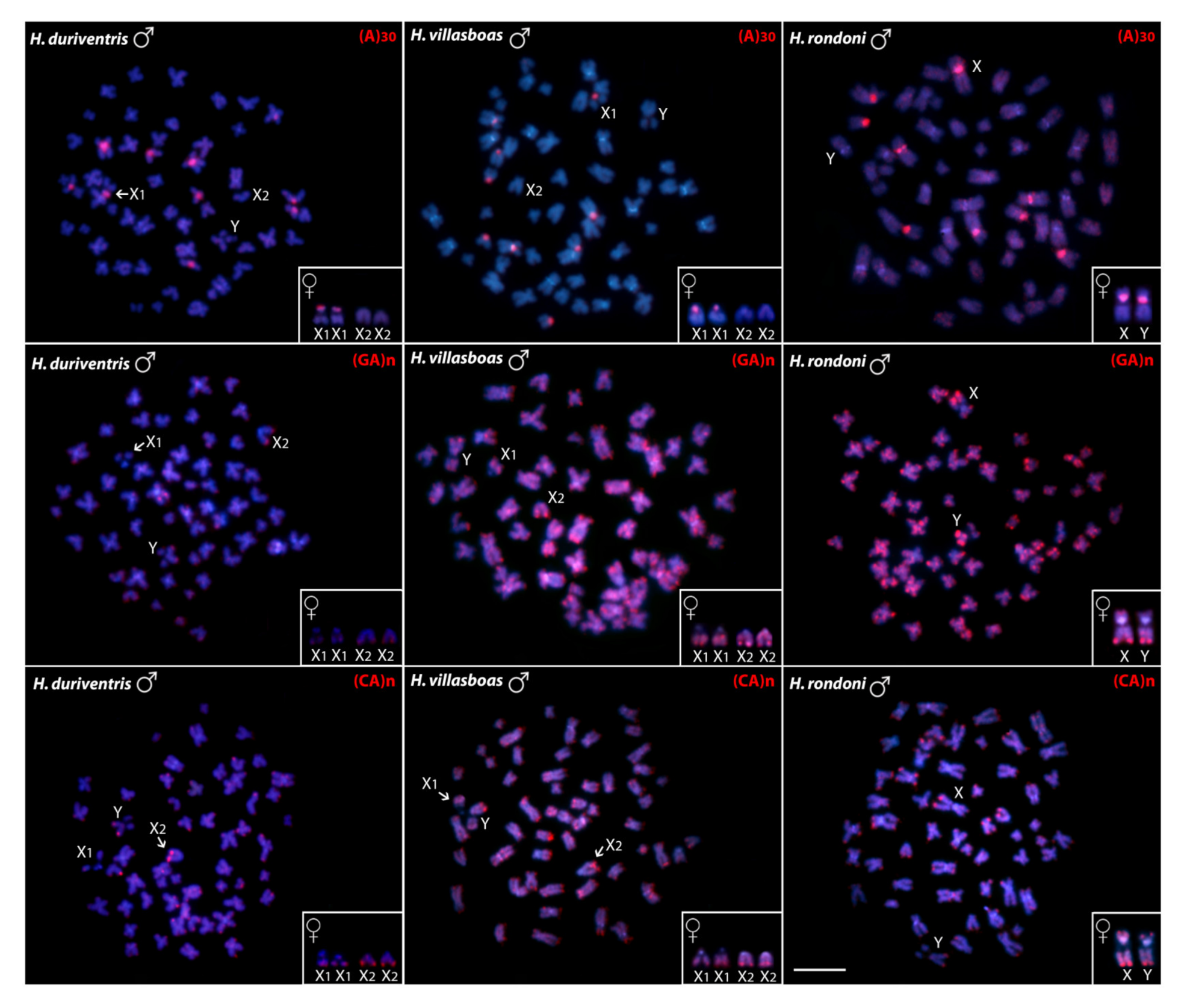
3.1. Karyotypes, C-banding and Sex Chromosomes

Harttia rondoni has 2n = 54 chromosomes (20m + 26sm + 4st + 4a) in both sexes. In turn, H. villasboas and H. duriventris have 2n = 56 chromosomes in the female specimens (18m+24sm+6st+8a in H. villasboas and 16m + 16sm + 16st + 8a in H. duriventris), but 2n = 55 chromosomes in the male specimens (19m + 24sm + 6st + 6a in H. villasboas and 17m + 16sm + 16st + 6a in H. duriventris). These specific male karyotypes are due to characteristics X1X1X2X2/X1X2Y multiple sex chromosome systems, where the Y corresponds to a medium-sized m chromosome (Figure 2).
In all species, C-positive heterochromatic bands are found in the pericentromeric region of all chromosomes and in the telomeric region of the acrocentric pairs (Figure 2). In general, C-banding does not identify any chromosome heteromorphism in H. rondoni females. However, in males, a slight heteromorphic pattern occurs at the proximal C-positive bands on the long arms of the pair 11 (Figure 2). This points to a likely XX/XY sex chromosome system in this species. On the other hand, no conspicuous heterochromatin accumulation is observed in the Y chromosome of H. duriventris and H. villasboas.

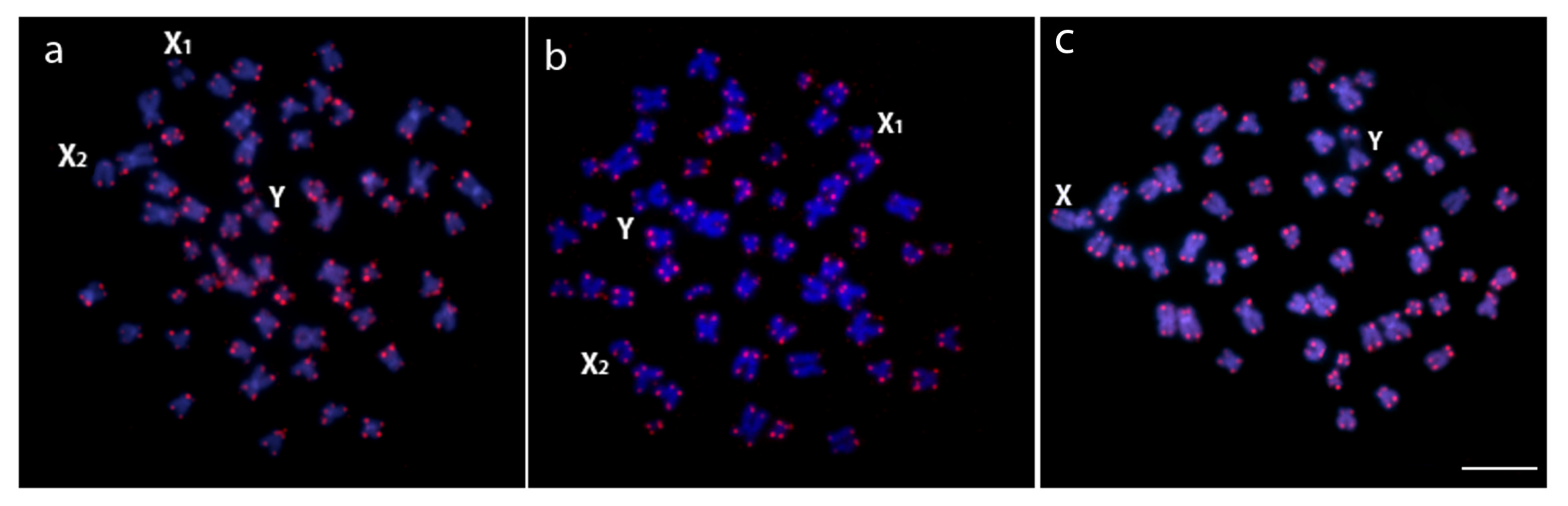
3.2. Chromosomal Distribution of rDNAs, Microsatellite Motifs and Telomeric Repeats

Cytogenetic mapping of the 18S and 5S rDNA sequences showed single sites in corresponding chromosomes of all three analyzed species. Specifically, the 18S rDNA sequences are mapped in the pericentromeric region of the XY chromosomes of H. rondoni, where a clear polymorphic state occurs in males (Figure 2). In both H. duriventris and H. villasboas, these sequences are located in both X1 (in females) and in the X1 and Y (in males). The statement that these rDNA loci are located on the X1 chromosome instead of in the X2 is based on available data showing that these sequences are also found in the corresponding X chromosome of the sister species, H. rondoni.
The A(30) motif showed hybridization in nine male chromosomes and in ten female chromosomes in all three species, this difference being due to the absence of signals in the Y chromosomes. Besides, H. rondoni also presents some small scattered marks in all other chromosomes for this same microsatellite. For the GA(15) probe, small scattered signals occur in the chromosomes of all species, with accumulation on telomeric regions. However, the sex chromosomes also present different accumulation patterns for this microsatellite: on the telomeric region of the X2 and the Y chromosomes of H. duriventris, on the telomeric regions of the X1, X2 and Y of H. villasboas, and strongly accumulated in the telomeric region of both X and Y chromosomes of H. rondoni. Additionally, CA(15) were identified in the telomeric regions of almost all chromosomes, of H. villasboas, including the sex ones (Figure 3).
FISH with telomeric (TTAGGG)n probe applied to male metaphases of H. duriventris, H. villasboas and H. rondoni revealed hybridization signals on the telomeres of all chromosomes in both species, with no additional interstitial telomeric sites (ITS) (Figure 4).

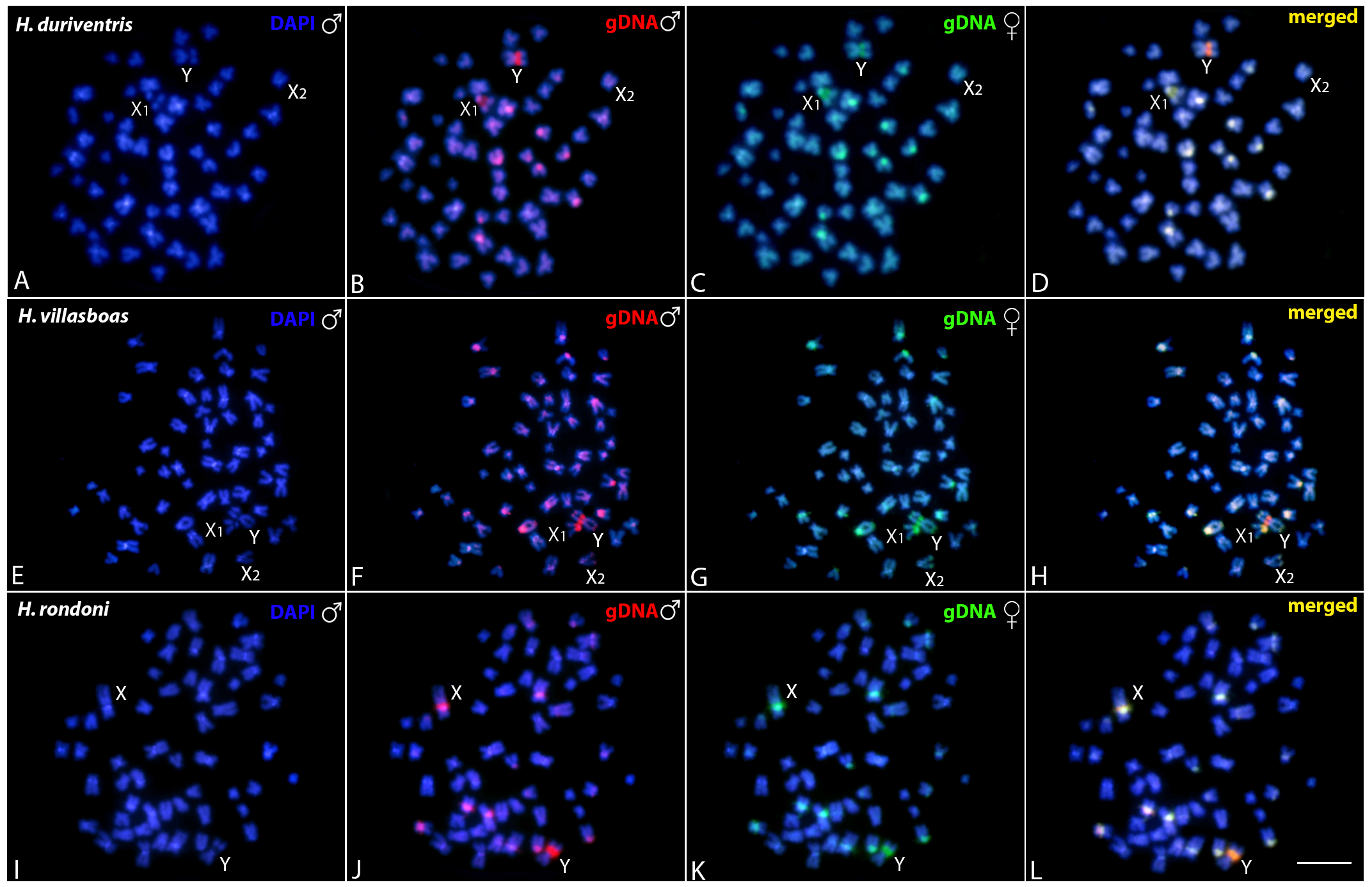
3.3. Comparative Genomic Hybridization (CGH)

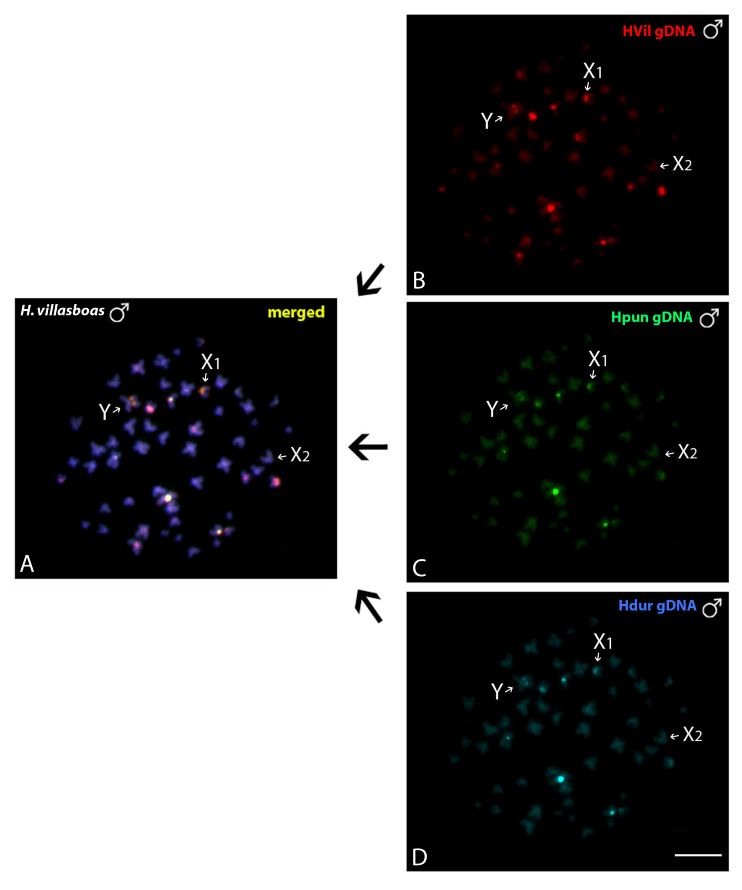
3.3.1. Intraspecific Genomic Relationships: Detecting Male-Specific Regions

In all species, CGH procedure failed to detect any conspicuous sex-specific region on male chromosomes (Figure 5). However, a slight binding preference for the male-derived probe to the pericentromeric region of the Y chromosome of H. villasboas and H. duriventris, and to the Y chromosome of H. rondoni was evidenced (Figure 5). Female-derived probe produced only a faint hybridization signal in such regions, while both probes matched equally the large heterochromatic pericentromeric segment in the X1 chromosome of H. villasboas and H. duriventris, and in the X chromosome of H. rondoni (Figure 5).

3.3.2. Interspecific Genomic Relationships, Focusing on the Multiple X1X2Y Sex System

When the gDNA of H. duriventris, H. villasboas and H. punctata (all X1X2Y-species) was compared, no species-specific region in the sex-chromosomes were observed, thus pointing to their common genomic content (Figure 6).

4. Discussion

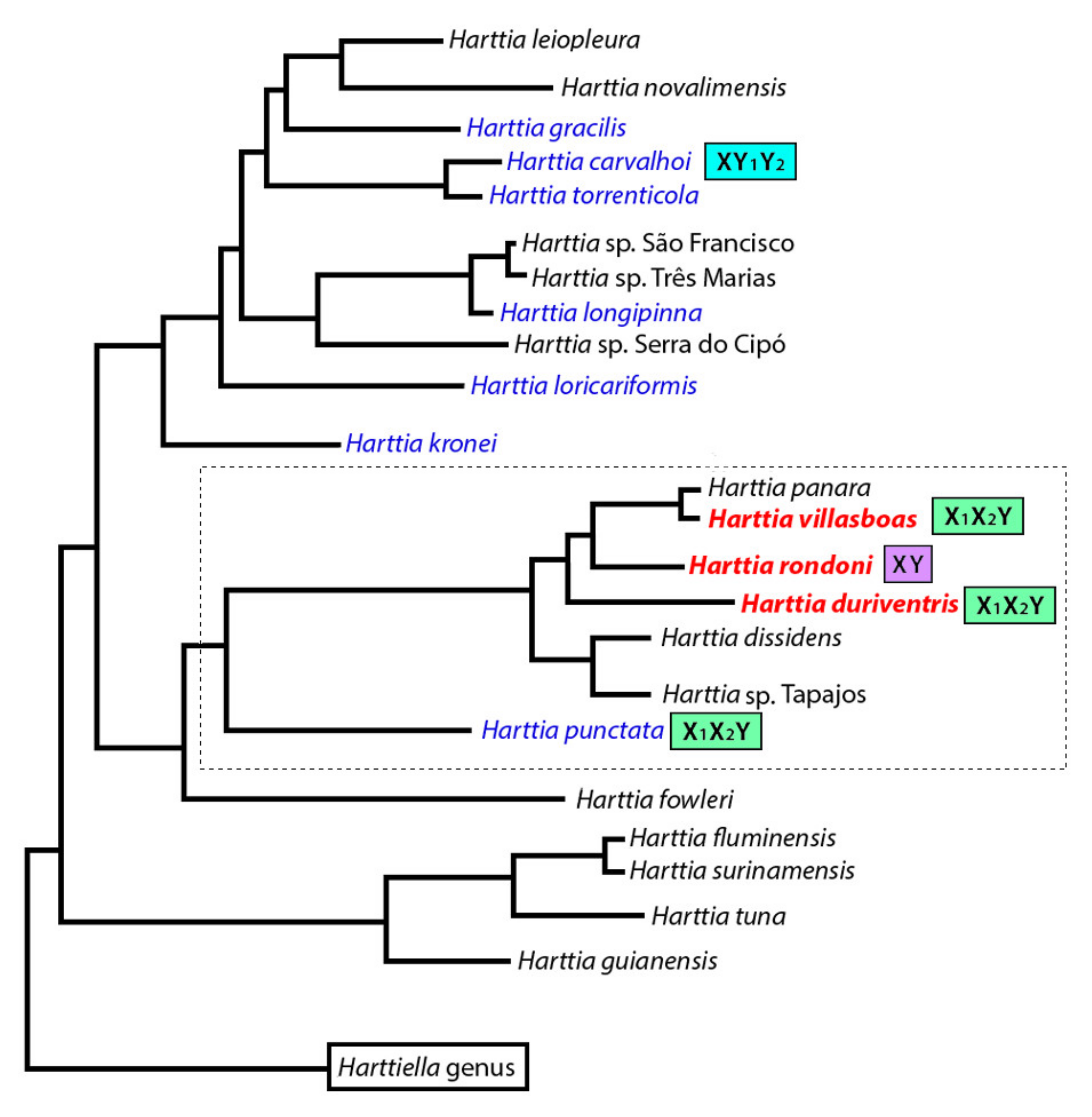
4.1. Evolutionary Relationships among Harttia Species

Chromosome data have shown that Harttia species, as a whole, presents an extensive variation in the diploid number, karyotype composition, in addition to multiple sex chromosomes in some species [5,6,7,8,10,11,12], the latter corresponding to an uncommon condition among fishes. Here we brought new data for three yet unexplored Amazonian species. If we consider previous available data, it is noticeable that the chromosome numbers of all species now investigated fit into the range of variation previously described for the genus, i.e., 2n = 52♀/53♂ in H. carvalhoi [8], and 2n = 62♀♂ in H. absaberi [9]. In addition, the general trend of single rDNA locus occurring in rearranged chromosomes is maintained within the genus [13]. However, a striking feature is that they all present new cases of sex chromosomes: an X1X1X2X2/X1X2Y system in H. duriventris and H. villasboas and a proto or a neo-XY system in H. rondoni (discussed below).
The karyotype diversification and morphological patterns are often indicators of the lifestyle of a species [32,33]. Although Harttia have a wide geographic distribution in many South American rivers and small streams, their low vagility fosters the fixation of chromosomal rearrangements into small populations, thus promoting chromosomal diversity. Indeed, several other fish species also presenting the same above characteristics, have been evidenced as carriers of a wide variety of karyotypes: A. fasciatus, for example, presents karyotypes with 2n = 45, 46, 47, 48 and 50 chromosomes [34,35,36] and Hoplias malabaricus, with seven main karyotypes including multiple and simple sex chromosome systems [23,37,38,39]. In Late Cretaceous and Cenozoic, large-scale tectonic events led to changes in river courses and watershed limits, resulting in complex river dynamisms [40], and affecting the distribution of fish populations. For example, the Serra do Cachimbo region (Pará-Brazil), presents highlands that reach 740 m of altitude, divided between Xingu and Tapajós river basins [41]. Located at the northern border of the Brazilian shield, this region has a high number of endemic species such as Leporinus guttatus [42], three species of Lebiasina genus [43], and Harttia villasboas and H. panara [41]. This high number of endemic species can be attributed by the high number of headwaters of the Xingu and Tapajós rivers [41]. There, H. rondoni and H. villasboas have a probable geographic barrier due to a series of waterfalls 40–60 m height, over a 50 m stretch, where H. rondoni and H. villasboas occur below and above this set of waterfalls, respectively [41,44]. Despite some karyotype similarities that they share, such as the higher number of m and sm chromosomes, a vicariance event may have facilitated the fixation of another series of chromosomal rearrangements in both groups, including the origin of two distinct, but related sex chromosome systems (a proto or- neo-XY system in H. rondoni and a multiple X1X1X2X2/X1X2Y system in H. villasboas). The generation of multiple sex chromosome systems usually involves centric fusions or fissions events and may retain vestiges of interstitial telomeric sequences (ITS) [45]. Our chromosomal mapping of telomeric sequences only reveal the expected terminal signals, without any ITS in the three species. However, it is also known that Robertsonian rearrangements can also lead to the loss or reduction of sequences close to the chromosomal breakpoints [45], and it is likely that this particular condition is responsible for the absence of ITS, as well as of microsatellite sequences and C-positive heterochromatin that, although identified on the X1 and X2 chromosomes of H. duriventris and H. villasboas, they are missing in the Y chromosome.
Blanco [13] discussed the split of H. punctata from a southeast clade with the origin of its multiple sex chromosome system being a characteristic determinant, probably originated from an ancestor with 2n = 58 chromosomes without a differentiated sex chromosome system. Now, one specific chromosomal pair present in H. rondoni shares similar patterns to the X1X2Y sex chromosomes present in the other closely related species, opening two main scenarios: (i) a proto-XY hypothesis: The X and Y chromosomes differ only slightly due to the amplification of repetitive sequences (major rDNAs) on the X chromosome, thus representing an early stage of differentiation. As all its closely related species until now studied have sex chromosomes (Figure 7, dotted line), we could hypothesize that this 2n = 58 ancestor have also carried a proto-XY sex system, as is the case of H. rondoni. Afterwards, chromosomal fissions on the X chromosomes may have created the X1 and X2 chromosomes present in H. punctata, H. duriventris and H. villasboas. (ii) A Neo-XY hypothesis: chromosomal fusions involving the ancestral X and Y chromosomes and a pair of autosomes could create such a neo-XY sex system, leading to the reduction of the 2n from 56 (present in the sister species) to 54 in H. rondoni. Besides, the bigger size of the neo-X chromosome in comparison with the Y and the loss of a A(n) rich region closer to the ribosomal loci in the later support this hypothesis. Among fishes, some cases of neo-XY sex chromosomes emerging from X and/or Y-autosomal translocations have been also observed [46,47,48]. Neo-sex chromosomes may not necessarily lead to the emergence of multiple sex chromosomes, as equal addition of autosomal segments to both sex chromosomes generates neo-XY or neo-ZW systems. Complementary studies, focusing on whole chromosome painting and in the genomic organization of these sex chromosomes, are necessary to clarify these two abovementioned hypotheses.

4.2. The Genus Harttia as A Repository of Multiple Sex Chromosome Systems

Teleost fishes represent one of the most diverse groups in terms of sex determination and differentiation [49,50]. Heteromorphic sex chromosomes are identified in about 5% of so far analyzed species [51], mostly corresponding to simple systems, the XX/XY being the most frequent one [52]. Multiple sex chromosomes occurs in a much lower number and, according to Pennel [53], 47 of such occurrences had been so far recorded, encompassing five different kinds of systems, named ♀X1X1X2X2/♂X1X2Y, ♀XX/♂XY1Y2, ♀X1X1X2X2/♂X1Y1X2Y2, ♂ZZ/♀ZW1W2 and ♂Z1Z1Z2Z2/♀Z1W1Z2W2, the first of them being the most prevalent one [54]. Here, a marked feature of both H. villasboas and H. duriventris karyotypes is an X1X2Y sex chromosome system. Such occurrences, together with the two previous multiple systems described for H. punctata (X1X2Y) and H. carvalhoi (XY1Y2), makes Harttia the genus with the most abundant number of multiple sex chromosome systems up to now identified among fishes [53].
It is known that Y-A fusions are the most common rearrangements related to the origin of X1X2Y systems [53] and it is likely that this is also true for both H. duriventris and H. villasboas cases. But the find of the concomitant proto/neo XY-system in the sister species, H. rondoni, adds a relevant question in this evolutionary puzzle. In such system the sex pair differ only slightly by the accumulation of repetitive sequences in only one of the chromosomes, i.e., the Y one. This heteromorphism was not always clearly detected on the pattern of the C-positive heterochromatin, but it was evident after the 18S rDNA mapping and the intraspecific CGH experiments. This scenario alone could raise doubts about the existence of such a proto-XY system, reinforcing the probable neo-origin to this chromosomal sex system. However, when the chromosomal pattern of H. rondoni is compared with those of H. villasboas and H. duriventris, it is possible to establish the karyological relationships among these species supporting the real occurrence of the putative sex chromosomes in H. rondoni, and the origin of the multiple X1X2Y sex chromosome systems in the two latter species. In fact, comparative genomic hybridization (CGH), plus chromosomal mapping of microsatellites and rDNAs repeats highlight that these three Harttia species share the same distribution pattern in the sex chromosomes. Although H. punctata also presents an X1X2Y system, its rDNA distribution includes both 5S and 18S sequences on the sex chromosomes [11,13], which is different from H. duriventris and H. villasboas where only 18S motifs occur on them, probably by a translocation of the 5S rDNA motif, present in the X1 of H. punctata, to autosomal chromosomes during lineage diversification in H. duriventris, H. villasboas and H. rondoni.
Meaningly, a close association between microsatellites, rDNAs and multiple sex chromosomes has been reported for many fish taxa [55,56,57,58,59], which highlights the probable role of these sequences in the genesis of such systems. Despite our CGH data do not reveal any conspicuous Y-specific region, neither in both Y chromosomes of H. duriventris and H. villasboas, nor in the proto/neo-Y of H. rondoni, a slight binding preference for the male-derived probe occur at the pericentromeric region of all these chromosomes (Figure 5). Potential effects of repetitive DNA accumulation on recombination rate have been considered [60], what could explain their initial accumulation on the sex chromosomes. When sex chromosomes stop recombination, repetitive sequences are predicted to have a rapid accumulation on them [61], and microsatellite repeats seem to play a key role as “early colonizers” in their differentiation [24,62]. Numerous examples in animals and plants document a massive and differential accumulation of such small motifs in sex-specific chromosomes, particularly in simple (XY or ZW) systems [24,56,63,64,65,66,67]. However, most X1X2Y systems lack substantial differentiation in the neo-Y, since the accumulation of repetitive DNAs (= large blocks of heterochromatin) would impair the proper pairing of the neo-sex chromosomes into a stable trivalent form, thus disturbing the meiotic process [68]. Here, no significant differences in microsatellites distribution could be identified concerning autosomes and sex chromosomes of the Harttia species. In general, while (A)30 sequences presented a strong accumulation pattern in the pericentromeric regions of some chromosomal pairs, both (CA)15 and (GA)15 displayed a widespread distribution pattern, with preferential accumulation in some telomeric regions (Figure 3). Besides, as evidenced by C-banding and CGH experiments, all the X1, X2 and Y chromosomes do not accumulate repetitive sequences, and the Y chromosome of H. rondoni is even missing (A)30 sequences probably lost after the chromosomal rearrangements related to its genesis. Thus, the recombination suppression in multiple sex chromosome system is a question that deserves to be better clarified.
According to the most updated phylogeny proposed for the genus Harttia [19]. H. duriventris, H. villasboas, and H. punctata, all harboring multiple X1X2Y sex system, are evolutionary related and belong to the same major clade. In accordance with, our CGH experiments showed that they sex chromosomes share genomic content, thus pointing to their relatedness and to a probable common origin. This is a characteristic that, in general, is not found among teleosts species. Although some Megaleporinus [69], Parodon [70] and Characidium species [71], as well as the whole genus Triportheus [66,72], have their sex chromosomes with a common origin and differentiation, this is an exceptional scenario [73]. In fact, an independent origin is more commonly found, even among congeneric species [49,52,74,75,76,77,78]. According to Schartl [79], such a situation could be explained by the emergence of sex-determining genes on different chromosomes which, by mutation, would promote male or female development and giving rise to new sex chromosomes. Next steps, implying finer-scale approaches such as Zoo-FISH experiments with whole chromosome painting (WCP), coupled with recent genome sequencing procedures, will shed more light on this issue, especially in the recent or ancient origin of the XY system of H. rondoni and its significance to the group.

5. Conclusions

Chromosomal data of the Brazilian northern Harttia species (H. duriventris, H. villaboas, H. rondoni and H. punctata) support the common origin for their sex chromosome systems. Additionally, the results also allowed us: (i) to track their evolutionary relationships with other Harttia species, adding new light on their relatedness inside the genus; (ii) to describe two new multiple sex chromosomes systems of the X1X2Y type; and (iii) to highlight a proto or neo-XY system in H. rondoni and its close association with the aforementioned multiple sex systems. The remarkable variation on karyotypic organization, in addition to the frequency and different types of sex chromosomes systems inside the genus, makes Harttia an useful model for evolutionary studies among fish focusing on karyotype differentiation and sex chromosomes evolution.

Author Contributions

Conceptualization, F.d.M.C.S., G.A.D., O.M.-F., M.R.V., M.B.C.; Formal analysis, F.d.M.C.S., G.A.D., O.M.-F., M.R.V., T.L., E.A.d.O., M.B.C.; Funding acquisition, O.M.-F., L.A.C.B., M.B.C.; Investigation, F.d.M.C.S., O.M.-F., M.B.C.; Methodology, F.d.M.C.S., G.A.D., E.A.d.O; Project administration, L.A.C.B., T.L., M.B.C.; Resources, T.L.; Supervision, O.M.-F., M.B.C; Validation, T.L., E.A.O.; Visualization, F.d.M.C.S., G.A.D., O.M.-F., M.R.V., L.A.C.B., T.L., E.A.d.O., M.B.C.; Writing—original draft, F.d.M.C.S.; G.A.D., M.R.V., M.B.C.; Writing—review and editing, F.d.M.C.S., G.A.D., O.M.-F., M.R.V., L.A.C.B., T.L., E.A.d.O., M.B.C. All authors have read and agreed to the published version of the manuscript.

Funding

MBC was supported by the Conselho Nacional de Desenvolvimento Científico e Tecnológico (CNPq) (302449/2018-3) and Fundação de Amparo à Pesquisa do Estado de São Paulo (FAPESP) (2018/22033-1). FMCS was supported by the Fundação de Amparo à Pesquisa do Estado de São Paulo (FAPESP) (2020/02681-9). This study was financed in part by the Coordenação de Aperfeiçoamento de Pessoal de Nível Superior, Brasil (CAPES), Finance Code 001.

Acknowledgments

The authors are grateful to Osvaldo Oyakawa (curator of the fish collection of the Museu de Zoologia da Universidade de São Paulo (MZUSP) for the proper specimens’ identification.

Conflicts of Interest

The authors declare no conflict of interest. The funders had no role in the design of the study; in the collection, analyses, or interpretation of data; in the writing of the manuscript, or in the decision to publish the results.

References

  1. Ferraris, C.J. Checklist of Catfishes, Recent and Fossil (Osteichthyes: Siluriformes), and Catalogue of Siluriform Primary Types; Magnolia Press: Auckland, New Zealand, 2007; ISBN 9781869770587. [Google Scholar]
  2. Fricke, R.; Eschmeyer, W.N.; van der Laan, R. Eschmeyer’s Catalog of Fishes: Genera, Species. Available online: http://researcharchive.calacademy.org/research/ichthyology/catalog/fishcatmain.asp (accessed on 12 February 2020).
  3. Traldi, J.B.; Lui, R.L.; Martinez, J.d.F.; Vicari, M.R.; Nogaroto, V.; Filho, O.M.; Blanco, D.R. Chromosomal distribution of the retroelements Rex1, Rex3 and Rex6 in species of the genus Harttia and Hypostomus (Siluriformes: Loricariidae). Neotrop. Ichthyol. 2019, 17, 1–10. [Google Scholar] [CrossRef] [Green Version]
  4. De Oliveira, J.C.; Oyakawa, O.T. New loricariid fishes from headwaters on Serra da Mantiqueira and Complexo do Espinhaço, Minas Gerais state, Brazil (Teleostei: Siluriformes: Loricariidae). Zootaxa 2019, 4586, 401–424. [Google Scholar] [CrossRef] [PubMed]
  5. Alves, A.L.; Oliveira, C.; Foresti, F. Karyotype variability in eight species of the subfamilies Loricariinae and Ancistrinae (Teleostei, Siluriformes, Loricariidae). Caryologia 2003, 56, 57–63. [Google Scholar] [CrossRef] [Green Version]
  6. Kavalco, K.F.; Pazza, R.; Bertollo, L.A.C.; Moreira-Filho, O. Heterochromatin characterization of four fish species of the family Loricariidae (Siluriformes). Hereditas 2004, 141, 237–242. [Google Scholar] [CrossRef] [PubMed]
  7. Kavalco, K.F.; Pazza, R.; Bertollo, L.A.C.; Moreira-Filho, O. Karyotypic diversity and evolution of Loricariidae (Pisces, Siluriformes). Heredity 2005, 94, 180–186. [Google Scholar] [CrossRef] [PubMed] [Green Version]
  8. Centofante, L.; Bertollo, L.A.C.; Moreira-Filho, O. Cytogenetic characterization and description of an XX/XY1Y2 sex chromosome system in catfish Harttia carvalhoi (Siluriformes, Loricariidae). Cytogenet. Genome Res. 2006, 112, 320–324. [Google Scholar] [CrossRef] [PubMed]
  9. Rodrigues, R.M. Estudos Cromossômicos e Moleculares em Loricariinae Com Ênfase em Espécies de Rineloricaria (Siluriformes, Loricariidae): Uma Perspectiva Evolutiva; Universidade de São Paulo: São Paulo, Brazil, 2010. [Google Scholar]
  10. Blanco, D.R.; Vicari, M.R.; Artoni, R.F.; Traldi, J.B.; Moreira-Filho, O. Chromosomal Characterization of Armored Catfish Harttia longipinna (Siluriformes, Loricariidae): First Report of B Chromosomes in the Genus. Zool. Sci. 2012, 29, 604–609. [Google Scholar] [CrossRef]
  11. Blanco, D.R.; Vicari, M.R.; Lui, R.L.; Bertollo, L.A.C.; Traldi, J.B.; Moreira-Filho, O. The role of the Robertsonian rearrangements in the origin of the XX/XY1Y2 sex chromosome system and in the chromosomal differentiation in Harttia species (Siluriformes, Loricariidae). Rev. Fish Biol. Fish. 2013, 23, 127–134. [Google Scholar] [CrossRef]
  12. Blanco, D.R.; Vicari, M.R.; Lui, R.L.; Artoni, R.F.; de Almeida, M.C.; Traldi, J.B.; Margarido, V.P.; Moreira-Filho, O. Origin of the X1X1X2X2/X1X2Y sex chromosome system of Harttia punctata (Siluriformes, Loricariidae) inferred from chromosome painting and FISH with ribosomal DNA markers. Genetica 2014, 142, 119–126. [Google Scholar] [CrossRef]
  13. Blanco, D.R.; Vicari, M.R.; Lui, R.L.; Traldi, J.B.; Bueno, V.; Martinez, J.d.F.; Brandão, H.; Oyakawa, O.T.; Moreira Filho, O. Karyotype Diversity and Evolutionary Trends in Armored Catfish Species of the Genus Harttia (Siluriformes: Loricariidae). Zebrafish 2017, 14, 169–176. [Google Scholar] [CrossRef]
  14. Cioffi, M.B.; Bertollo, L.A.C.; Garrido-Ramos, M.A.; Genome Dynamics, V. Chromosomal Distribution and Evolution of Repetitive DNAs in Fish. Genome Dyn. 2012, 7, 197–221. [Google Scholar] [PubMed]
  15. Barby, F.F.; Bertollo, L.A.C.; de Oliveira, E.A.; Yano, C.F.; Hatanaka, T.; Ráb, P.; Sember, A.; Ezaz, T.; Artoni, R.F.; Liehr, T.; et al. Emerging patterns of genome organization in Notopteridae species (Teleostei, Osteoglossiformes) as revealed by Zoo-FISH and Comparative Genomic Hybridization (CGH). Sci. Rep. 2019, 9, 1112. [Google Scholar] [CrossRef] [PubMed] [Green Version]
  16. de Menezes Cavalcante Sassi, F.; Aguiar de Oliveira, E.; Bertollo, L.A.C.; Nirchio, M.; Hatanaka, T.; Marinho, M.M.F.; Moreira-Filho, O.; Aroutiounian, R.; Liehr, T.; Al-Rikabi, A.B.H.; et al. Chromosomal evolution and evolutionary relationships of Lebiasina species (Characiformes, Lebiasinidae). Int. J. Mol. Sci. 2019, 20, 2944. [Google Scholar]
  17. de Moraes, R.L.R.; Sember, A.; Bertollo, L.A.; De Oliveira, E.A.; Rab, P.; Hatanaka, T.; Marinho, M.M.; Liehr, T.; Al-Rikabi, A.B.; Feldberg, E.; et al. Comparative cytogenetics and neo-Y formation in small-sized fish species of the genus Pyrrhulina (Characiformes, Lebiasinidae). Front. Genet. 2019, 10, 1–13. [Google Scholar] [CrossRef] [Green Version]
  18. Toma, G.A.; De Moraes, R.L.R.; Sassi, F.D.M.C.; Bertollo, L.A.C.; De Oliveira, E.A.; Rab, P.; Sember, A.; Liehr, T.; Hatanaka, T.; Viana, P.F.; et al. Cytogenetics of the small-sized fish, Copeina guttata (Characiformes, Lebiasinidae): Novel insights into the karyotype differentiation of the family. PLoS ONE 2019, 14, 1–13. [Google Scholar] [CrossRef]
  19. Covain, R.; Fisch-Muller, S.; Oliveira, C.; Mol, J.H.; Montoya-Burgos, J.I.; Dray, S. Molecular phylogeny of the highly diversified catfish subfamily Loricariinae (Siluriformes, Loricariidae) reveals incongruences with morphological classification. Mol. Phylogenetics Evol. 2016, 94, 492–517. [Google Scholar] [CrossRef]
  20. Bertollo, L.A.C.; Cioffi, M.d.B.; Galetti, P.M.; Filho, O.M. Contributions to the cytogenetics of the Neotropical fish fauna. Comp. Cytogenet. 2017, 11, 665–690. [Google Scholar] [CrossRef] [Green Version]
  21. Sumner, A.T. A simple technique for demonstrating centromeric heterochromatin. Exp. Cell Res. 1972, 75, 304–306. [Google Scholar] [CrossRef]
  22. Pendás, A.M.; Móran, P.; Freije, J.P.; Garcia-Vásquez, E. Chromosomal location and nucleotide sequence of two tandem repeats of the Atlantic salmon 5S rDNA. Cytogenet. Genome Res. 1994, 67, 31–36. [Google Scholar] [CrossRef]
  23. Cioffi, M.d.B.; Martins, C.; Centofante, L.; Jacobina, U.P.; Bertollo, L.A.C. Chromosomal variability among allopatric populations of Erythrinidae fish Hoplias malabaricus: Mapping of three classes of repetitive DNAs. Cytogenet. Genome Res. 2009, 125, 132–141. [Google Scholar] [CrossRef]
  24. Kubat, Z.; Hobza, R.; Vyskot, B.; Kejnovský, E. Microsatellite accumulation in the Y chromosome of Silene latifolia. Genome 2008, 51, 350–356. [Google Scholar] [CrossRef] [PubMed] [Green Version]
  25. Yano, C.F.; Bertollo, L.A.C.; Cioffi, M.B. Fish-FISH: Molecular cytogenetics in fish species. In Fluorescence in Situ Hybridization (FISH)- Application Guide; Liehr, T., Ed.; Springer: Berlin, Germany, 2017; pp. 429–444. [Google Scholar]
  26. Sambrook, J.; Russell, D.W. Molecular Cloning, A Laboratory Manual; Cold Spring Harbor Laboratory Press: New York, NY, USA, 2001. [Google Scholar]
  27. Zwick, M.S.; Hanson, R.E.; Mcknight, T.D.; Islam-Faridi, M.H.; Stelly, D.M.; Wing, R.A.; Price, H.J. A rapid procedure for the isolation of C 0 t-1 DNA from plants. Genome 1997, 40, 138–142. [Google Scholar] [CrossRef] [PubMed] [Green Version]
  28. de Moraes, R.L.R.; Bertollo, L.A.C.; Marinho, M.M.F.; Yano, C.F.; Hatanaka, T.; Barby, F.F.; Troy, W.P.; Cioffi, M.D.B. Evolutionary relationships and cytotaxonomy considerations in the genus Pyrrhulina (Characiformes, Lebiasinidae). Zebrafish 2017, 14, 536–546. [Google Scholar] [CrossRef] [PubMed]
  29. Sassi, F.M.C.; Hatanaka, T.; Moraes, R.L.R.; Toma, G.A.; Oliveira, E.A.; Liehr, T.; Rab, P.; Bertollo, L.A.C.; Viana, P.F.; Feldberg, E.; et al. An insight into the chromosomal evolution of Lebiasinidae (Teleostei, Characiformes). Genes 2020, 11, 365. [Google Scholar]
  30. Symonová, R.; Sember, A.; Majtánová, Z.; Ráb, P. Characterization of fish genome by GISH and CGH. In Fish Cytogenetic Techniques. Ray-Fin Fishes and Chondrichthyans; Ozouf-Costaz, C., Pisano, E., Foresti, F., de Almeida-Toledo, L.F., Eds.; CRC Press: Boca Raton, FL, USA, 2015; pp. 118–131. [Google Scholar]
  31. Levan, A.; Fredga, K.; Sandberg, A.A. Nomenclature for centromeric position on chromosomes. Hereditas 1964, 52, 201–220. [Google Scholar] [CrossRef]
  32. Karr, J.; James, F.C. Eco-morphological configurations and convergent evolution in species and communities. In Ecology and Evolution of Communities; Cody, M.L., Diamond, J.M., Eds.; Harvard University Press: Cambridge, UK, 1975; pp. 258–291. [Google Scholar]
  33. Wainwright, P.C.; Reilly, S.M. Ecological Morphology: Integrative Organismal Biology; University of Chicago Press: Chicago, IL, USA, 1994. [Google Scholar]
  34. Pazza, R.; Kavalco, K.F.; Bertollo, L.A.C. Chromosome polymorphism in Astyanax fasciatus (Teleostei, Characidae). 1. Karyotype analysis, Ag-NORs and mapping of the 18S and 5S ribosomal genes in sympatric karyotypes and their possible hybrid forms. Cytogenet. Genome Res. 2006, 112, 313–319. [Google Scholar] [CrossRef]
  35. Ferreira-Neto, M.; Artoni, R.; Vicari, M.; Moreira Filho, O.; Camacho, J.; Bakkali, M.; Oliveira, C.; Foresti, F. Three sympatric karyomorphs in the fish Astyanax fasciatus (Teleostei, Characidae) do not seem to hybridize in natural populations. Comp. Cytogenet. 2012, 6, 29–40. [Google Scholar] [CrossRef] [Green Version]
  36. Pazza, R.; Dergam, J.A.; Kavalco, K.F. Trends in karyotype evolution in Astyanax (Teleostei, Characiformes, Characidae): Insights from molecular data. Front. Genet. 2018, 9, 131. [Google Scholar] [CrossRef]
  37. Bertollo, L.A.C.; Born, G.G.; Dergam, J.A.; Fenocchio, A.S.; Moreira-Filho, O. A biodiversity approach in the neotropical Erythrinidae fish, Hoplias malabaricus. Karyotypic survey, geographic distribution of cytotypes and cytotaxonomic considerations. Chromosome Res. 2000, 8, 603–613. [Google Scholar] [CrossRef]
  38. Vicari, M.R.; Artoni, R.F.; Bertollo, L.A.C. Comparative cytogenetics of Hoplias malabaricus (Pisces, Erythrinidae): A population analysis in adjacent hydrographic basins. Genet. Mol. Biol. 2005, 110, 103–110. [Google Scholar] [CrossRef] [Green Version]
  39. Ibagón, N.; Maldonado-Ocampo, J.A.; Cioffi, M.d.B.; Dergam, J.A. Chromosomal Diversity of Hoplias malabaricus (Characiformes, Erythrinidae) Along the Magdalena River (Colombia—Northern South America) and Its Significance for the Neotropical Region. Zebrafish 2020, 17, 211–219. [Google Scholar] [CrossRef]
  40. Lundberg, J.G.; Marshall, L.G.; Guerrero, J.; Horton, B.; Malabarba, M.; Wesselingh, F. The stage for Neotropical fish diversification: A history of tropical South American rivers. In Phylogeny and Classification of Neotropical Fishes; Malabarba, L.R., Reis, R.E., Vari, R.P., Lucena, C.A.S., Eds.; Edipucrs Porto Alegre: Porto Alegre, Brazil, 1998; Volume 27, pp. 13–48. [Google Scholar]
  41. Oyakawa, O.T.; Fichberg, I.; Py-Daniel, L.R. Three new species of Harttia (Loricariidae: Loricariinae) from Serra do Cachimbo, Rio Xingu basin, Pará, Northern Brazil. Zootaxa 2018, 4387, 75–90. [Google Scholar] [CrossRef] [PubMed]
  42. Birindelli, J.L.O.; Britski, H.A. New species of the genus Leporinus Agassiz (Characiformes: Anostomidae) from the rio Curuá, rio Xingu basin, Serra do Cachimbo, Brazil, with comments on Leporinus reticulatus. Neotrop. Ichthyol. 2009, 7, 1–10. [Google Scholar] [CrossRef] [Green Version]
  43. Netto-Ferreira, A.L. Three new species of Lebiasina (Characiformes: Lebiasinidae) from the Brazilian shield border at Serra do Cachimbo, Pará, Brazil. Neotrop. Ichthyol. 2012, 10, 487–498. [Google Scholar] [CrossRef] [Green Version]
  44. Varella, H.R.; Sabaj Pérez, M.H. A titan among dwarfs: Apistogramma kullanderi, new species (Teleostei: Cichlidae). Ichthyol. Explor. Freshw. 2014, 25, 243–258. [Google Scholar]
  45. Meyne, J.; Ratliff, R.L.; Moyzis, R.K. Conservation of the human telomere sequence (TTAGGG)n among vertebrates. Proc. Natl. Acad. Sci. USA 1989, 86, 7049–7053. [Google Scholar] [CrossRef] [PubMed] [Green Version]
  46. de Freitas, N.L.; Al-Rikabi, A.B.H.; Bertollo, L.A.C.; Ezaz, T.; Yano, C.F.; de Oliveira, E.A.; Hatanaka, T.; de Bello Cioffi, M. Early Stages of XY Sex Chromosomes Differentiation in the Fish Hoplias malabaricus (Characiformes, Erythrinidae) Revealed by DNA Repeats Accumulation. Curr. Genom. 2017, 19, 216–226. [Google Scholar] [CrossRef]
  47. Gammerdinger, W.J.; Kocher, T.D. Unusual Diversity of Sex Chromosomes in African Cichlid Fishes. Genes 2018, 9, 480. [Google Scholar] [CrossRef] [Green Version]
  48. Kottler, V.; Schartl, M. The Colorful Sex Chromosomes of Teleost Fish. Genes 2018, 9, 233. [Google Scholar] [CrossRef] [Green Version]
  49. Schartl, M.; Schmid, M.; Nanda, I. Dynamics of vertebrate sex chromosome evolution: From equal size to giants and dwarfs. Chromosoma 2016, 125, 553–571. [Google Scholar] [CrossRef]
  50. Guiguen, Y.; Pasquier, J.; Fostier, A.; Bobe, J. Evolution of Sex Hormone Binding Globulins Reveals Early Gene Duplication at the Root of Vertebrates. bioRxiv 2020. [Google Scholar] [CrossRef]
  51. Arai, R. Fish Karyotypes: A Check List; Springer Science & Business Media: Heidelberg, Germany, 2011. [Google Scholar]
  52. Devlin, R.H.; Nagahama, Y. Sex determination and sex differentiation in fish: An overview of genetic, physiological, and environmental influences. Aquaculture 2002, 208, 191–364. [Google Scholar] [CrossRef]
  53. Pennell, M.W.; Kirkpatrick, M.; Otto, S.P.; Vamosi, J.C.; Peichel, C.L.; Valenzuela, N.; Kitano, J. Y Fuse? Sex Chromosome Fusions in Fishes and Reptiles. PLoS Genet. 2015, 11, 1–17. [Google Scholar] [CrossRef] [PubMed] [Green Version]
  54. Kitano, J.; Peichel, C.L. Turnover of sex chromosomes and speciation in fishes. Environ. Biol. Fishes 2012, 94, 549–558. [Google Scholar] [CrossRef] [Green Version]
  55. Cioffi, M.B.; Bertollo, L.A.C. Initial steps in XY chromosome differentiation in Hoplias malabaricus and the origin of an X1X2Y sex chromosome system in this fish group. Heredity 2010, 105, 554–561. [Google Scholar] [CrossRef] [Green Version]
  56. Cioffi, M.B.; Camacho, J.P.M.; Bertollo, L.A.C. Repetitive DNAs and differentiation of sex chromosomes in neotropical fishes. Cytogenet. Genome Res. 2011, 132, 188–194. [Google Scholar] [CrossRef]
  57. Bitencourt, J.A.; Sampaio, I.; Ramos, R.T.C.; Vicari, M.R.; Affonso, P.R.A.D.M. First Report of Sex Chromosomes in Achiridae (Teleostei: Pleuronectiformes) with Inferences about the Origin of the Multiple X1X1X2X2/X1X2Y System and Dispersal of Ribosomal Genes in Achirus achirus. Zebrafish 2017, 14, 90–95. [Google Scholar] [CrossRef]
  58. Ferreira, M.; Garcia, C.; Matoso, D.A.; de Jesus, I.S.; Feldberg, E. A new multiple sex chromosome system X1X1X2X2/X1Y1X2Y2 in Siluriformes: Cytogenetic characterization of Bunocephalus coracoideus (Aspredinidae). Genetica 2016, 144, 591–599. [Google Scholar] [CrossRef]
  59. Xu, D.; Sember, A.; Zhu, Q.; De Oliveira, E.A.; Liehr, T.; Al-Rikabi, A.B.H.; Xiao, Z.; Song, H.; De Bello Cioffi, M. Deciphering the Origin and Evolution of the X1X2Y System in Two Closely-Related Oplegnathus Species (Oplegnathidae and Centrarchiformes). Int. J. Mol. Sci. 2019, 20, 3571. [Google Scholar] [CrossRef] [Green Version]
  60. Reed, K.M.; Phillips, R.B. Polymorphism of the nucleolus organizer region (NOR) on the putative sex chromosomes of Arctic char (Salvelinus alpinus) is not sex related. Chromosome Res. 1997, 5, 221–227. [Google Scholar] [CrossRef]
  61. Charlesworth, D.; Charlesworth, B.; Marais, G. Steps in the evolution of heteromorphic sex chromosomes. Heredity 2005, 95, 118–128. [Google Scholar] [CrossRef] [PubMed] [Green Version]
  62. Cioffi, M.B.; Moreira-Filho, O.; Almeida-Toledo, L.F.; Bertollo, L.A.C. The contrasting role of heterochromatin in the differentiation of sex chromosomes: An overview from Neotropical fishes. J. Fish Biol. 2012, 80, 2125–2139. [Google Scholar] [CrossRef]
  63. Phillips, B.C.; Edmands, S. Does the speciation clock tick more slowly in the absence of heteromorphic sex chromosomes? BioEssays 2012, 34, 166–169. [Google Scholar] [CrossRef] [PubMed]
  64. Poltronieri, J.; Marquioni, V.; Bertollo, L.A.C.; Kejnovsky, E.; Molina, W.F.; Liehr, T.; Cioffi, M.B. Comparative chromosomal mapping of microsatellites in Leporinus species (Characiformes, Anostomidae): Unequal accumulation on the W chromosomes. Cytogenet. Genome Res. 2013, 142, 40–45. [Google Scholar] [CrossRef] [PubMed]
  65. Sabath, N.; Itescu, Y.; Feldman, A.; Meiri, S.; Mayrose, I.; Valenzuela, N. Sex determination, longevity, and the birth and death of reptilian species. Ecol. Evol. 2016, 6, 5207–5220. [Google Scholar] [CrossRef] [PubMed] [Green Version]
  66. Yano, C.F.; Bertollo, L.A.C.; Liehr, T.; Troy, W.P.; Cioffi, M.D.B. W Chromosome Dynamics in Triportheus Species (Characiformes, Triportheidae): An Ongoing Process Narrated by Repetitive Sequences. J. Hered. 2016, 107, 342–348. [Google Scholar] [CrossRef] [PubMed] [Green Version]
  67. Viana, P.F.; Ezaz, T.; de Bello Cioffi, M.; Liehr, T.; Al-Rikabi, A.; Goll, L.G.; Rocha, A.M.; Feldberg, E. Landscape of snake’ sex chromosomes evolution spanning 85 MYR reveals ancestry of sequences despite distinct evolutionary trajectories. Sci. Rep. 2020, 10, 1–14. [Google Scholar] [CrossRef] [PubMed]
  68. Moreira-Filho, O.; Bertollo, L.A.C.; Galetti, P.M. Distribution of sex chromosome mechanisms in neotropical fish and description of a ZZ/ZW system in Parodon hilarii (Parodontidae). Caryologia 1993, 46, 115–125. [Google Scholar] [CrossRef] [Green Version]
  69. Ramirez, J.L.; Birindelli, J.L.; Carvalho, D.C.; Affonso, P.R.; Venere, P.C.; Ortega, H.; Carrillo-Avila, M.; Rodríguez-Pulido, J.A.; Galetti, P.M., Jr. Revealing hidden diversity of the underestimated neotropical ichthyofauna: DNA barcoding in the recently described genus Megaleporinus (Characiformes: Anostomidae). Front. Genet. 2017, 8, 149. [Google Scholar] [CrossRef] [Green Version]
  70. Schemberger, M.O.; Bellafronte, E.; Nogaroto, V.; Almeida, M.C.; Schühli, G.S.; Artoni, R.F.; Moreira-Filho, O.; Vicari, M.R. Differentiation of repetitive DNA sites and sex chromosome systems reveal closely related group in Parodontidae (Actinopterygii: Characiformes). Genetica 2011, 139, 1499–1508. [Google Scholar] [CrossRef]
  71. Pucci, M.B.; Nogaroto, V.; Bertollo, L.A.C.; Moreira-Filho, O.; Vicari, M.R. The karyotypes and evolution of ZZ/ZW sex chromosomes in the genus Characidium (Characiformes, Crenuchidae). Comp. Cytogenet. 2018, 12, 421. [Google Scholar] [CrossRef] [PubMed]
  72. Yano, C.F.; Bertollo, L.A.C.; Ezaz, T.; Trifonov, V.; Sember, A.; Liehr, T.; Cioffi, M.B. Highly conserved Z and molecularly diverged W chromosomes in the fish genus Triportheus (Characiformes, Triportheidae). Heredity 2017, 118, 276–283. [Google Scholar] [CrossRef] [PubMed] [Green Version]
  73. Cioffi, M.D.B.; Yano, C.F.; Sember, A.; Bertollo, L.A.C. Chromosomal evolution in lower vertebrates: Sex chromosomes in neotropical fishes. Genes 2017, 8, 258. [Google Scholar] [CrossRef] [PubMed]
  74. Takehana, Y.; Naruse, K.; Hamaguchi, S.; Sakaizumi, M. Evolution of ZZ/ZW and XX/XY sex-determination systems in the closely related medaka species, Oryzias hubbsi and O. dancena. Chromosoma 2007, 116, 463–470. [Google Scholar] [CrossRef] [PubMed]
  75. Tanaka, K.; Takehana, Y.; Naruse, K.; Hamaguchi, S.; Sakaizumi, M. Evidence for different origins of sex chromosomes in closely related oryzias fishes: Substitution of the master sex-determining gene. Genetics 2007, 177, 2075–2081. [Google Scholar] [CrossRef] [PubMed] [Green Version]
  76. Mank, J.E.; Avise, J.C. Evolutionary diversity and turn-over of sex determination in teleost fishes. Sex. Dev. 2009, 3, 60–67. [Google Scholar] [CrossRef]
  77. Schultheis, C.; Böhne, A.; Schartl, M.; Volff, J.N.; Galiana-Arnoux, D. Sex determination diversity and sex chromosome evolution in poeciliid fish. Sex. Dev. 2009, 3, 68–77. [Google Scholar] [CrossRef]
  78. Cioffi, M.B.; Liehr, T.; Trifonov, V.; Molina, W.F.; Bertollo, L.A.C. Independent sex chromosome evolution in lower vertebrates: A molecular cytogenetic overview in the erythrinidae fish family. Cytogenet. Genome Res. 2013, 141, 186–194. [Google Scholar] [CrossRef]
  79. Schartl, M. Sex chromosome evolution in non-mammalian vertebrates. Curr. Opin. Genet. Dev. 2004, 14, 634–641. [Google Scholar] [CrossRef]
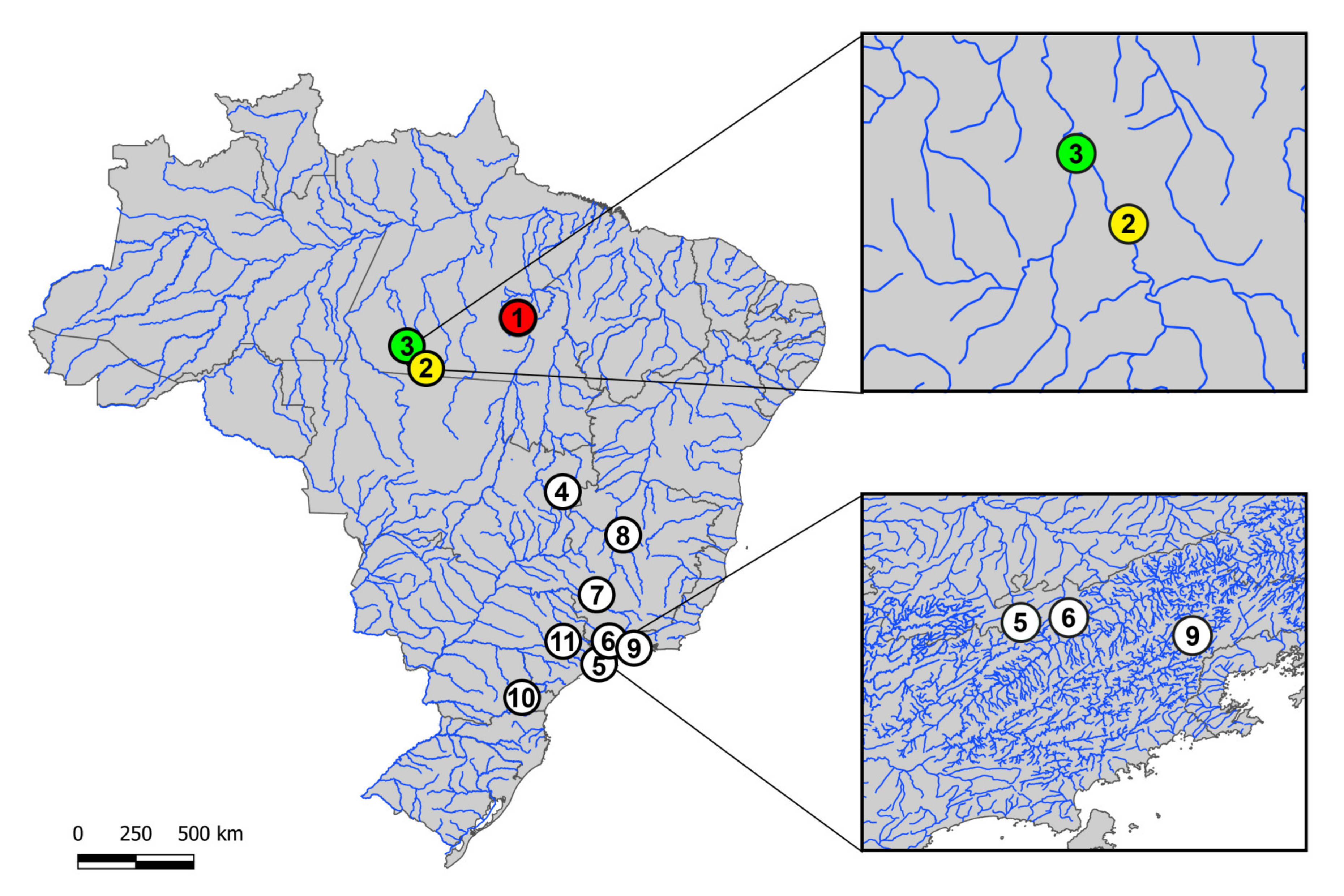
Figure 1. Brazilian collection sites of the three Harttia species cytogenetically investigated in the present study (colored circles) and the ones previously cytogenetically analyzed (white circles: data from [9,13]). 1. H. duriventris (red circle); 2. H. villasboas (yellow circle); 3. H. rondoni (3-green circle); 4. H. punctata; 5. H. gracilis; 6. H. carvalhoi; 7. H. torrenticola; 8. H. longipinna; 9. H. loricariformis; 10. H. kronei; 11. H. absaberi. The boxes highlight the non-sympatric distribution of some species.
Genes 11 01179 g001
Figure 2. Karyotypes of H. duriventris (a–c) H. villasboas (d–f) and H. rondoni (g–i) arranged by sequentially Giemsa-stained (a, d and g) and C-banded chromosomes (b, e and h) and hybridized with 5S rDNA (red) and 18S rDNA (green) probes after a double-FISH analysis (c, f, and i). Boxes depict the male sex chromosomes. Bar = 5 μm.
Genes 11 01179 g002
Figure 3. Metaphase chromosomes of males H. duriventris, H. villasboas and H. rondoni hybridized with microsatellite-containing oligonucleotides. Chromosomes were counterstained with DAPI (blue) and microsatellite probes were directly labeled with Cy3 during synthesis (red signals). The female sex chromosomes are shown in boxes. Bar = 5 µm.
Genes 11 01179 g003
Figure 4. Male metaphase plates of H. duriventris (a), H. villasboas (b) and H. rondoni (c) showing telomeric hybridization signals on both telomeres of all chromosomes. Bar = 5 μm.
Genes 11 01179 g004
Figure 5. Mitotic chromosome spreads of males H. duriventris (A–D), H. villasboas (E–H) and H. rondoni (I–L) after intraspecific CGH procedures. Male- and female-derived genomic probes were hybridized together for each species. First column (A, E and I): DAPI images (blue); Second column (B, F, and J): hybridization pattern of the male-derived probe (red); Third column (C, G, and K): hybridization pattern of the female-derived probe (green). The fourth column (D, H, and L): merged images of both genomic probes and DAPI staining. The common genomic regions for males and females are depicted in yellow. Sex chromosomes are indicated. Bar = 10 μm.
Genes 11 01179 g005
Figure 6. Comparative Genomic Hybridization (CGH) of Harttia species bearing X1X2Y sex chromosome system. Mitotic male chromosome spreads of H. villasboas (A) hybridized against male-derived genome probes of H. villasboas (B), H. punctata (C) and H. duriventris (D). Sex chromosomes are indicated and the common genomic regions for the three species are depicted in yellow on (A). Bar = 10 μm.
Genes 11 01179 g006
Figure 7. Adapted phylogenetic tree for the genus Harttia, based on the molecular-phylogenetic data generated by Covain [19]. Previous and now cytogenetic analyzed species are indicated in blue and red, respectively. The multiple X1X2Y (green boxes) and XY1Y2 (blue box) sex chromosome found in Harttia is also indicated, together with the putative proto or neo XY system (purple box). Species from northern Brazilian region are highlighted with the dotted line.
Genes 11 01179 g007
Table 1. Collection sites and samples size (N) of the species analyzed.
SpeciesLocalityN
1. H. duriventris Parauapebas River, Canaã dos Carajás-PA (Brazil) (6°30’06.5’’ S 50°02’35.3’’ W)08♀, 07♂
2. H. villasboasCuruá River, Cachoeira da Serra-PA (Brazil) (8°44’09.0’’ S 54°57’46.0’’ W)34♀, 38♂
3. H. rondoni13 de Maio River, Cachoeira da Serra-PA (Brazil) (8°38’53.0’’ S 55°01’41.0’’ W)15♀, 14♂
4. H. punctataItiquira river, Formosa—GO (Brazil) (15°19’25’’ S 47°25’26’’ W)10♀, 12♂

